

Review
doi: 10.1038/ncb3191.
Epub 2015 Jun 29.
Integration of actin dynamics and cell adhesion by a three-dimensional, mechanosensitive molecular clutch
Affiliations
- PMID: 26121555
- PMCID: PMC6300998
- DOI: 10.1038/ncb3191
Item in Clipboard
Review
Integration of actin dynamics and cell adhesion by a three-dimensional, mechanosensitive molecular clutch
Nat Cell Biol.
2015 Aug.
Abstract
During cell migration, the forces generated in the actin cytoskeleton are transmitted across transmembrane receptors to the extracellular matrix or other cells through a series of mechanosensitive, regulable protein-protein interactions termed the molecular clutch. In integrin-based focal adhesions, the proteins forming this linkage are organized into a conserved three-dimensional nano-architecture. Here we discuss how the physical interactions between the actin cytoskeleton and focal-adhesion-associated molecules mediate force transmission from the molecular clutch to the extracellular matrix.
Conflict of interest statement
COMPETING FINANCIAL INTERESTS
The authors declare no competing financial interests.
Figures
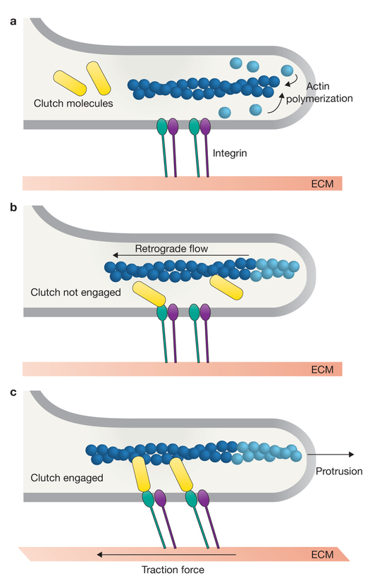
The molecular clutch hypothesis. (a) New actin monomers (light blue) are incorporated on to the barbed end of a pre-existing actin filament (dark blue) facing the leading edge of the lamellipodia. Transmembrane integrin dimers (green and purple) are bound to the extracellular matrix (ECM). (b) If the clutch (yellow) is not engaged to connect actin to the ECM, then actin polymerization results in rapid retrograde cytoskeletal flow, no net leading edge protrusion and no traction force on the ECM. (c) If the clutch is engaged, the forces generated by polymerization of the actin cytoskeleton are physically transmitted to the ECM, resulting in slowing of retrograde flow, traction force on the ECM and a net edge protrusion.
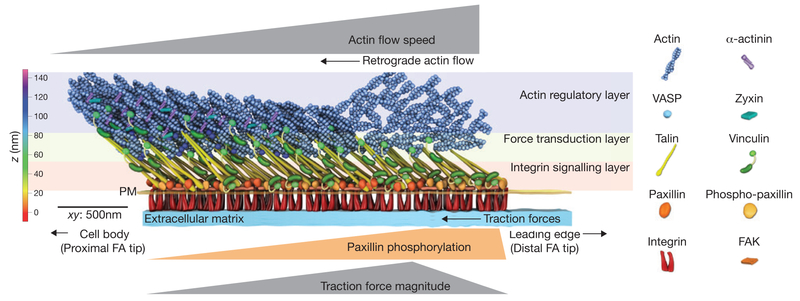
Nano-scale architecture of the focal adhesion clutch. Focal adhesions (FAs) are organized into 3D ‘nano-domains’ with unique protein compositions and mechanical signatures. The distal tip of the FA facing the leading edgeis where lamellipodial dendritic actin interacts with the FA, and contains an enrichment of phosphorylated paxillin, rapid retrograde flow and high traction forces. The proximal tip of the FA interacts with the actin stress fibre and is enriched with the actin binding proteins α-actinin, zyxin and VASP, and is characterized by slow retrograde flow and low traction forces. Additionally, proteins are stratified in the axis perpendicular to the cell plasma membrane (PM). Paxillin, FAK and the talin head domain are co-localized with integrin cytoplasmic tails near the plasma membrane in the integrin signalling layer. Actin and actin-binding proteins are localized >50 nm above the plasma membrane in the actin regulatory layer. Talin and vinculin reside in the force transduction layer that spans between the integrin signalling and actin regulatory layers. Talin is oriented with the N-terminus near the plasma membrane and the C-terminus ~30 nm higher and extended towards the FA proximal tip. The colour bar shows the vertical distance from the extracellular matrix, whereas the scale bar denotes the distance across the xy plane.
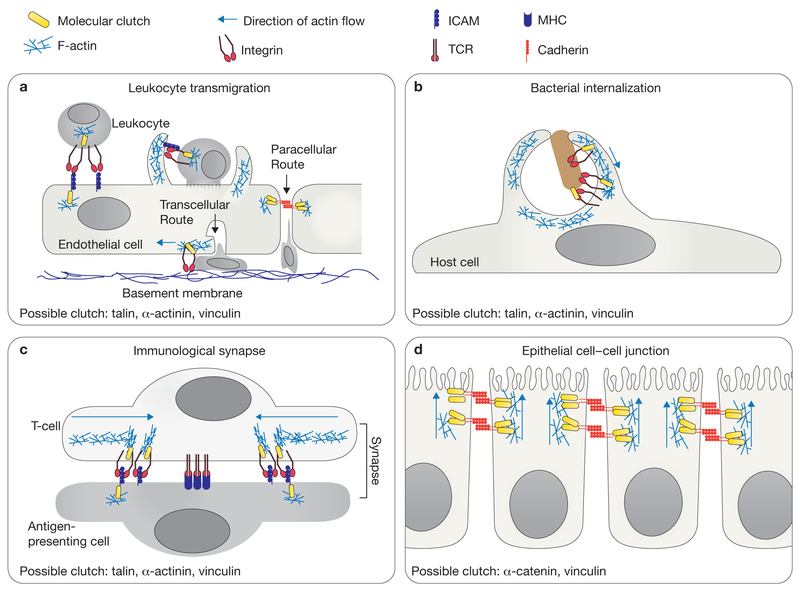
Molecular clutches may mediate diverse cell adhesive interactions. (a) During leukocyte diapedesis, initial cell–cell adhesion is mediated by the interactions of the LFA−1 integrin and its ligand ICAM−1. Paracellular migration occurs when the endothelial cells temporarily disassemble cell–cell junctions, allowing the leukocyte to migrate between two endothelial cells. Transcellular migration occurs when the leukocyte migrates through a single endothelial cell. The migrating leukocyte extends invasive protrusions into the endothelial cell, and the endothelial cell forms a transmigratory cup around the leukocyte. Following successful transmigration, the transmigratory pore is closed by integrin-dependent ventral lamellipodia to restore endothelial barrier integrity. (b) Pathogens often seek entry into host cells by co-opting the integrin or cadherin adhesion machinery. Bacteria can bind to these adhesion receptors, stimulate actin polymerization and activate clutch molecules to promote the formation of a phagocytic cup. (c) The T-cell immunological synapse requires centripetal actin flow to organize adhesion receptors into distinct domains. Rapid retrograde flow organizes and potentially activates LFA−1 integrins in the actin-rich regions. In contrast, the T-cell receptors (TCR) cluster in the actin-free centre. MHC, major histocompatibility complex. (d) Cadherins mediate cell–cell adhesion and connect indirectly to the actin cytoskeleton through β-catenin, α-catenin and vinculin. Cadherins have been observed to undergo actin-dependent basal-to-apical flow that could generate force for epithelial morphogenesis. Active polymerization of the actin cytoskeleton is depicted as a blue mesh and the direction of actin flow is indicated with a blue arrow (a–d).
References
Publication types
MeSH terms
Substances
Grants and funding
LinkOut - more resources
Full Text Sources
Other Literature Sources
