. 2015 Aug 3;125(8):3027-36.
doi: 10.1172/JCI79514. Epub 2015 Jun 29.

Flow-dependent expression of ectonucleotide tri(di)phosphohydrolase-1 and suppression of atherosclerosis

Flow-dependent expression of ectonucleotide tri(di)phosphohydrolase-1 and suppression of atherosclerosis

Yogendra Kanthi et al. J Clin Invest. .

Abstract

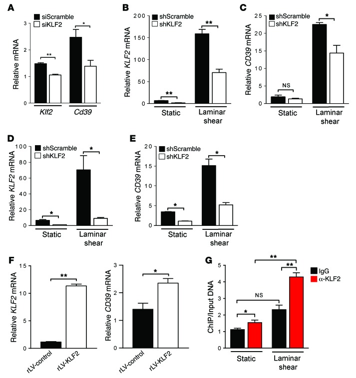
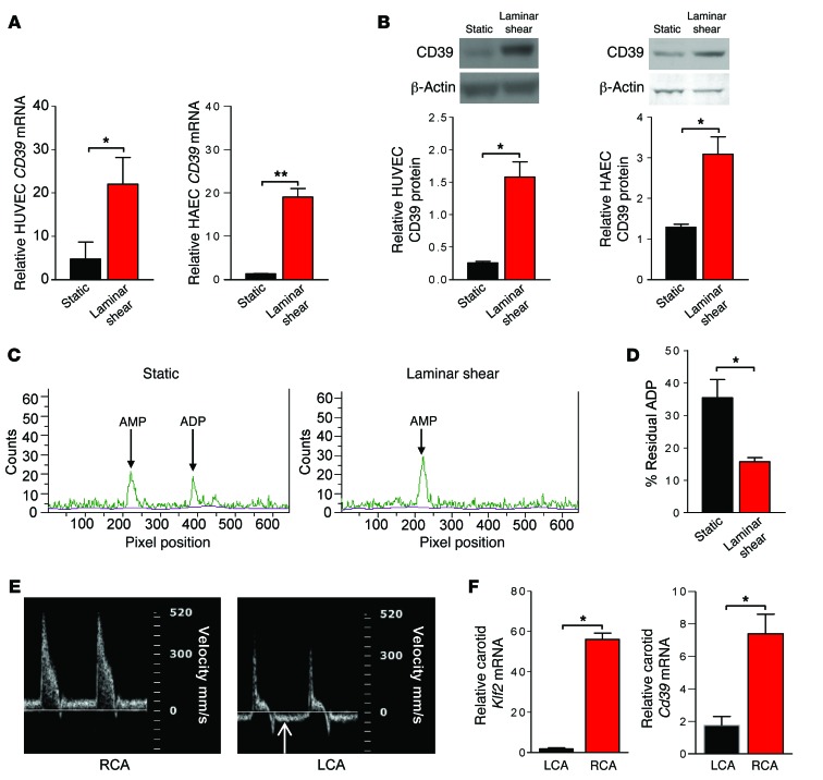
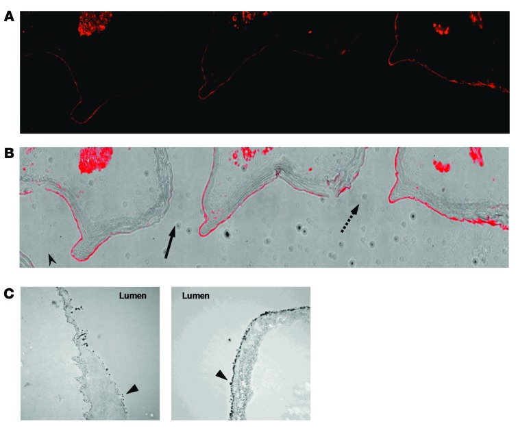
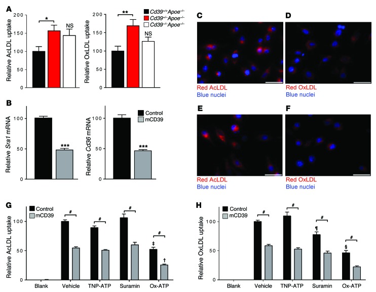
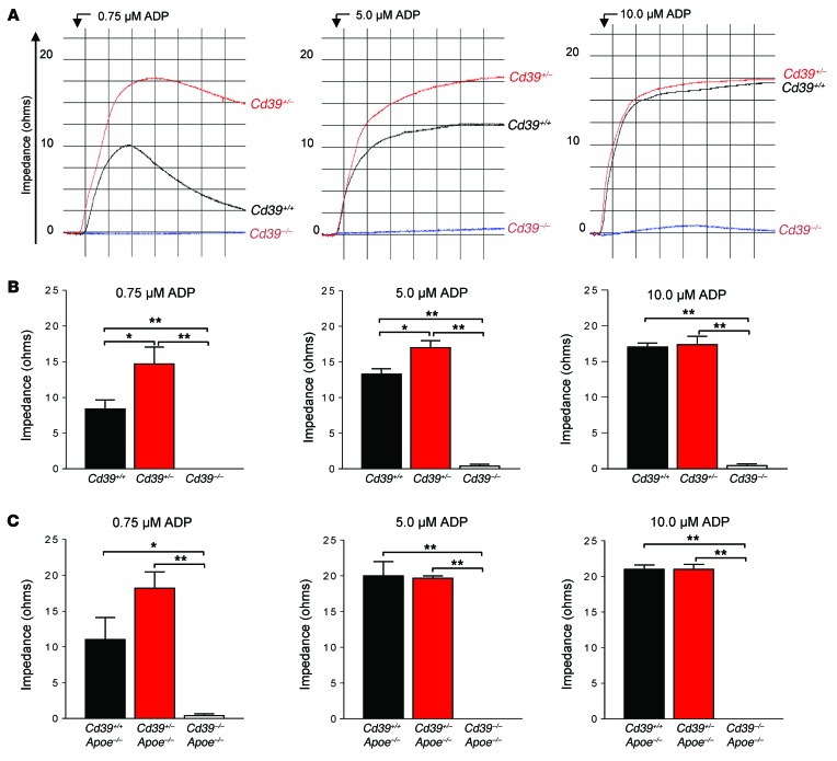
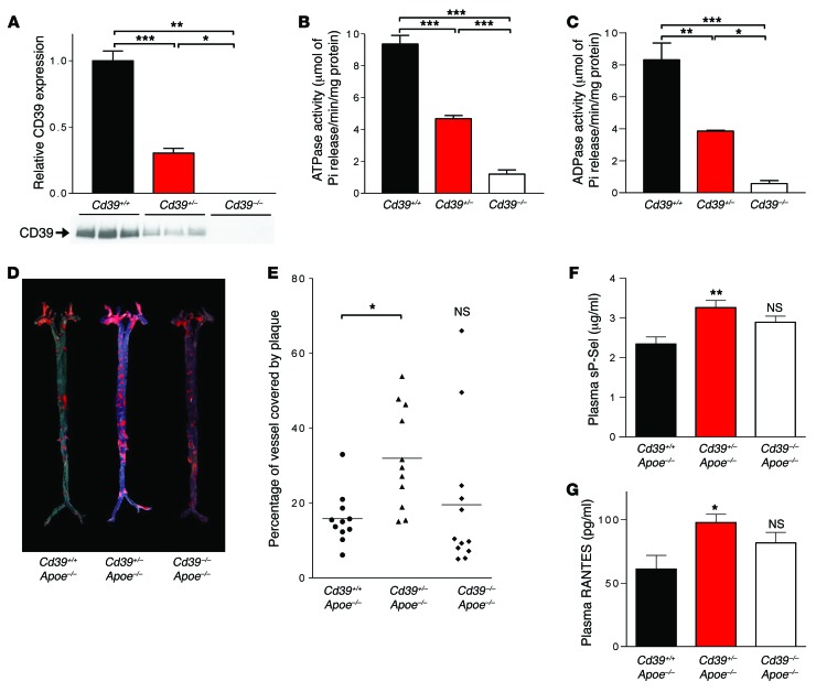
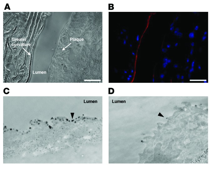
The ability of cells to detect and respond to nucleotide signals in the local microenvironment is essential for vascular homeostasis. The enzyme ectonucleotide tri(di)phosphohydrolase-1 (ENTPD1, also known as CD39) on the surface of leukocytes and endothelial cells metabolizes locally released, intravascular ATP and ADP, thereby eliminating these prothrombotic and proinflammatory stimuli. Here, we evaluated the contribution of CD39 to atherogenesis in the apolipoprotein E-deficient (ApoE-deficient) mouse model of atherosclerosis. Compared with control ApoE-deficient animals, plaque burden was markedly increased along with circulating markers of platelet activation in Cd39+/-Apoe-/- mice fed a high-fat diet. Plaque analysis revealed stark regionalization of endothelial CD39 expression and function in Apoe-/- mice, with CD39 prominently expressed in atheroprotective, stable flow regions and diminished in atheroprone areas subject to disturbed flow. In mice, disturbed flow as the result of partial carotid artery ligation rapidly suppressed endothelial CD39 expression. Moreover, unidirectional laminar shear stress induced atheroprotective CD39 expression in human endothelial cells. CD39 induction was dependent upon the vascular transcription factor Krüppel-like factor 2 (KLF2) binding near the transcriptional start site of CD39. Together, these data establish CD39 as a regionalized regulator of atherogenesis that is driven by shear stress.


Figures

Figure 7
Figure 7. Cd39 expression is regulated by KLF2 via direct interaction with the CD39 promoter.
(A) Cd39 expression is decreased by silencing Klf2 in WT bone marrow–derived macrophages using siRNA (siKLF2) versus controls (siScramble) (n = 3 per group). *P < 0.05; **P < 0.01. (BE) Silencing KLF2 with shRNA targeting KLF2 (shKLF2) decreased KLF2 gene transcript by more than 65% in static conditions and more than 65% under laminar shear stress versus controls (shScramble) in HUVECs (B) and HAECs (D) (n = 3–4 per group). *P < 0.05; **P < 0.005. KLF2 silencing abrogated laminar shear stress–induced expression of CD39 versus nonsilenced controls in HUVECs (C) and HAECs (E) (n = 3–4 per group). *P < 0.05. (F) CD39 induction by KLF2 overexpression. HUVECs transduced with a recombinant lentivirus (rLV-KLF2) to overexpress human KLF2 or vector alone (rLV-Control). Induction of KLF2 (left) and CD39 (right) transcripts were measured with quantitative RT-PCR. (n = 3 per group). *P < 0.05; **P < 0.001. (G) ChIP of HUVECs treated with static or laminar shear stress using anti-KLF2 antibody or nonspecific IgG revealed KLF2 binding to the CD39 promoter in static conditions, which was significantly enhanced under laminar shear stress conditions (n = 3 per group) *P < 0.05; **P < 0.01. Data are expressed as mean ± SEM. Student’s t test was used when comparing 2 variables. One-way ANOVA and Tukey’s multiple comparison tests were used when comparing more than 2 variables.
Figure 6
Figure 6. Laminar shear stress induces endothelial CD39 expression and nucleotidase activity in vitro and in vivo.
HUVECs and HAECs were exposed to laminar shear stress (15 dynes/cm2) for 48 hours and then assessed for (A) CD39 transcript expression, as measured by quantitative RT-PCR (n = 3–6 per group), and (B) protein expression, as measured by immunoblotting (n = 3–6 per group). Samples exposed to shear stress were compared with static controls. *P < 0.05; **P < 0.005. TLC demonstrated (C, representative plots, and D) enhanced CD39-mediated phosphohydrolysis of radiolabeled nucleotides by intact endothelial cells following laminar shear stress versus static conditions (n = 3–6 per group). *P < 0.05. Partial ligation of left common carotid artery in mice causes disturbed flow and reduced endothelial CD39 expression. Representative ultrasound demonstrating flow velocity profiles in the right (RCA) and left (LCA) common carotid artery revealing (E) disturbed flow (right) 48 hours following partial LCA ligation with diastolic flow reversal in the LCA (indicated by arrow), while contralateral RCA flow remains stable (left) (n = 3 mice). (F) Carotid endothelial expression of Klf2 and Cd39 is decreased under disturbed flow following partial carotid ligation (n = 3 mice) *P < 0.05, Student’s t test. Data are expressed as mean ± SEM.
Figure 5
Figure 5. Regional expression of CD39 in the murine aorta.
Coronal sections of a WT aorta. (A) Analysis of the WT aortic arch showed a stark regionalization of CD39 expression (red) and a consistent pattern centering on aortic branch points. (B) The same image is overlaid with bright-field imaging of the region. Original magnification, ×20. Arrowhead designates innominate artery ostium. Arrow designates left common carotid artery ostium. Dashed arrow designates left subclavian artery ostium. n = 3 mice. Transmission electron micrographs show cerium chloride staining for ATPase activity in the luminal face of the left common carotid artery branch (C, left) proximal to the carotid ostium, where disturbed flow predominates and (C, right) distal to the carotid ostium where blood flow is stable. Black cerium precipitate (denoted by arrowheads) identifies sites of ATPase activity. Original magnification, ×19,000. n = 5 mice, representative images shown.
Figure 4
Figure 4. CD39 suppresses lipid uptake in macrophages.
Peritoneal macrophages isolated from Cd39+/+ Apoe–/–, Cd39+/– ApoE–/–, and Cd39–/– Apoe–/– mice were exposed to (A, left) DiI-AcLDL or (A, right) DiI-OxLDL, and lipoprotein uptake was measured as a corollary for foam cell formation (n = 12–20 per group). *P < 0.05; **P < 0.005. The RAW 264.7 macrophage cell line was transfected to overexpress murine CD39 (mCD39) or “empty” pcDNA3. Quantitative RT-PCR was used to measure the effect CD39 has on the expression of the scavenger receptors (B, left) Sra1 and (B, right) Cd36 (n = 6 per group). ***P < 0.0005. Representative images (C and E) of pcDNA3-transfected (n = 6) and (D and F) mCD39–transfected RAW cells (n = 6) 2 hours after exposure to (C and D) DiI-AcLDL and (E and F) DiI-OxLDL. Scale bars: 20 μm. (G) DiI-AcLDL and (H) DiI-OxLDL uptake were measured in pcDNA3-transfected and mCD39-transfected RAW macrophages (n = 12–20 per group). #P < 0.001; P < 0.05 versus groups of the same genotype; P < 0.01 versus groups of the same genotype; §P < 0.01 versus vehicle and TNP-ATP of the same genotype; P < 0.05 versus vehicle and TNP-ATP of the same genotype. Data are expressed as mean ± SEM. Student’s t test was used when comparing 2 variables. One-way ANOVA and Tukey’s multiple comparison tests were used when comparing more than 2 variables.
Figure 3
Figure 3. Modulation of whole blood aggregometry by CD39.
Whole blood was drawn from Cd39+/+, Cd39+/–, and Cd39–/– mice to test for ADP sensitivity. (A) Representative aggregometry responses were taken at different concentrations of ADP stimulation (n = 3–9 per group). Summed aggregometry data are shown for (B) normolipidemic (n = 3–9 per group) and (C) hyperlipidemic Cd39+/+, Cd39+/–, and Cd39–/– mice (n = 3–9 per group). *P < 0.05; **P < 0.01. Data are expressed as mean ± SEM. One-way ANOVA and Tukey’s multiple comparison tests were used.
Figure 2
Figure 2. CD39 deficiency alters the course of atherogenesis.
The membrane protein fraction was isolated from tissue taken from Cd39+/+, Cd39+/–, and Cd39–/– mice. Isolated protein was immunoblotted to determine (A) CD39 expression and was also examined with a malachite green assay for (B) ATPase activity (n = 3–4 per group) and (C) ADPase activity (n = 3–4 mice per group). *P < 0.05; **P < 0.005; ***P < 0.001. Aortas from Cd39+/+, Cd39+/–, and Cd39–/– mice crossed to an Apoe–/– background and fed a fat-enriched diet for 16 weeks were stained with oil red O and pinned en face to quantify aortic plaque burden. (D) Representative vessels and (E) summed quantifications are shown (n = 11–12 mice per group). *P < 0.05. Circulating levels of platelet-related biomarkers (F) sP-Sel and (G) RANTES were measured in the blood of fasting Cd39+/+ Apoe–/–, Cd39+/– Apoe–/–, and Cd39–/– Apoe–/– mice via ELISA (n = 7–17 mice per group). *P < 0.05; **P < 0.01. NS, no statistical significance compared with Cd39+/+ Apoe–/–. Data are expressed as mean ± SEM. One-way ANOVA and Tukey’s multiple comparison tests were used.
Figure 1
Figure 1. CD39 function and expression over atherosclerotic plaque.
Coronal sections of an Apoe–/– aortic arch with atherosclerotic plaque are shown via (A) bright-field and (B) immunofluorescent microscopy. CD39 is shown in red and nuclei are blue (n = 3 mice). Scale bars: 25 μm. Transmission electron micrographs of cerium chloride–stained ATPase activity in Apoe–/– aorta at sites of (C) healthy and (D) atherosclerotic vascular wall. Black cerium precipitate denotes sites of ATPase activity (arrowheads). n = 3 mice, representative images shown. Original magnification, ×34,000.

References

    1. Lozano R, et al. Global and regional mortality from 235 causes of death for 20 age groups in 1990 and 2010: a systematic analysis for the Global Burden of Disease Study 2010. Lancet. 2012;380(9859):2095–2128. doi: 10.1016/S0140-6736(12)61728-0. - DOI - PMC - PubMed
    1. von Hundelshausen P, et al. RANTES deposition by platelets triggers monocyte arrest on inflamed and atherosclerotic endothelium. Circulation. 2001;103(13):1772–1777. doi: 10.1161/01.CIR.103.13.1772. - DOI - PubMed
    1. Koltsova EK, et al. Dynamic T cell-APC interactions sustain chronic inflammation in atherosclerosis. J Clin Invest. 2012;122(9):3114–3126. doi: 10.1172/JCI61758. - DOI - PMC - PubMed
    1. Dunn J, et al. Flow-dependent epigenetic DNA methylation regulates endothelial gene expression and atherosclerosis. J Clin Invest. 2014;124(7):3187–3199. doi: 10.1172/JCI74792. - DOI - PMC - PubMed
    1. Huo Y, et al. Circulating activated platelets exacerbate atherosclerosis in mice deficient in apolipoprotein E. Nat Med. 2003;9(1):61–67. - PubMed

Publication types

MeSH terms