. 2015 Jun 30:5:11840.
doi: 10.1038/srep11840.

Acceleration of protein folding by four orders of magnitude through a single amino acid substitution

Affiliations

Acceleration of protein folding by four orders of magnitude through a single amino acid substitution

Daniel J A Roderer et al. Sci Rep. .

Abstract

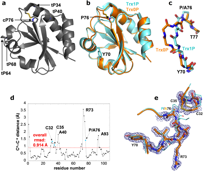
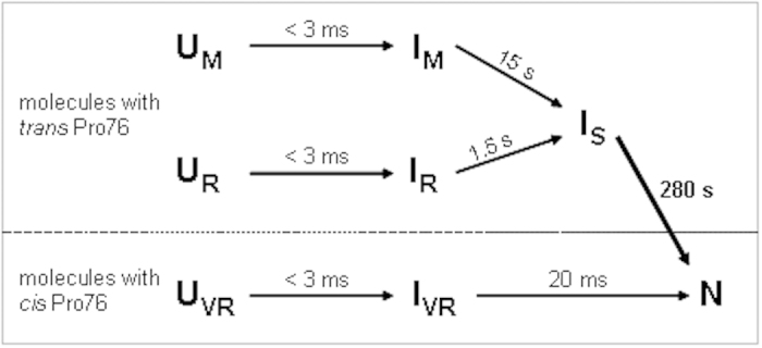
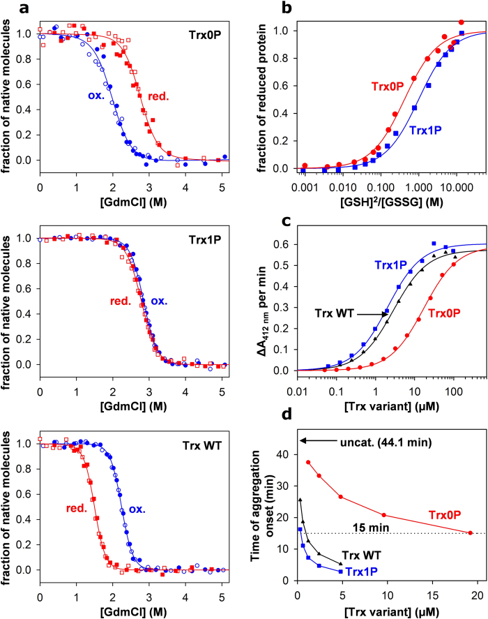
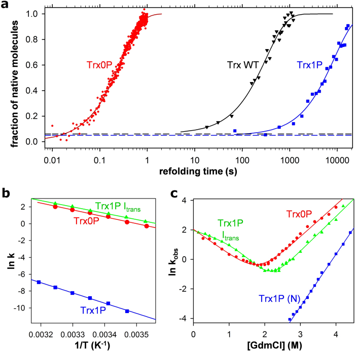
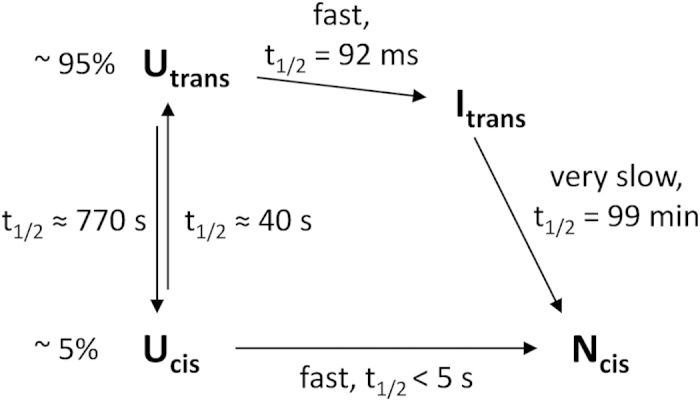
Cis prolyl peptide bonds are conserved structural elements in numerous protein families, although their formation is energetically unfavorable, intrinsically slow and often rate-limiting for folding. Here we investigate the reasons underlying the conservation of the cis proline that is diagnostic for the fold of thioredoxin-like thiol-disulfide oxidoreductases. We show that replacement of the conserved cis proline in thioredoxin by alanine can accelerate spontaneous folding to the native, thermodynamically most stable state by more than four orders of magnitude. However, the resulting trans alanine bond leads to small structural rearrangements around the active site that impair the function of thioredoxin as catalyst of electron transfer reactions by more than 100-fold. Our data provide evidence for the absence of a strong evolutionary pressure to achieve intrinsically fast folding rates, which is most likely a consequence of proline isomerases and molecular chaperones that guarantee high in vivo folding rates and yields.


Figures

Figure 1
Figure 1. Comparison of the 1.65 Å X-ray structure of oxidized Trx0P (orange) with that of oxidized Trx1P (cyan; pdb ID 4HU7).
a: Ribbon diagram of the X-ray structure of oxidized Trx WT. The side chains of the active-site cysteine pair (Cys32 and Cys35), the four trans prolines (tP34, 40, 64 and 68) and the single cis proline (cP76) are indicated in stick representation. b: Superposition of chain A of Trx0P with chain A of Trx1P, showing the conformational rearrangement in the loop segment 70–75 of Trx0P. The side chains of cis Pro76 and Tyr70 of Trx1P and the catalytic disulfide bond (Cys32–Cys35) of Trx0P are shown as stick representations. c: Comparison of the main chain segments 70–77 of Trx0P and Trx1P. The side chains of Pro76/Ala76 are also indicated. d: Plot of Cα-Cα distances of the Trx0P/Trx1P pair against the residue number. e: Stick representations of the active-site segments C32–C35 and the segments Y70–A/P76 of Trx0P (thick orange lines) and Trx1P (thin cyan lines) in atom-specific colors. Segments C32–C35 and Y70–A76 of Trx0P are surrounded by 2mFo-DFc density, contoured at 1 σ.
Figure 2
Figure 2. Proposed in vitro folding mechanism of E. coli thioredoxin, with half-lives of the individual reaction steps.
(adapted from Georgescu et al. 1998).
Figure 3
Figure 3. Stability and functional properties of Trx0P relative to Trx WT and Trx1P at 25 °C and pH 7.0.
a: GdmCl-dependent equilibrium unfolding and refolding transitions (open and closed symbols, respectively) of the oxidized (blue circles) and reduced (red squares) proteins. Transitions were followed via the CD signal at 220 nm, fitted to the two-state model of folding (solid lines) and normalized. The deduced parameters D1/2 and meq with errors from the fits are given in Table 1. b: Redox potentials of Trx0P (red circles) and Trx1P (blue squares), determined via their equilibrium constants with GSH/GSSG. The fractions of reduced protein were obtained from fluorimetric data, plotted against the [GSH]2/[GSSG] ratio and fitted according to a disulfide exchange equilibrium (equation (3), solid lines), yielding equilibrium constants of 0.42 ± 0.05 M and 0.99 ± 0.11 M, and redox potentials (Eo’) of −0.229 ± 2 mV and −0.240 ± 2 mV for Trx0P and Trx1P, respectively. The indicated errors result from fits according to equation (3). c: Kinetic analyses of Trx variants as substrates of thioredoxin reductase (TrxR). All variants show similar values of vmax, but the KM value for Trx0P (red circles) is increased 6.0 and 8.4-fold compared to that of TrxWT (black triangles) and Trx1P (blue squares), respectively (cf. Supplementary Table 3). The indicated errors are from fits according to the Michaelis Menten equation. d: The insulin reductase activity of Trx0P (red circles) is dramatically diminished relative to Trx1P and Trx WT (blue squares and black triangles, respectively). Kinetics of insulin aggregation as a result of Trx-catalyzed reduction by DTT were followed via the increase in optical density at 600 nm. The time of aggregation onset was plotted against catalyst concentration. Data were empirically fitted to a double exponential decay to identify the Trx concentration required to reduce the time of aggregation onset from 44.1 ± 0.3 min (uncatalyzed reaction) to 15 min (dashed line). The estimated error of the deduced Trx concentrations is 10% (see Supplementary Table 3).
Figure 4
Figure 4. Folding kinetics of the oxidized forms of Trx WT, Trx0P, Trx1P and its trans-Pro76 intermediate (Itrans) at 25 °C and pH 7.0.
a: Kinetics of formation of N during refolding by dilution from 4.0 to 0.2 M GdmCl. Folding of Trx0Pox (red dots) was consistent with a two-state mechanism (cf. panel C). A monoexponential fit (solid line) yielded an apparent rate constant of folding of 3.49 ± 0.01 s−1. Formation of N during refolding of Trx WT and Trx1P was recorded with interrupted refolding experiments (cf. Fig. S3). Trx WT (black triangles) showed 6% fast folding molecules (black dotted line), and 94% of the molecules reached N at a single rate of 3.10 ± 0.18 · 10−3 s−1 (solid line). Trx1P (blue squares) showed 5% fast folders (blue dotted line), and the residual 95% folded very slowly with a single rate of 1.16 ± 0.15 · 10−4 s−1. The indicated errors are standard errors from monoexponential fits. b: Arrhenius plot for folding of Trx0P (red circles), formation of Itrans from unfolded Trx1P (green triangles), and formation of native Trx1P from Itrans (blue squares), yielding activation energies of 67.6 ± 3.2, 62.4 ± 1.4 and 101.0 ± 2.8 kJ mol−1, respectively. The indicated errors are standard errors from Arrhenius fits. c: Chevron Plots showing the dependence of the apparent rate constant of unfolding/refolding (kobs) on [GdmCl] for Trx0P (red circles) and Trx1P Itrans (green triangles). Data were fitted according to a two-state model of protein folding in the case of Trx0P (solid, red line). Unfolding/refolding of Itrans was evaluated according to a three-state model with a high-energy on-pathway intermediate (solid, green line) (see legend to Table 1 for the deduced kinetic parameters). The unfolding branch of Trx1P is shown for comparison.
Figure 5
Figure 5. Minimal folding mechanism of Trx1P at pH 7.0 and 25 °C.
Potential fast phases in the dead time of stopped flow mixing on the pathway from Utrans to Itrans and from Ucis to Ncis were not considered.
Figure 6
Figure 6. Biophysical and functional properties of Trx1P Itrans compared to Trx0P and native Trx1P at 0.2 M GdmCl, 25 °C and pH 7.0.
a: Fluorescence spectra of Trx0P, native Trx1P and Trx1P Itrans. b: Reduction of Trx0P, Trx1P, and Trx1P Itrans (2 μM each; red, blue and green dots, respectively) by 20 μM DTT, fitted according to second-order kinetics (solid lines) (see Supplementary Table 3 for the deduced rate constants and errors). Reduction of unfolded Trx1P with 100 μM DTT (black dots) revealed a rate constant of 10.3 ± 1.48 M−1 s−1) and was thus more than 100-fold slower than reduction of native Trx1P or Itrans (cf. Supplementary Table 3). c: Kinetic analysis documenting the properties of Trx0P (red circles), Trx1P (blue squares) and Trx1P Itrans (green triangles) as substrate of TrxR in the presence of 20 mM GdmCl at pH 8.0. Similar to Trx0P, Itrans showed an about 10-fold increase in KM relative to that of Trx1P, but its catalytic parameters could not be determined accurately due to unspecific aggregation at higher concentrations (cf. Supplementary Table 3 for the parameters and errors deduced from Michaelis Menten fits). d: Itrans has a compact tertiary structure with a buried trans Ile75-Pro76 peptide bond that is inaccessible to catalysis by the PPIase trigger factor. Formation of native Trx1P from Itrans (90 μM) in the absence (black circles) and presence (red squares) of excess trigger factor (150 μM) was monitored by the increase in TrxR substrate activity and fitted monoexponentially (solid lines). Inset: Size exclusion chromatography on Superdex 75 of Itrans, native Trx1P and native Trx0P (oxidized forms), showing that Itrans has same hydrodynamic volume as native Trx1P. The retention time of all proteins at ~26.5 min is about ¼ of the half-life of Itrans folding (99 min), guaranteeing that only a minor fraction of Itrans reacted to N during the chromatography. Retention times of molecular mass standard proteins (in kDa) are indicated on the top.
Figure 7
Figure 7. Energy diagrams of folding of Trx0P and Trx1P.

References

    1. Schmid F. X. Prolyl isomerase: enzymatic catalysis of slow protein-folding reactions. Annual review of biophysics and biomolecular structure 22, 123–142, 10.1146/annurev.bb.22.060193.001011 (1993). - DOI - PubMed
    1. Jabs A., Weiss M. S. & Hilgenfeld R. Non-proline cis peptide bonds in proteins. Journal of molecular biology 286, 291–304, 10.1006/jmbi.1998.2459 (1999). - DOI - PubMed
    1. Reimer U. et al. Side-chain effects on peptidyl-prolyl cis/trans isomerisation. J Mol Biol 279, 449–460, 10.1006/jmbi.1998.1770 (1998). - DOI - PubMed
    1. Schmidpeter P. A., Koch J. R. & Schmid F. X. Control of protein function by prolyl isomerization. Biochimica et biophysica acta, 10.1016/j.bbagen.2014.12.019 (2014). - DOI - PubMed
    1. Susanti D. et al. Thioredoxin targets fundamental processes in a methane-producing archaeon, Methanocaldococcus jannaschii. Proceedings of the National Academy of Sciences of the United States of America 111, 2608–2613, 10.1073/pnas.1324240111 (2014). - DOI - PMC - PubMed

LinkOut - more resources