Review
. 2015 Aug:74:125-38.
doi: 10.1016/j.yhbeh.2015.06.010. Epub 2015 Jun 27.

Estrogen receptors in the central nervous system and their implication for dopamine-dependent cognition in females

Affiliations
Review

Estrogen receptors in the central nervous system and their implication for dopamine-dependent cognition in females

Anne Almey et al. Horm Behav. 2015 Aug.

Abstract

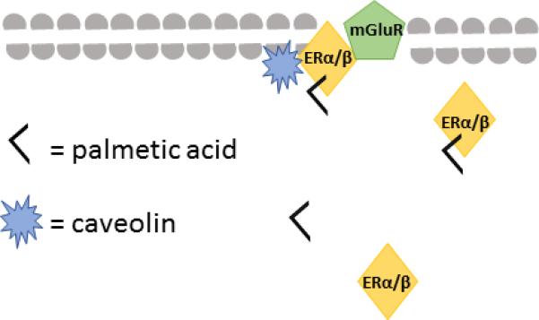
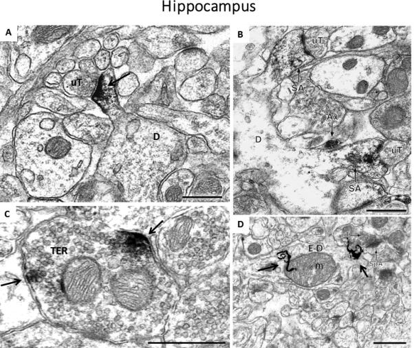
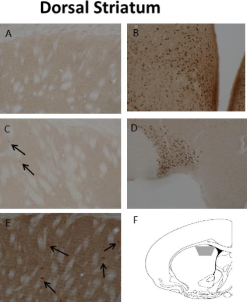
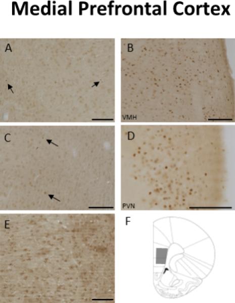
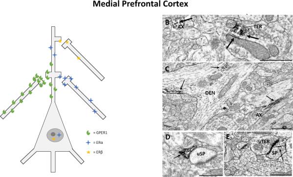
This article is part of a Special Issue "Estradiol and cognition". Over the past 30 years, research has demonstrated that estrogens not only are important for female reproduction, but also play a role in a diverse array of cognitive functions. Originally, estrogens were thought to have only one receptor, localized exclusively to the cytoplasm and nucleus of cells. However, it is now known that there are at least three estrogen receptors (ERs): ERα, ERβ and G-protein coupled ER1 (GPER1). In addition to being localized to nuclei, ERα and ERβ are localized to the cell membrane, and GPER1 is also observed at the cell membrane. The mechanism through which ERs are associated with the membrane remains unclear, but palmitoylation of receptors and associations between ERs and caveolin are implicated in membrane association. ERα and ERβ are mostly observed in the nucleus using light microscopy unless they are particularly abundant. However, electron microscopy has revealed that ERs are also found at the membrane in complimentary distributions in multiple brain regions, many of which are innervated by dopamine inputs and were previously thought to contain few ERs. In particular, membrane-associated ERs are observed in the prefrontal cortex, dorsal striatum, nucleus accumbens, and hippocampus, all of which are involved in learning and memory. These findings provide a mechanism for the rapid effects of estrogens in these regions. The effects of estrogens on dopamine-dependent cognition likely result from binding at both nuclear and membrane-associated ERs, so elucidating the localization of membrane-associated ERs helps provide a more complete understanding of the cognitive effects of these hormones.

Keywords: Dopamine; Electron microscopy; Estrogen receptor alpha; Estrogen receptor beta; G protein coupled estrogen receptor 1.


Figures

Fig 1
Fig 1
Schematic representation of how ERα or ERβ may be bound to the neuronal membrane via associations with caveolin following palmitoylation of the receptor.
Fig 2
Fig 2
Images of labeling for estrogen receptors and estradiol in the hippocampus of female rats. A) Immunoperoxidase labeling for ERβ in a dendritic spine (D) is contacted by an unlabeled terminal (uT) in CA1 stratum radiatum, B) Immunoperoxidase labeling for ERα is found in two dendritic spines identifiable by the presence of spine apparati (SA), which arise from the same dendrite (D). Both labeled spines are contacted by unlabeled terminals (uT), and an ERα-labeled axon (Ax) is found nearby, C) Clusters of immunoperoxidase labeling for GPER1 are found in small synaptic vesicles near the plasma membrane of a terminal, D) Autoradiographic silver grains (black squiggly lines) denoting 125I-estradiol binding in stratum radiatum of the CA1 region of the hippocampus in a dendrite (E-D) overlying a mitochondrion (m). Black arrows = immunoperoxidase labeling/radioactive marker, Scale Bar = 500nm. The image in A) is taken from: Milner, T.A., Ayoola, K., Drake, C.T., Herrick, S.P., Tabori, N.E., et al. 2005. Ultrastructural localization of estrogen receptor beta immunoreactivity in the rat hippocampal formation. J. Comp. Neurol. 491, 81-95, B) is taken from: Milner, T.A., McEwen, B.S., Hayashi, S., Li, C.J., Reagan, L.P., Alves, S.E. 2001. Ultrastructural evidence that hippocampal alpha estrogen receptors are located at extranuclear sites. J. Comp. Neurol. 429, 355-71., C) is taken from: Waters, E.M., Thompson,, L.I., Paterl, P., Gonzalez, A.D., Ye, H., Filardo, E.J., Clegg, D.J., Goreka, J., Akama, K.T., McEwen, B.S., Milner, T.A. 2015. G-Protein-Coupled Estrogen Receptor 1 Is Anatomically Positioned to Modulate Synaptic Plasticity in the Mouse Hippocampus. J. Neurosci. 35., D) is taken from: Milner, T.A., Lubbers, L.S., Alves, S.E., McEwen, B.S. 2008. Nuclear and extranuclear estrogen binding sites in the rat forebrain and autonomic medullary areas. Endocrinology 149, 3306-12.
Fig 3
Fig 3
Light microscopic localization of ERs in the dSTR. A) Neither nuclear nor extranuclear ERα-IR is detected. B) Dense nuclear ERα-IR in the ventromedial and arcuate nuclei of the hypothalamus. C) No extranuclear ERβ-IR is detected; however, rarely a nucleus with ERβ-IR (arrow) is detected. D) Dense nuclear ERβ-IR in the supraoptic nucleus. E) Dense extranuclear GPER-1-IR is detected in the neuropil; moreover, several cells with GPER-1-IR (arrows) are seen. F) A coronal schematic of the striatum [atlas level 14; AP +1.00mm from bregma (Paxinos and Watson, 1998)] indicated the region (grey trapezoid) analysed by EM. Images taken from Almey, A., Filardo, E.J., Milner, T.A., Brake, W.G. 2012. Estrogen receptors are found in glia and at extranuclear neuronal sites in the dorsal striatum of female rats: evidence for cholinergic but not dopaminergic colocalization. Endocrinology 153, 5373-83.
Fig 4
Fig 4
A) Representation of estrogen receptor localization within pre- and post-synaptic profiles in the dorsal striatum. Although estrogens are known to increase dopamine release in this area, almost no estrogen receptors were observed on dopamine terminals. Thus, estrogens are likely to affect dopamine release via changes in presynaptic transmission of GABA or cholinergic neurons. B) ERα-immunoreactivity (IR) in a dendritic spine (SP) that is contacted by an unlabeled axon terminal (uTER), and an axon terminal (TER) that forms an asymmetric synapses with an unlabeled dendritic spine (uSP); C) ERα-IR in a dendritic shaft (DEN) where it is affiliated with the plasma membrane and a mitochondrion (mit), and in a glial process (GL); D) ERβ-IR in an axon terminal where it is associated with the plasma membrane and with a mitochondrion (mit) (TER), and an unmyelinated axon (AX); E) GPER1-IR is found in Golgi bodies (Golgi) in a soma (SOM). Black arrow, immunoperoxidase labeling for estrogen receptors; Scale Bar, 500nm. Images taken from Almey, A., Filardo, E.J., Milner, T.A., Brake, W.G. 2012. Estrogen receptors are found in glia and at extranuclear neuronal sites in the dorsal striatum of female rats: evidence for cholinergic but not dopaminergic colocalization. Endocrinology 153, 5373-83.
Fig 5
Fig 5
Light microscopic examination of ER localization in the mPFC. A) No extranuclear immunoreactivity (IR) for ERα, but there were very low levels of nuclear ERα-IR (arrows), B) Dense ERα-IR in the ventromedial hypothalamus, C) No extranuclar ERβ-IR was observed in the mPFC, but a couple of labeled nuclei were observed (arrows), D) Dense nuclear ERβ-IR in the hypothalamus, E) Dense extranuclear GPER1-IR is detected in the neuropil, F) A coronal schematic depicting the area of the mPFC (grey trapezoid) analyzed by electron microscopy. Scale Bar, 100μm. Taken from Almey, A., Cannell, E., Bertram, K., Filardo, E., Milner, T.A., Brake, W.G.. 2014. Medial prefrontal cortical estradiol rapidly alters memory system bias in female rats: ultrastructural analysis reveals membrane-associated estrogen receptors as potential mediators. Endocrinology 155, 4422-32.
Fig 6
Fig 6
A) Representation of estrogen receptor localization to pre- and post-synaptic profiles in the prefrontal cortex. Estrogen receptors were most commonly localized to axons and terminals in the prefronal cortex, depicted alongside the pyramidal neuron, and were also observed in dendrites and dendritic spines at lower levels, depicted on the apical dendrite of the pyramidal neuron. Low levels of nuclear labelling for ERα and ERβ were observed via light microscopy. B) ERα in an axon (AX) and in a terminal (TER), where immunoreactivity (IR) is observed at small synaptic vesicles and on the membrane of a mitochondrion (mit), C) GPER1-IR in an axon (AX) and in a dendrite (DEN),where it is associated with microtubules and the cell membrane, D) ERβ-IR in a glial cell (GL) that is in apposition to an unlabeled dendritic spine (uSP), E) ERβ-IR in a dendritic spine (SP) that forms an asymmetrical synapse with an unlabeled terminal (uTER). Black arrow, immunoperoxidase labeling for estrogen receptors; Scale Bar, 500nm. Taken from Almey, A., Cannell, E., Bertram, K., Filardo, E., Milner, T.A., Brake, W.G.. 2014. Medial prefrontal cortical estradiol rapidly alters memory system bias in female rats: ultrastructural analysis reveals membrane-associated estrogen receptors as potential mediators. Endocrinology 155, 4422-32.
Fig 7
Fig 7
A) Representation of estrogen receptor localization in pre- and post-synaptic regions in the nucleus accumbens. There are low levels of estrogen receptors associated with dopaminergic terminals, so estrogens could affect dopamine transmission directly by binding at these receptors. Additionally, a moderate proportion of ERα and GPER1 are observed in GABAergic terminals and dendrites; estrogens could indirectly affect dopamine release through pre- or post-synaptic changes in GABAergic transmission. B) ERα-immunoreactivity (IR) associated with small synaptic vesicles and a mitochondrion (mit) in an axon terminal (TER) and in an axon (AX), C) GPER1-IR in a large dendrite, where it is associated with microtubules, D) ERβ-IR in two adjacent axons. Black arrow, immunoperoxidase labeling for estrogen receptors; Scale Bar, 500nm.

References

    1. Adams MM, Fink SE, Shah RA, Janssen WG, Hayashi S, et al. Estrogen and aging affect the subcellular distribution of estrogen receptor-alpha in the hippocampus of female rats. J. Neurosci. 2002;22:3608–14. - PMC - PubMed
    1. Akama KT, Thompson LI, Milner TA, McEwen BS. Post-synaptic density-95 (PSD-95) binding capacity of G-protein-coupled receptor 30 (GPR30), an estrogen receptor that can be identified in hippocampal dendritic spines. J. Bio. Chem. 2013;288:6438–50. - PMC - PubMed
    1. Akhondzadeh S, Nejatisafa AA, Amini H, Mohammadi MR, Larijani B, et al. Adjunctive estrogen treatment in women with chronic schizophrenia: a double-blind, randomized, and placebo-controlled trial. Prog. Neuropsychopharmacol. Biol. Psychiatry. 2003;27:1007–12. - PubMed
    1. Almey A, Cannell E, Bertram K, Filardo E, Milner TA, Brake WG. Medial prefrontal cortical estradiol rapidly alters memory system bias in female rats: ultrastructural analysis reveals membrane-associated estrogen receptors as potential mediators. Endocrinology. 2014;155:4422–32. - PMC - PubMed
    1. Almey A, Filardo EJ, Milner TA, Brake WG. Estrogen receptors are found in glia and at extranuclear neuronal sites in the dorsal striatum of female rats: evidence for cholinergic but not dopaminergic colocalization. Endocrinology. 2012;153:5373–83. - PMC - PubMed

Publication types