

Subcortical brain alterations in major depressive disorder: findings from the ENIGMA Major Depressive Disorder working group
- PMID: 26122586
- PMCID: PMC4879183
- DOI: 10.1038/mp.2015.69
Subcortical brain alterations in major depressive disorder: findings from the ENIGMA Major Depressive Disorder working group
Abstract
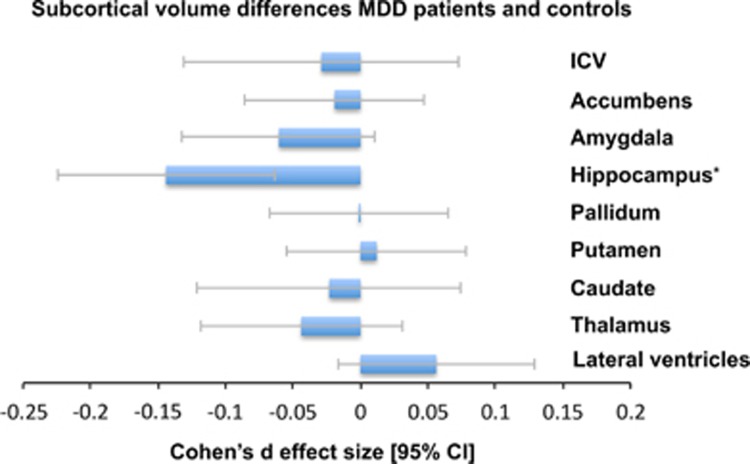
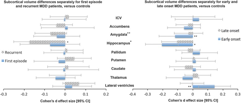
The pattern of structural brain alterations associated with major depressive disorder (MDD) remains unresolved. This is in part due to small sample sizes of neuroimaging studies resulting in limited statistical power, disease heterogeneity and the complex interactions between clinical characteristics and brain morphology. To address this, we meta-analyzed three-dimensional brain magnetic resonance imaging data from 1728 MDD patients and 7199 controls from 15 research samples worldwide, to identify subcortical brain volumes that robustly discriminate MDD patients from healthy controls. Relative to controls, patients had significantly lower hippocampal volumes (Cohen's d=-0.14, % difference=-1.24). This effect was driven by patients with recurrent MDD (Cohen's d=-0.17, % difference=-1.44), and we detected no differences between first episode patients and controls. Age of onset ⩽21 was associated with a smaller hippocampus (Cohen's d=-0.20, % difference=-1.85) and a trend toward smaller amygdala (Cohen's d=-0.11, % difference=-1.23) and larger lateral ventricles (Cohen's d=0.12, % difference=5.11). Symptom severity at study inclusion was not associated with any regional brain volumes. Sample characteristics such as mean age, proportion of antidepressant users and proportion of remitted patients, and methodological characteristics did not significantly moderate alterations in brain volumes in MDD. Samples with a higher proportion of antipsychotic medication users showed larger caudate volumes in MDD patients compared with controls. This currently largest worldwide effort to identify subcortical brain alterations showed robust smaller hippocampal volumes in MDD patients, moderated by age of onset and first episode versus recurrent episode status.
Figures
Comment in
-
The volumes of subcortical regions in depressed and healthy individuals are strikingly similar: a reinterpretation of the results by Schmaal et al.Mol Psychiatry. 2016 Jun;21(6):724-5. doi: 10.1038/mp.2015.199. Epub 2015 Dec 15. Mol Psychiatry. 2016. PMID: 26666203 Free PMC article. No abstract available.
-
Size matters; but so does what you do with it!Mol Psychiatry. 2016 Jun;21(6):725-6. doi: 10.1038/mp.2015.200. Epub 2016 Jan 5. Mol Psychiatry. 2016. PMID: 26728567 No abstract available.
-
Response to Dr Fried & Dr Kievit, and Dr Malhi et al.Mol Psychiatry. 2016 Jun;21(6):726-8. doi: 10.1038/mp.2016.9. Epub 2016 Feb 23. Mol Psychiatry. 2016. PMID: 26903270 Free PMC article. No abstract available.
References
-
- Murray CJL, Vos T, Lozano R, Naghavi M, Flaxman AD, Michaud C et al. Disability-adjusted life years (DALYs) for 291 diseases and injuries in 21 regions, 1990-2010: a systematic analysis for the Global Burden of Disease Study 2010. Lancet 2012; 380: 2197–2223. - PubMed
-
- Arnone D, McIntosh AM, Ebmeier KP, Munafò MR, Anderson IM. Magnetic resonance imaging studies in unipolar depression: systematic review and meta-regression analyses. Eur Neuropsychopharmacol 2012; 22: 1–16. - PubMed
-
- Lorenzetti V, Allen NB, Fornito A, Yücel M. Structural brain abnormalities in major depressive disorder: a selective review of recent MRI studies. J Affect Disord 2009; 117: 1–17. - PubMed
Publication types
MeSH terms
Grants and funding
LinkOut - more resources
Full Text Sources
Other Literature Sources
Medical
Miscellaneous
