Review
. 2015 Jul;16(7):406-16.
doi: 10.1038/nrm4009.

Specifying and protecting germ cell fate

Affiliations
Review

Specifying and protecting germ cell fate

Susan Strome et al. Nat Rev Mol Cell Biol. 2015 Jul.

Abstract

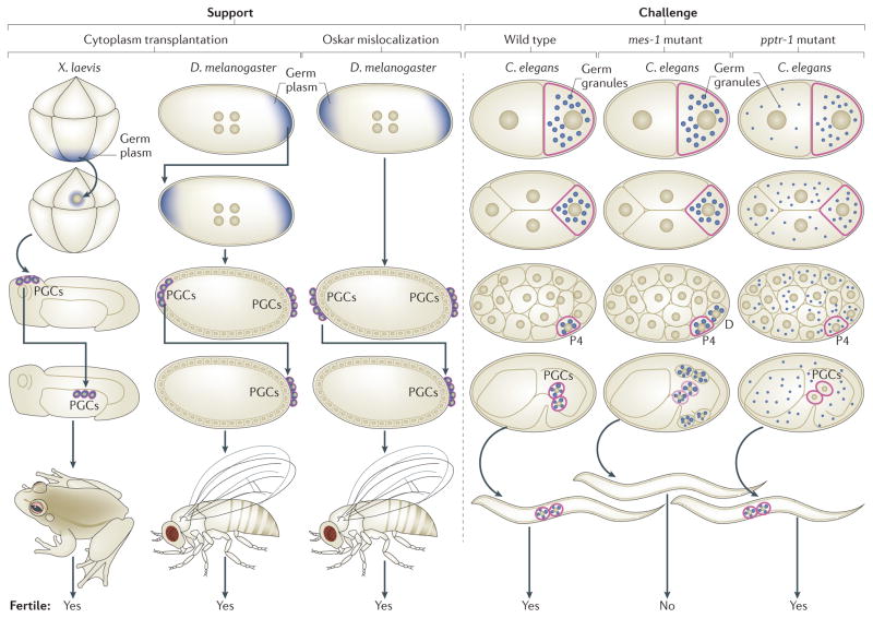
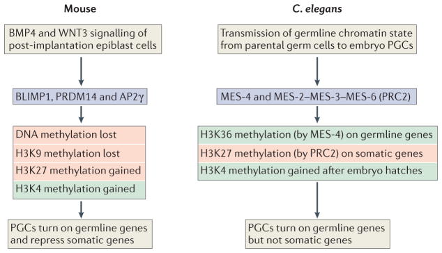
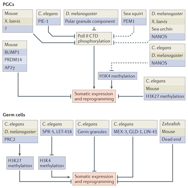
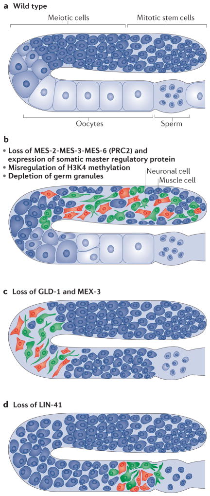
Germ cells are the special cells in the body that undergo meiosis to generate gametes and subsequently entire new organisms after fertilization, a process that continues generation after generation. Recent studies have expanded our understanding of the factors and mechanisms that specify germ cell fate, including the partitioning of maternally supplied 'germ plasm', inheritance of epigenetic memory and expression of transcription factors crucial for primordial germ cell (PGC) development. Even after PGCs are specified, germline fate is labile and thus requires protective mechanisms, such as global transcriptional repression, chromatin state alteration and translation of only germline-appropriate transcripts. Findings from diverse species continue to provide insights into the shared and divergent needs of these special reproductive cells.


Conflict of interest statement

Competing interests statement

The authors declare no competing interests.

Figures

Figure 1
Figure 1. Findings that support or challenge the notion that germ granules are ‘determinants’ of PGC fate
Germline cells are outlined in red. Support: cytoplasm-transfer experiments in Xenopus laevis and Drosophila melanogaster demonstrated that the cytoplasm containing germ granules (germ plasm; shown in blue) can cause formation of primordial gem cells (PGCs) at ectopic sites. Those ectopic PGCs were functional and able to generate a fertile germline after they were transferred to the normal PGC location in embryos. Mislocalization of the germ-granule component Oskar to the wrong (anterior) end of D. melanogaster embryos caused induction of anterior PGCs that, after transfer to the normal PGC location, were functional and capable of generating a fertile germline. Challenges: in Caenorhabditis elegans maternal effect sterile 1 (mes 1)-mutant embryos, germ granules (shown in blue) are mis-segregated to both the PGC (P4) and its somatic sister cell (D). Despite containing germ granules, both cells develop as muscle (the normal fate of D) instead of germline (the normal fate of the PGC). In C. elegans protein phosphatase 2A regulatory subunit 1 (pptr 1)-mutant embryos, maternal germ granules do not become enriched in embryonic PGCs. The PGCs nevertheless turn on a germline programme, including zygotic synthesis of germ granules, and can develop into a fertile germline.
Figure 2
Figure 2. Chromatin regulation in the PGCs of mice and C. elegans
In mice, bone morphogenetic protein 4 (BMP4) signalling from extra-embryonic tissue causes a small cluster of WNT3-primed post-implantation epiblast cells to express the transcription factors BLIMP1 (also known as PRDM1), PR domain zinc-finger protein 14 (PRDM14) and activating enhancer-binding protein 2γ (AP2γ). This set of three transcription factors is necessary and sufficient to cause the indicated chromatin changes, turn on germline genes and repress somatic genes in the newly induced primordial germ cells (PGCs). In Caenorhabditis elegans, the memories of germline gene expression and repression are transmitted from parent germ cells to progeny germ cells by maternal effect sterile 4 (MES-4) and by MES-2 MES-3 MES-6, the worm version of polycomb repressive complex 2 (PRC2), respectively. MES-4 achieves this via methylation of Lys36 on histone H3 (H3K36), and MES-2 MES-3 MES-6 operates via methylation of H3K27 PGCs are kept in a relatively quiescent chromatin state, lacking H3K4 methylation, until after the embryo hatches and starts feeding. Chromatin marks associated with gene expression are in green, and those associated with repression are in red.
Figure 3
Figure 3. Diverse factors and mechanisms that repress expression of somatic genes and protect germ cells from reprogramming towards somatic cells
In embryos, a common target of regulation in the primordial germ cells (PGCs) is the carboxy-terminal domain (CTD) of RNA Polymerase II (Pol II). Phosphorylation of the Pol II CTD is required for transcription elongation, and organisms use diverse methods to prevent CTD phosphorylation in PGCs. Inhibition of Pol II keeps early PGCs transcriptionally silent. At later stages, repression is more selective, to keep somatic genes switched off while germline genes are being expressed. In adults, this selective repression is achieved at the level of chromatin regulation by histone modifiers and remodellers (for example, polycomb repressive complex 2 (PRC2) promotes methylation of Lys27 on histone H3 (H3K27) and gene repression, and suppressor of presenilin defect 5 (SPR-5) and lethal 418 (LET-418) remove H3K4 methylation, which is associated with gene activation), as well as at the level of translational regulation by germ granules, translational regulators (for example, muscle excess 3 (MEX-3), defective in germline development 1 (GLD-1) and abnormal cell lineage 41 (LIN-41)) and proteins that stabilize germline-specific RNA (such as dead end). C. elegans, Caenorhabditis elegans; D. melanogaster, Drosophila melanogaster; PEM1, posterior end mark 1; PIE-1, pharynx intestine in excess 1; PRDM14, PR domain zinc-finger protein 14; X. laevis, Xenopus laevis.
Figure 4
Figure 4. Conditions that cause adult germ cells to reprogramme towards somatic cells in C. elegans
a | Wild-type Caenorhabditis elegans germlines contain a progression of mitotic stem cells, meiotic cells, oocytes and sperm. b | Somatic cell types most commonly observed after germ soma reprogramming are neuronal (shown in green) and muscle (shown in red). Loss of the complex of maternal effect sterile 2 (MES-2), MES-3 and MES-6 (the worm version of polycomb repressive complex 2 (PRC2)), mis-regulation of methylation of Lys4 on histone H3 (H3K4), and depletion of germ granules cause mitotic stem cells to reprogramme towards somatic lineages. c | Loss of defective in germline development 1 (GLD-1) and muscle excess 3 (MEX-3) causes meiotic cells to reprogramme towards soma. d | Loss of abnormal cell lineage 41 (LIN-41) causes oocytes to reprogramme towards somatic cells, owing to the embryonic programme being turned on prematurely.

References

    1. Seydoux G, Braun RE. Pathway to totipotency: lessons from germ cells. Cell. 2006;127:891–904. - PubMed
    1. Hegner RW. Effects of removing the germ-cell determinants from the eggs of some chrysomelid beetles. Biol Bull. 1908;16:19–26.
    1. Eddy EM. Germ plasm and the differentiation of the germ cell line. Int Rev Cytol. 1975;43:229–280. - PubMed
    1. Strome S, Wood WB. Generation of asymmetry and segregation of germ-line granules in early C elegans embryos. Cell. 1983;35:15–25. - PubMed
    1. Hay B, Jan LY, Jan YN. A protein component of Drosophila polar granules is encoded by vasa and has extensive sequence similarity to ATP-dependent helicases. Cell. 1988;55:577–587. - PubMed