

Comment
doi: 10.1038/mt.2015.97.
Macrophage-Derived IGF-1 Is a Potent Coordinator of Myogenesis and Inflammation in Regenerating Muscle
Affiliations
- PMID: 26122828
- PMCID: PMC4817792
- DOI: 10.1038/mt.2015.97
Item in Clipboard
Comment
Macrophage-Derived IGF-1 Is a Potent Coordinator of Myogenesis and Inflammation in Regenerating Muscle
Mol Ther.
2015 Jul.
No abstract available
Figures
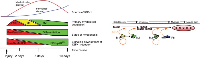
Schematic of temporal relationships between stages of myogenesis, inflammation, and IGF-1 release following muscle injury. Acute injury induces an initial invasion of muscle by neutrophils (PMNs), proinflammatory M1 macrophages, and anti-inflammatory M2 macrophages that coincides with increased expression of insulin-like growth factor-1 (IGF-1) in myeloid cells. Myeloid cell–derived IGF-1 (solid orange arrows) can drive proliferation of activated satellite cells through a pathway mediated by Ras/Raf/MAP kinase signaling in muscle. Myeloid cell–derived IGF-1 also promotes a shift in macrophage phenotype from an M1-biased to an M2-biased population. At approximately 5 days after injury, myeloid cell numbers decline and there is a shift in the primary site of IGF-1 expression from the myeloid compartment to nonmyeloid, nonmuscle cells that are probably fibroblasts (Fb). Muscle cell differentiation to myotubes and subsequent growth to muscle fibers may be increased by IGF-1 produced by fibroblasts or M2 macrophages (dashed orange arrows). IGF-1-mediated muscle differentiation is signaled via a PI3-kinase/p70S6k path during later stages of muscle regeneration.
Comment on
-
Monocyte/Macrophage-derived IGF-1 Orchestrates Murine Skeletal Muscle Regeneration and Modulates Autocrine Polarization.Mol Ther. 2015 Jul;23(7):1189-1200. doi: 10.1038/mt.2015.66. Epub 2015 Apr 21. Mol Ther. 2015. PMID: 25896247 Free PMC article.
References
-
- Powell-Braxton, L, Hollingshead, P, Warburton, C, Dowd, M, Pitts-Meek, S, Dalton, D et al. (1993). IGF-I is required for normal embryonic growth in mice. Genes Dev 7: 2609–2617. - PubMed
-
- Chargé, SB and Rudnicki, MA (2004). Cellular and molecular regulation of muscle regeneration. Physiol Rev 84: 209–238. - PubMed
Publication types
MeSH terms
Substances
Grants and funding
LinkOut - more resources
Full Text Sources
Other Literature Sources
Miscellaneous
