. 2015 Jul;146(1):65-77.
doi: 10.1085/jgp.201411336.

Transient receptor potential melastatin 3 is a phosphoinositide-dependent ion channel

Affiliations

Transient receptor potential melastatin 3 is a phosphoinositide-dependent ion channel

Doreen Badheka et al. J Gen Physiol. 2015 Jul.

Abstract

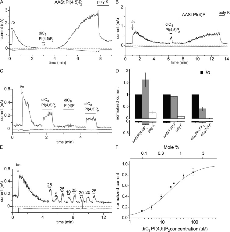
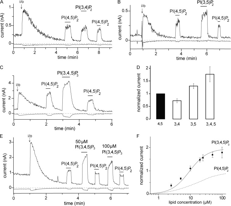
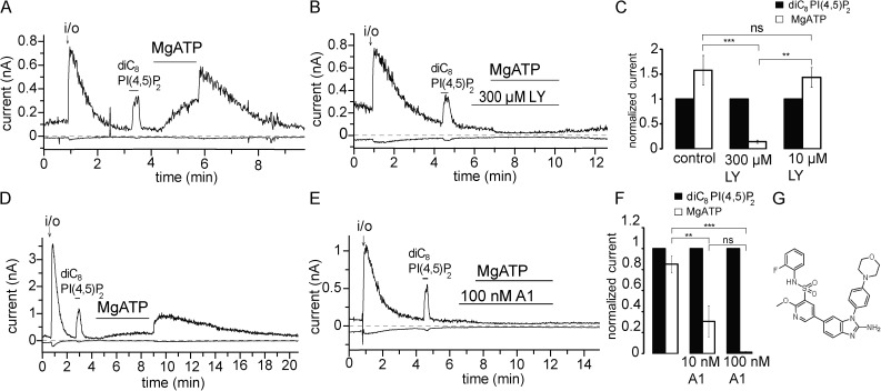
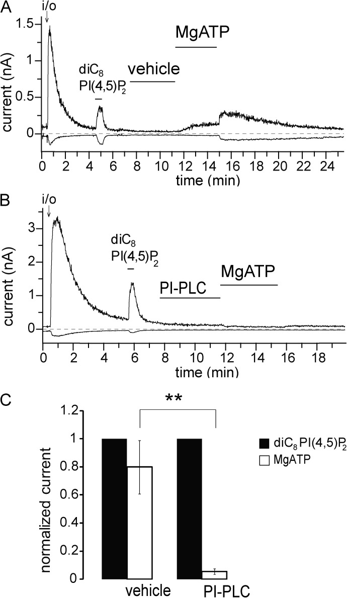
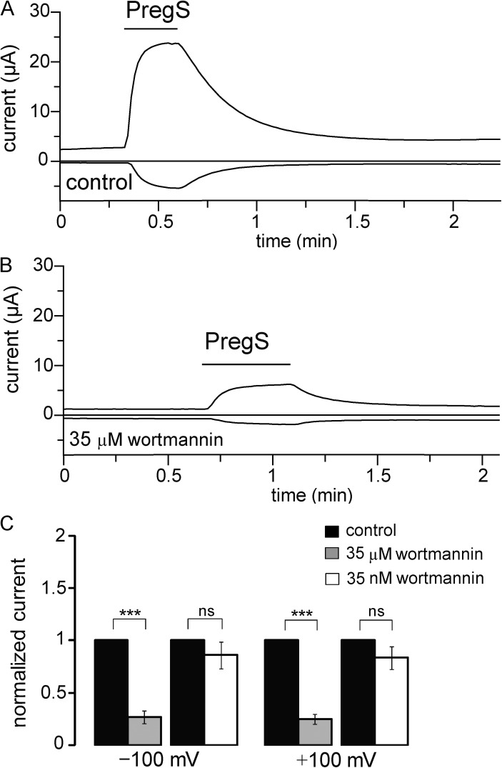
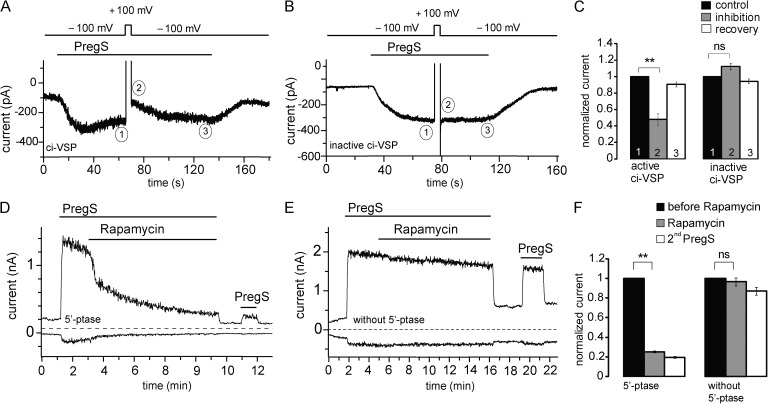
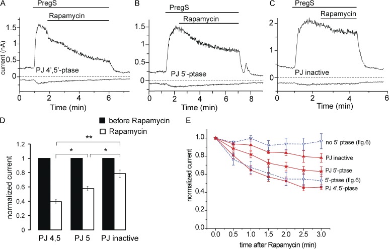
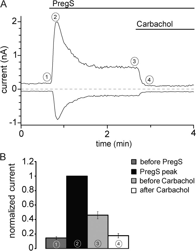
Phosphoinositides are emerging as general regulators of the functionally diverse transient receptor potential (TRP) ion channel family. Phosphatidylinositol 4,5-bisphosphate (PI(4,5)P2) has been reported to positively regulate many TRP channels, but in several cases phosphoinositide regulation is controversial. TRP melastatin 3 (TRPM3) is a heat-activated ion channel that is also stimulated by chemical agonists, such as pregnenolone sulfate. Here, we used a wide array of approaches to determine the effects of phosphoinositides on TRPM3. We found that channel activity in excised inside-out patches decreased over time (rundown), an attribute of PI(4,5)P2-dependent ion channels. Channel activity could be restored by application of either synthetic dioctanoyl (diC8) or natural arachidonyl stearyl (AASt) PI(4,5)P2. The PI(4,5)P2 precursor phosphatidylinositol 4-phosphate (PI(4)P) was less effective at restoring channel activity. TRPM3 currents were also restored by MgATP, an effect which was inhibited by two different phosphatidylinositol 4-kinase inhibitors, or by pretreatment with a phosphatidylinositol-specific phospholipase C (PI-PLC) enzyme, indicating that MgATP acted by generating phosphoinositides. In intact cells, reduction of PI(4,5)P2 levels by chemically inducible phosphoinositide phosphatases or a voltage-sensitive 5'-phosphatase inhibited channel activity. Activation of PLC via muscarinic receptors also inhibited TRPM3 channel activity. Overall, our data indicate that TRPM3 is a phosphoinositide-dependent ion channel and that decreasing PI(4,5)P2 abundance limits its activity. As all other members of the TRPM family have also been shown to require PI(4,5)P2 for activity, our data establish PI(4,5)P2 as a general positive cofactor of this ion channel subfamily.


Figures

Figure 1.
Figure 1.
PI(4,5)P2 reactivates hTRPM3 in excised inside-out patches after rundown. (A–C) Representative traces at 100 and −100 mV; experiments were performed on hTRPM3-expressing Xenopus oocytes with 100 µM PregS in the patch pipette as described in Materials and methods. The measurements start in the cell-attached mode; the establishment of the inside-out configuration (i/o) is indicated with an arrow. The applications of 25 µM diC8 PI(4,5)P2, 10 µM AASt PI(4,5)P2, 10 µM AASt PI(4)P, 30 µg/ml Poly-Lys (poly K), and 25 µM diC8 PI(4)P are indicated with the horizontal lines. (D) Statistical summary; the data are normalized to the TRPM3 current immediately after the establishment of the inside-out configuration at 100 mV (n = 6–7). (E) Representative trace showing the effects of different concentrations (µM) of diC8 PI(4,5)P2 applied to an excised inside-out patch; 25 µM diC8 PI(4,5)P2 was applied repetitively to normalize the effect of other concentrations and control for any time-dependent decrease in the responsiveness of TRPM3. (F) Summary of the dose–response relationship of diC8 PI(4,5)P2 (n = 4–10 for the individual concentrations), EC50 = 18.01 µM. Current values were initially normalized to the effect of the repetitively applied 25 µM diC8 PI(4,5)P2 and then renormalized for plotting to the maximal value obtained by the curve fitting. The top horizontal axis shows the mole percentage corresponding to the diC8 PI(4,5)P2 concentrations calculated using the formula from Collins and Gordon (2013). Error bars represent SEM.
Figure 2.
Figure 2.
The effects of 3-phosphorylated phosphoinositides on hTRPM3. (A–C) Representative traces at 100 and −100 mV; experiments were performed on hTRPM3-expressing Xenopus oocytes with 100 µM PregS in the patch pipette as described in Materials and methods. The establishment of the inside-out (i/o) configuration is indicated with an arrow. The applications of 25 µM diC8 PI(4,5)P2, 25 µM diC8 PI(3,4)P2, 25 µM diC8 PI(3,5)P2, and 25 µM diC8 PI(3,4,5)P3 are indicated with the horizontal lines. (D) Statistical summary of current amplitudes at 100 mV; the data are normalized to the TRPM3 current evoked by diC8 PI(4,5)P2 (n = 6–7). (E) Representative measurement for a concentration–response relationship for PI(3,4,5)P3. (F) Statistical summary compared with PI(4,5)P2, which is replotted from Fig. 1 F (dashed line). Error bars represent SEM.
Figure 3.
Figure 3.
MgATP reactivates hTRPM3 through PI4Ks. Excised inside-out patch measurements have been performed on TRPM3-expressing Xenopus oocytes with 100 µM PregS in the patch pipette as described in Materials and methods; data are plotted at 100 and −100 mV. (A and B) Representative traces for the effects of MgATP in the absence and presence of LY294002 (LY); the applications of 2 mM MgATP, 300 µM LY294002, and 25 µM diC8 PI(4,5)P2 are indicated by the horizontal lines. (C) Summary of the data for control and 10 and 100 µM LY294002 (n = 5 for control and 300 µM LY and n = 3 for 10 nM LY). (D and E) Representative traces for the effects of MgATP in the absence and presence of the PI4K inhibitor A1; the applications of 2 mM MgATP 100 nM A1 and 25 µM diC8 PI(4,5)P2 are indicated by the horizontal lines. (F) Summary of the data for control and 10 and 100 nM A1 (n = 5–6). (G) Chemical formula for compound A1. Error bars represent SEM. **, P < 0.01; ***, P < 0.005.
Figure 4.
Figure 4.
PI-PLC eliminates the effect of MgATP. Excised inside-out patch measurements have been performed on hTRPM3-expressing Xenopus oocytes with 100 µM PregS in the patch pipette as described in Materials and methods; data are plotted at 100 and −100 mV. (A and B) Representative traces for the effects of MgATP in the absence and presence of PI-PLC; the applications of 2 mM MgATP, 1 U/ml PI-PLC, and 25 µM diC8 PI(4,5)P2 are indicated by the horizontal lines. Vehicle denotes standard bath solution with 0.5% glycerol. (C) Summary of the data for control and PI-PLC–treated patches (n = 6). Error bars represent SEM. **, P < 0.01.
Figure 5.
Figure 5.
Inhibiting PI4K decreases hTRPM3 current. (A and B) Representative data from two TEVC measurements performed on the same hTRPM3-expressing Xenopus oocyte before and after incubation in 35 µM wortmannin for 2 h. Measurements are shown at 100 and −100 mV; the application of 50 µM PregS is shown by horizontal lines. (C) Statistical summary of the data at both positive and negative voltages for the effects of 35 nM (n = 17) and 35 µM wortmannin (n = 14). Error bars represent SEM. ***, P < 0.005.
Figure 6.
Figure 6.
Rapidly inducible 5′-phosphatases inhibit mTRPM3α2. (A) Representative trace of mTRPM3 current recorded from a HEK cell transfected with the active ci-VSP at a holding potential of −100 mV followed by short depolarizing pulse of 100 mV to activate the phosphatase. (B) Representative trace from a HEK cell transfected with the phosphatase-inactive mutant of ci-VSP (C363S) using the same voltage protocol as in A. (C) Summary of the inhibition of PregS-induced TRPM3 current plotted by comparing the current at −100 mV before and immediately after the depolarization pulse for the active (n = 6) and inactive phosphatases (n = 5). (D) Representative measurement in a HEK cell expressing the mTRPM3 and the components of the rapamycin-inducible 5-phosphatase. Measurements were performed using a ramp protocol from −100 to 100 mV, and current amplitudes are plotted at 100 and −100 mV. The applications of 25 µM PregS and 100 nM rapamycin are indicated by the horizontal lines. (E) Similar experiment as in D in a control cell, expressing TRPM3 and the components of the rapamycin-inducible system without the 5-phosphatase. (F) Statistical summary of the data (n = 6–7). Error bars represent SEM. **, P < 0.01.
Figure 7.
Figure 7.
The effects of the rapamycin-inducible 4′- and 5′-phosphatase pseudojanin. (A) Representative measurement in a HEK cell expressing the mTRPM3 and the components of the rapamycin-inducible 4′- and 5′-phosphatase pseudojanin. Measurements were performed using a ramp protocol from −100 to 100 mV, and current amplitudes are plotted at 100 and −100 mV. The applications of 25 µM PregS and 100 nM rapamycin are indicated by the horizontal lines. (B) Similar measurements as in A in a cell transfected with a pseudojanin construct in which the 4′-phosphatase was inactivated. (C) Similar measurements as in A in a cell in which both the 5′- and the 4′-phosphatase domains in pseudojanin were inactivated. (D) Summary of the extent of inhibition at the end of rapamycin application. (E) Time course of inhibition for the three pseudojanin constructs and for the 5′-phosphatase and its control construct from Fig. 6. Error bars represent SEM. *, P < 0.05; **, P < 0.01.
Figure 8.
Figure 8.
Activation of PLCβ by carbachol inhibits mTRPM3α2 currents. (A) Representative whole-cell patch clamp experiment in a HEK cell expressing mTRPM3α2 and hM1 in the presence of 2 mM extracellular Ca2+. The applications of 50 µM PregS and 100 µM carbachol are indicated by the horizontal lines. (B) Summary of data normalized to the peak of the PregS-induced current at the time points indicated. Error bars represent SEM.

Comment in

References

    1. Balla T. 2001. Pharmacology of phosphoinositides, regulators of multiple cellular functions. Curr. Pharm. Des. 7:475–507. 10.2174/1381612013397906 - DOI - PubMed
    1. Balla T. 2013. Phosphoinositides: tiny lipids with giant impact on cell regulation. Physiol. Rev. 93:1019–1137. 10.1152/physrev.00028.2012 - DOI - PMC - PubMed
    1. Bennett T.M., Mackay D.S., Siegfried C.J., and Shiels A.. 2014. Mutation of the melastatin-related cation channel, TRPM3, underlies inherited cataract and glaucoma. PLoS ONE. 9:e104000 10.1371/journal.pone.0104000 - DOI - PMC - PubMed
    1. Bojjireddy N., Botyanszki J., Hammond G., Creech D., Peterson R., Kemp D.C., Snead M., Brown R., Morrison A., Wilson S., et al. . 2014. Pharmacological and genetic targeting of the PI4KA enzyme reveals its important role in maintaining plasma membrane phosphatidylinositol 4-phosphate and phosphatidylinositol 4,5-bisphosphate levels. J. Biol. Chem. 289:6120–6132. 10.1074/jbc.M113.531426 - DOI - PMC - PubMed
    1. Cao C., Zakharian E., Borbiro I., and Rohacs T.. 2013a. Interplay between calmodulin and phosphatidylinositol 4,5-bisphosphate in Ca2+-induced inactivation of transient receptor potential vanilloid 6 channels. J. Biol. Chem. 288:5278–5290. 10.1074/jbc.M112.409482 - DOI - PMC - PubMed

Publication types

MeSH terms

Substances