. 2015 Jul 14;112(28):8738-43.
doi: 10.1073/pnas.1510830112. Epub 2015 Jun 29.

Pre- and postexposure efficacy of fully human antibodies against Spike protein in a novel humanized mouse model of MERS-CoV infection

Affiliations

Pre- and postexposure efficacy of fully human antibodies against Spike protein in a novel humanized mouse model of MERS-CoV infection

Kristen E Pascal et al. Proc Natl Acad Sci U S A. .

Abstract

Traditional approaches to antimicrobial drug development are poorly suited to combatting the emergence of novel pathogens. Additionally, the lack of small animal models for these infections hinders the in vivo testing of potential therapeutics. Here we demonstrate the use of the VelocImmune technology (a mouse that expresses human antibody-variable heavy chains and κ light chains) alongside the VelociGene technology (which allows for rapid engineering of the mouse genome) to quickly develop and evaluate antibodies against an emerging viral disease. Specifically, we show the rapid generation of fully human neutralizing antibodies against the recently emerged Middle East Respiratory Syndrome coronavirus (MERS-CoV) and development of a humanized mouse model for MERS-CoV infection, which was used to demonstrate the therapeutic efficacy of the isolated antibodies. The VelocImmune and VelociGene technologies are powerful platforms that can be used to rapidly respond to emerging epidemics.

Keywords: DPP4; MERS-CoV; Spike; mouse model; neutralizing antibody.


Conflict of interest statement

Conflict of interest statement: K.E.P., A.O.M., V.K., A.B., J.F., C.H., J.S., R.B., G.C., K.-M.V.L., T.T.H., W.O., G.D.Y., N.S., and C.A.K. are employees of Regeneron Pharmaceuticals, Inc. The work was funded by Regeneron Pharmaceuticals, Inc.

Figures

Fig. S1.
Fig. S1.
Schematic of immunization and antibody selection process for anti-S antibodies.
Fig. S2.
Fig. S2.
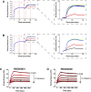
Label-free characterization of anti–MERS-CoV S monoclonal antibodies. (A and B) Competitive/noncompetitive behavior between REGN3048 and REGN3051 was determined using a premix assay format on Octet RED96 biosensor. REGN3048 (A) or REGN3051 (B) was first captured using AHC Octet biosensor (step 1), followed by a blocking step to saturate the unoccupied AHC sensors (step 2). Finally, MERS-CoV-RBD precomplexed with second antibody was allowed to bind to the AHC captured anti–MERS-CoV antibody (step 3), which is shown in the respective insets. An isotype control monoclonal antibody and MERS-CoV RBD sample prepared without any antibody were used as negative controls in the experiment. Black line: REGN3048; red line: REGN3051; green line: isotype control; blue line: no antibody. (C and D) Kinetics of REGN3048 and REGN3051 binding to MERS-CoV RBD At 37 °C were determined using Biacore T200 by first capturing the mAb on a CM5 chip immobilized with anti-human Fc at two different surface densities followed by 7-min injection of different concentrations of MERS-CoV RBD. The dissociation of bound MERS-CoV RBD from the captured mAb was monitored for 30 min. The real-time sensorgrams were globally fit to a 1:1 binding model with mass transport limitation using Biacore T200 evaluation software v1.0 and the values of kinetics parameters are tabulated in Table S2. The real-time binding of REGN3048 (C) and REGN3051 (D) to MERS-CoV RBD is plotted in black line and the resulted fits are plotted in red line.
Fig. 1.
Fig. 1.
MERS-CoV pseudotype and live MERS-CoV is neutralized by REGN3048 and REGN3051 in vitro. Luciferase expressing particles pseudotyped with either MERS-CoV S (A) or VSV-G (B) were incubated with either antibody at the indicated concentrations and the mix was used to transduce Huh-7 cells. At 72 h postinfection, luciferase was measured. Percent neutralization was calculated as the ratio of luciferase signal relative to no nontransduced and nonantibody-treated controls. (C) Live MERS-CoV was premixed with each noted antibody and used to infect cells across a dose range of each antibody. At 48 h postinfection, cells were analyzed for viability. Percent survival is graphed as a measure of MERS-CoV replication leading to cell death.
Fig. S3.
Fig. S3.
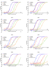
Neutralization of particles pseudotyped with MERS-CoV S protein encoded by clinical isolates by REGN3048, REGN3051, 3B12, MERS-4, and MERS-17. Luciferase-expressing particles pseudotyped with the indicated variants of the MERS-CoV S protein were incubated with antibodies at the various concentrations and the mix was used to transduce Huh-7 cells. At 72 h postinfection, luciferase was measured. Percent neutralization was calculated as the ratio of luciferase signal relative to no nontransduced and nonantibody-treated controls.
Fig. S4.
Fig. S4.
Expression of hPP4 in huDPP4 mice. (A) Schematic of the mouse chromosome with huDPP4 gene knocked-in (KI) to replace the mouse DPP4 coding region while maintaining mouse upstream and downstream regions. (B) Paraffin-embedded lungs from huDPP4 mice were sectioned and stained with anti-DPP4 antibody. In lungs, clear expression is seen in club cells in airways (black arrowheads, Lower Left) and type I and type 2 cells in the interstitium (black arrowheads, Lower Right) and alveolar macrophages (white arrowhead). Secondary antibody alone showed no staining (Upper). Magnification: 40×.
Fig. 2.
Fig. 2.
Transgenic huDPP4-containing mice and pathogenesis characterization. (A and B) Quantitative PCR of MERS-CoV transcript (A is MERS-CoV transcribed mRNA, B is MERS-CoV genome) in infected mice at days 2 and 4 postinfection (n = 5 mice per time point). (C) MERS-CoV viral titer quantitation of infected mouse lung at day 4 postinfection. Mouse lung MERS-CoV levels quantified by TCID50 assay and expressed as plaque-forming unit per gram of homogenized mouse lung (n = 5 mice per time point).
Fig. 3.
Fig. 3.
MERS-CoV pathogenesis in huDPP4 mice. H&E staining of mouse lung from MERS-CoV infected mice. Airway (10× magnification), vasculature (10× magnification), and interstitium (40× magnification) are shown for PBS, day 2 and day 4 postinfection mice (representative images of five mice at each time point).
Fig. S5.
Fig. S5.
Histological analysis of brain tissue from MERS-CoV infected huDPP4 mice at 4 dpi. Paraffin-embedded sections of mouse brain were stained with H&E: 10× and 40× magnification are shown. Notice no inflammation is found around either capillaries or neurons in infected mice.
Fig. 4.
Fig. 4.
In vivo treatment with anti-S antibodies one day before infection protects huDPP4 mice from MERS-CoV infection. Mice were intraperitoneally injected with a dose range of REGN3051 and REGN3048 monoclonal antibodies at 24 h preinfection with MERS-CoV. At 4 dpi, lungs were harvested and viral RNA and virus titer quantified. (A and B) Quantitative PCR of MERS-CoV transcript (A is MERS-CoV genome, B is MERS-CoV transcribed mRNA) from infected lungs was quantified using primers directed against the MERS-CoV genome and compared with hIgG1 isotype control-treated mice. All samples were compared with hIgG1 control (n = 5 mice per time point and quantitative PCR performed in triplicate). (C) Analysis of viral titer in the lungs quantitated by plaque assay reported as plaque-forming unit per gram lung (n = 5 mice per time point and titers performed in triplicate). *P < 0.05 and **P < 0.01.
Fig. S6.
Fig. S6.
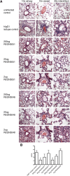
Histological analysis of MERS-CoV infected huDPP4 mice with antibody pretreatment. (A) H&E-stained sections of mouse lung showing airway, vasculature and interstitium of a representative mouse from each group. (B) Histological scoring of mouse lungs in A.
Fig. 5.
Fig. 5.
In vivo treatment with anti-S antibodies 1 d after infection protects huDPP4 mice from MERS-CoV infection. Mice were intraperitoneally injected with 200 μg or 500 μg of REGN3051 or an hIgG isotype control at 1 dpi or with 200 μg of REGN3051 at 24 h preinfection. At 4 dpi, lungs were harvested and viral RNA and virus titer quantified. (A and B) Quantitative PCR of MERS-CoV transcript (A is MERS-CoV genome, B is MERS-CoV transcribed mRNA) from infected lungs was quantified using primers directed against the MERS-CoV genome and compared with hIgG1 isotype control treated mice. All samples were compared with hIgG1 control. Notice the reduction in viral RNA levels for REGN3051 treated mice given 24 h after treatment compared with levels of isotype control treated mice (n = 5 mice per time point and quantitative PCR performed in triplicate). (C) Analysis of viral titer in the lungs quantitated by plaque assay reported as plaque-forming unit per gram lung (n = 5 mice per time point and titers performed in triplicate). **P < 0.01, and ***P < 0.001.
Fig. S7.
Fig. S7.
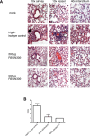
Histological analysis of MERS-CoV infected HuDPP4 mice with antibody treatment at 1 dpi. (A) H&E-stained sections of mouse lung showing airway, vasculature, and interstitium of a representative mouse from each group. Notice the reduction in pathology and inflammation of 200 μg and 500 μg REGN3051-treated groups compared with hIgG1 isotype control. Solid arrowhead denotes peribronchiolar cuffing. White arrowhead denotes perivascular cuffing. An asterisk denotes alveolar septa thickening. (B) Histological scoring of mouse lungs in A.

Comment in

References

    1. Zaki AM, van Boheemen S, Bestebroer TM, Osterhaus ADME, Fouchier RAM. Isolation of a novel coronavirus from a man with pneumonia in Saudi Arabia. N Engl J Med. 2012;367(19):1814–1820. - PubMed
    1. Coleman CM, Frieman MB. Emergence of the Middle East respiratory syndrome coronavirus. PLoS Pathog. 2013;9(9):e1003595. - PMC - PubMed
    1. Assiri A, et al. KSA MERS-CoV Investigation Team Hospital outbreak of Middle East respiratory syndrome coronavirus. N Engl J Med. 2013;369(5):407–416. - PMC - PubMed
    1. Cotten M, et al. Spread, circulation, and evolution of the Middle East respiratory syndrome coronavirus. MBio. 2014;5(1):e01062-13. - PMC - PubMed
    1. Perlman S, McCray PB., Jr Person-to-person spread of the MERS coronavirus—An evolving picture. N Engl J Med. 2013;369(5):466–467. - PubMed

MeSH terms

Substances