. 2015 Jul 14;112(28):E3699-708.
doi: 10.1073/pnas.1510329112. Epub 2015 Jun 29.

Massive accumulation of luminal protease-deficient axonal lysosomes at Alzheimer's disease amyloid plaques

Affiliations

Massive accumulation of luminal protease-deficient axonal lysosomes at Alzheimer's disease amyloid plaques

Swetha Gowrishankar et al. Proc Natl Acad Sci U S A. .

Abstract

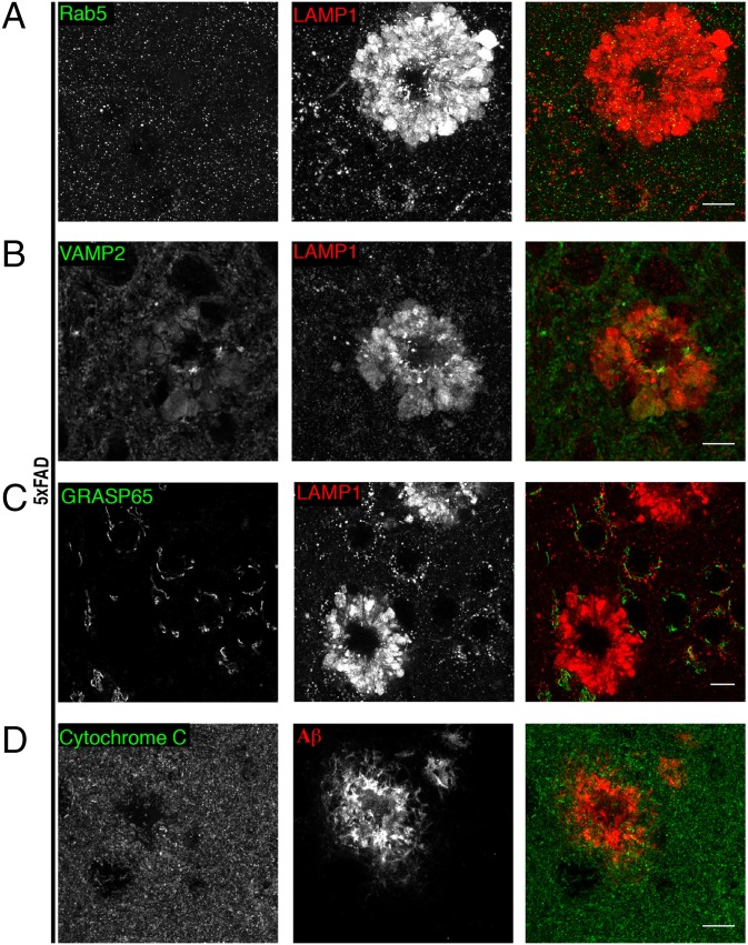
Through a comprehensive analysis of organellar markers in mouse models of Alzheimer's disease, we document a massive accumulation of lysosome-like organelles at amyloid plaques and establish that the majority of these organelles reside within swollen axons that contact the amyloid deposits. This close spatial relationship between axonal lysosome accumulation and extracellular amyloid aggregates was observed from the earliest stages of β-amyloid deposition. Notably, we discovered that lysosomes that accumulate in such axons are lacking in multiple soluble luminal proteases and thus are predicted to be unable to efficiently degrade proteinaceous cargos. Of relevance to Alzheimer's disease, β-secretase (BACE1), the protein that initiates amyloidogenic processing of the amyloid precursor protein and which is a substrate for these proteases, builds up at these sites. Furthermore, through a comparison between the axonal lysosome accumulations at amyloid plaques and neuronal lysosomes of the wild-type brain, we identified a similar, naturally occurring population of lysosome-like organelles in neuronal processes that is also defined by its low luminal protease content. In conjunction with emerging evidence that the lysosomal maturation of endosomes and autophagosomes is coupled to their retrograde transport, our results suggest that extracellular β-amyloid deposits cause a local impairment in the retrograde axonal transport of lysosome precursors, leading to their accumulation and a blockade in their further maturation. This study both advances understanding of Alzheimer's disease brain pathology and provides new insights into the subcellular organization of neuronal lysosomes that may have broader relevance to other neurodegenerative diseases with a lysosomal component to their pathology.

Keywords: Alzheimer's; axonal transport; cathepsin; lysosome; progranulin.


Conflict of interest statement

The authors declare no conflict of interest.

Figures

Fig. 1.
Fig. 1.
LAMP1 is massively enriched at amyloid plaques. (A) WT and 5xFAD (6-mo-old) mouse cerebral cortex stained for LAMP1 (composites assembled from multiple images acquired with a 20× objective). (B) Composite image showing double labeling for LAMP1 and Aβ (expanded view of 5xFAD sample from A). (C) LAMP1 localization in WT vs. 5xFAD mouse hippocampus (dentate gyrus). (D) LAMP1 staining of lysosomes within both WT and 5xFAD neuronal cell bodies (dashed outline, cerebral cortex). (E) Higher-magnification images of plaques and surrounding LAMP1 in 5xFAD cerebral cortex. [Scale bars: 200 μm (A and B), 20 μm (C), and 10 μm (D and E).] See also Fig. S1.
Fig. 2.
Fig. 2.
LAMP1 accumulations are present at all sizes and ages of amyloid plaques. (A) LAMP1 and Aβ staining in 8-mo-old APP/PS1 mouse cerebral cortex. (B and C) LAMP1 and Aβ staining in cerebral cortices of 6- (B) and 3-mo-old 5xFAD mice (C). Note that the changes in LAMP1 staining intensity/area at individual plaques change in parallel with the Aβ staining. (D) High-magnification image of LAMP1 and Aβ staining in 3-mo-old 5xFAD mice. (E) Quantification of the area of lysosome accumulation around each plaque plotted as data from individual plaques and mice (Left) and the mean of all data (Right), in 3- and 6-mo-old 5xFAD mice (n = 3 animals per age; mean ± SD). ***P < 0.001 (unpaired t test). [Scale bars; 20 μm (AC), 10 μm (D).]
Fig. 3.
Fig. 3.
Selective accumulation of LAMP1 at amyloid plaques. (A) Immunofluorescence staining for LAMP1 and Rab5 (early endosome marker). (B) Immunofluorescence staining for VAMP2 (synaptic vesicle marker) and LAMP1 costaining. (C) Immunofluorescence staining for GRASP65 (labels golgi) and LAMP1 double-labeling. (D) Mitochondria (Cytochrome C) are not enriched at amyloid plaques (Aβ labeling). (Scale bars: 10 μm.) All data in this figure were acquired from the cerebral cortices of 6-mo-old 5xFAD mice. See also Fig. S2.
Fig. S1.
Fig. S1.
Early endocytic proteins fail to coenrich with LAMP1 at amyloid plaques (related to Fig. 1). Representative images of mouse cortices from 6-mo-old 5xFAD mice costained for either LAMP1 or amyloid (Thioflavin S) along with the EEA1 (A), Dynamin 1 (B), BIN1 (C), Sortilin 1 (E), and PDI (F). (D) Representative images of mouse cortex of 8-mo-old APP/PS1 mice costained for LAMP1 and SORLA. (Scale bars: 10 μm.)
Fig. S2.
Fig. S2.
Investigation of the effect of amyloid plaques on vGLUT1 and cation-independent mannose-6-phosphate receptor (CI-M6PR) localization (related to Fig. 3). (A) Representative images of 5xFAD cortex (6-mo) costained for vGLUT1 and LAMP1. (B) Representative staining for M6PR and LAMP1 in WT cerebral cortices of 6-mo-old mice. (C) Representative staining for M6PR and LAMP1 (high-magnification image) and composite images of 5xFAD cerebral cortex and hippocampus. CI-M6PR shows limited enrichment at sites of major LAMP1 accumulation. (Scale bars: 10 µm.)
Fig. 4.
Fig. 4.
Ultrastructural analysis demonstrates the intracellular accumulation of organelles with lysosome-like morphology at amyloid plaques. (A) Electron micrograph of APP/PS1 cerebral cortex tissue showing a central extracellular amyloid deposit (outlined in dashed red line) surrounded by electron-dense organelles (outlined with dashed green line). (Scale bar: 10 µm.) (B) Higher magnification of the boxed region from A reveals the lysosome-like morphology of organelles that accumulate within cellular processes that contact the amyloid plaque. (Scale bar: 2 µm.) (C) Schematic diagram depicting lysosomal accumulation within swollen cellular processes that surround an extracellular β-amyloid deposit.
Fig. 5.
Fig. 5.
Lysosome accumulations at amyloid plaques are predominantly axonal in origin. (A) Confocal images of cortex from 3-mo-old 5xFAD mice stained for CD68 (microglial lysosome marker), Aβ (amyloid), and PGRN (lysosomes). (B) Quantification of the percentage of small Aβ deposits that have either CD68 and/or PGRN associated with them [mean ± SD, n = 3 (3-mo-old) 5xFAD mice per experiment, >40 plaques scored per mouse]. ***P < 0.001. (C) GFAP and LAMP1 staining in 5xFAD cerebral cortex. (D) 5xFAD cerebral cortex mice stained for MAP2B (dendritic marker) and LAMP1. (E) Neurofilament and LAMP1 staining in 5xFAD cerebral cortex. (Scale bars: 10 μm.) See also Fig. S3.
Fig. S3.
Fig. S3.
Lysosome accumulations at amyloid plaques predominantly reside within neuronal axons (related to Fig. 5). (A) Visualization of microglia (GFP signal) along with Aβ (plaque) and LAMP1 demonstrates that most LAMP1 around plaques is not of microglial origin. (B) Representative confocal images of cerebral cortex (3- and 6-mo-old 5xFAD mice as indicated) stained for Iba-1 (microglia), Thioflavin S (plaque), and LAMP1 (lysosomes). (C) Td-Tomato expressing AAV injected into 5xFAD hippocampus selectively colocalizes with the LAMP1 in swellings around amyloid plaques in the axonal, but not in the dendritic field. Insets from regions in the dendritic field (marked by dashed green line) and axonal field (marked by dashed blue line) are shown as enlarged panels. (Scale bars: 10 µm.)
Fig. 6.
Fig. 6.
RagC and PGRN are lysosome proteins that coenrich with LAMP1 at amyloid plaques. (A) WT and 5xFAD cerebral cortices stained for RagC and LAMP showing colocalization of the two proteins in neuronal cell bodies and around plaques. (B) PGRN and LAMP1 localization in WT cerebral cortex neuronal cell bodies. (C) Labeling for PGRN, LAMP1, and Aβ in the 6-mo-old 5xFAD cerebral cortex. (Scale bars: 10 μm.) See also Fig. S4.
Fig. S4.
Fig. S4.
Localization of RagC and PGRN in WT and APP/PS1 mice (related to Fig. 6). (A and B) Representative confocal images of cerebral cortex neuronal cell bodies and their processes stained for Rag C and LAMP1 (A), as well as PGRN and LAMP1 (B). (C) Staining of the APP/PS1 cerebral cortex (8 mo) for RagC and LAMP1. (D) PGRN, LAMP1, and Aβ staining in the APP/PS1 cerebral cortex (8 mo). (Scale bars: 10 µm.)
Fig. 7.
Fig. 7.
Multiple lysosomal luminal proteases fail to enrich at amyloid plaques. (AD) 5xFAD mouse (6 mo old) cerebral cortices double labeled for LAMP1 and either Cathepsin B (A) or Cathepsin L (B), AEP (asparaginyl endopeptidase) (C), or Cathespin D (D). (E) Quantification of the relative enrichment of lysosomal proteins in dystrophic axons compared with neuronal cell body lysosomes (mean ± SD). ***P < 0.001; ****P < 0.0001 (ANOVA with Dunnett’s posttest; n = 3 experiments, five dystrophies were analyzed per condition). (Scale bars: 10 μm.)
Fig. S5.
Fig. S5.
Proteases of the lysosomal lumen are preferentially enriched in neuronal cell bodies compared with the neuropil (related to Fig. 7). (A and B) WT (A) and 5xFAD (B) cerebral cortex stained for Cathepsin L and RagC showing the enrichment of RagC compared with Cathepsin L in dystrophies. (Scale bars: 10 μm.) (C and D) Immunofluorescence staining of the hippocampal CA1 region of WT mouse brain for LAMP1 with PGRN (C) and Cathepsin B (D) showing the preferential enrichment of Cathepsin B in pyramidal neuronal cell bodies, whereas LAMP1 is seen in all three regions. PGRN is present in stratum oriens and radiatum more often than Cathepsin B. (E) Primary cortical neuron culture labeled for AEP and LAMP1 showing AEP enrichment in lysosomes in neuronal cell body, whereas LAMP1 alone is observed in more peripheral lysosomes.
Fig. 8.
Fig. 8.
Luminal proteases are preferentially enriched in the lysosomes of neuronal cell bodies. (A and B) Labeling of LAMP1 along with either Cathepsin B (A) or Cathepsin L (B) in the WT mouse cerebral cortex. (C) Labeling of LAMP1 along with Cathepsin B in the hippocampal CA1 region showing cathepsin L enrichment in the cell bodies compared with the stratum oriens and stratum radiatum. (D) Primary cortical neuron culture labeled for cathepsin L and LAMP1 showing cathepsin L enrichment in lysosomes in neuronal cell body (highlighted by blue arrow), whereas LAMP1 alone in observed in more peripheral lysosomes (yellow arrowheads). (Scale bars: 10 μm.) See also Fig. S5.
Fig. 9.
Fig. 9.
BACE1 is enriched in LAMP1-positive axonal swellings at amyloid plaques. (A) WT cerebral cortex stained for BACE1 and LAMP1 shows their distinct patterns of localization. (B) 5xFAD cerebral cortex (in 3- and 6-mo-old animals) showing coaccumulation of LAMP1 and BACE1 (arrowheads, 3-mo-old sample) in dystrophic axons. (Scale bars: 10 μm.) (C) Schematic diagram that summarizes the distribution of lysosomes and their relative content of luminal proteases in healthy neurons as well as in AD neurons whose axons make contact with extracellular Aβ deposits.

References

    1. Selkoe DJ. SnapShot: Pathobiology of Alzheimer's disease. Cell. 2013;154(2):468–468.e1. - PubMed
    1. Karran E, Mercken M, De Strooper B. The amyloid cascade hypothesis for Alzheimer’s disease: An appraisal for the development of therapeutics. Nat Rev Drug Discov. 2011;10(9):698–712. - PubMed
    1. Tanzi RE, Bertram L. Twenty years of the Alzheimer’s disease amyloid hypothesis: a genetic perspective. Cell. 2005;120(4):545–555. - PubMed
    1. Holtzman DM, Morris JC, Goate AM. Alzheimer’s disease: The challenge of the second century. Sci Transl Med. 2011;3(77):sr1. - PMC - PubMed
    1. Jonsson T, et al. A mutation in APP protects against Alzheimer’s disease and age-related cognitive decline. Nature. 2012;488(7409):96–99. - PubMed

Publication types

Substances