. 2015 Jul 14;112(28):8744-9.
doi: 10.1073/pnas.1508432112. Epub 2015 Jun 29.

A circadian oscillator in the fungus Botrytis cinerea regulates virulence when infecting Arabidopsis thaliana

Affiliations

A circadian oscillator in the fungus Botrytis cinerea regulates virulence when infecting Arabidopsis thaliana

Montserrat A Hevia et al. Proc Natl Acad Sci U S A. .

Abstract

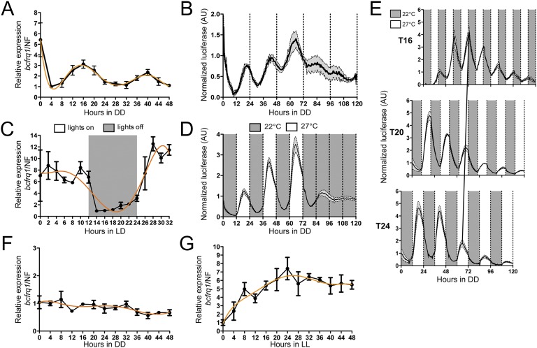
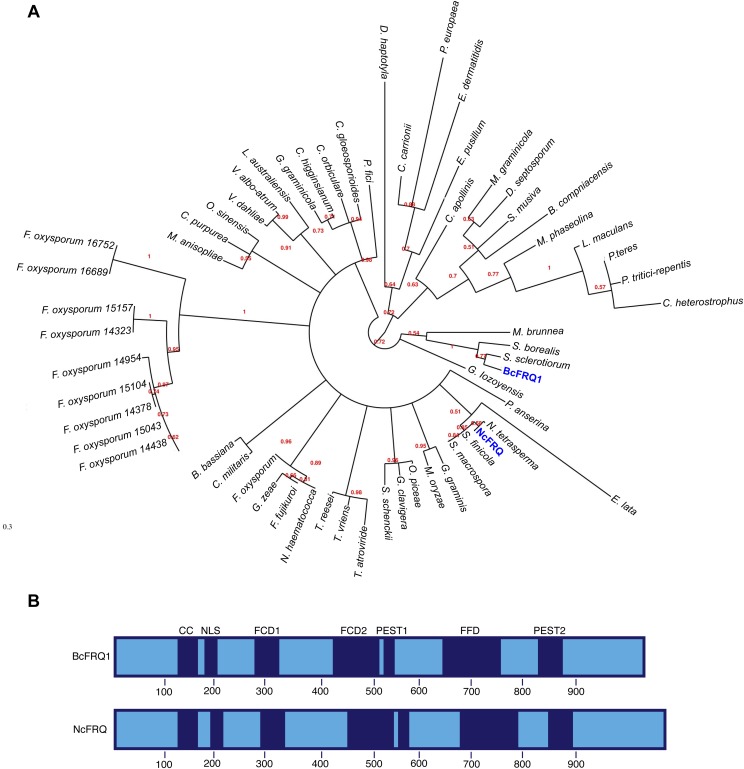
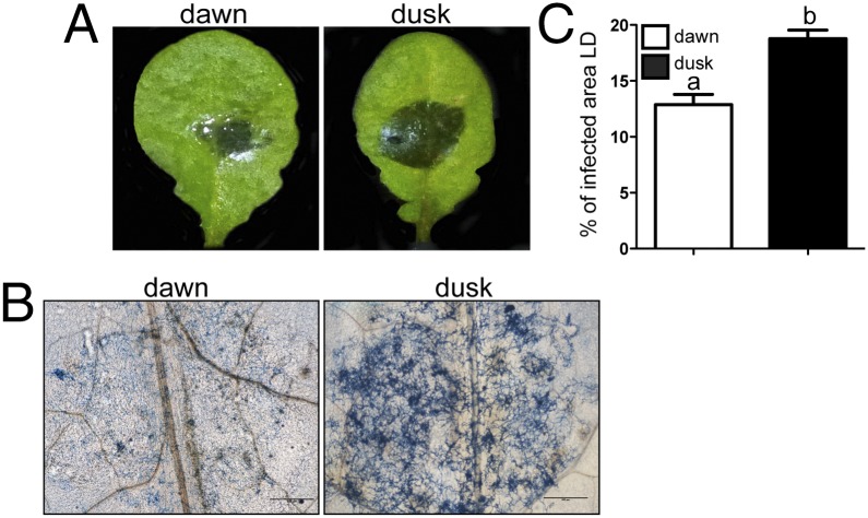
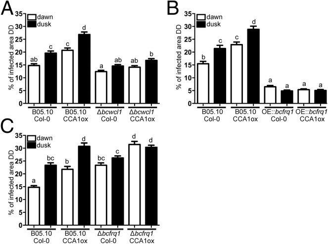
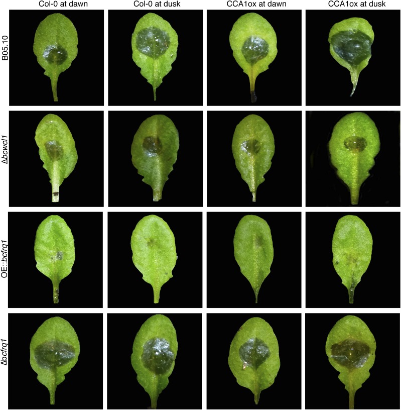
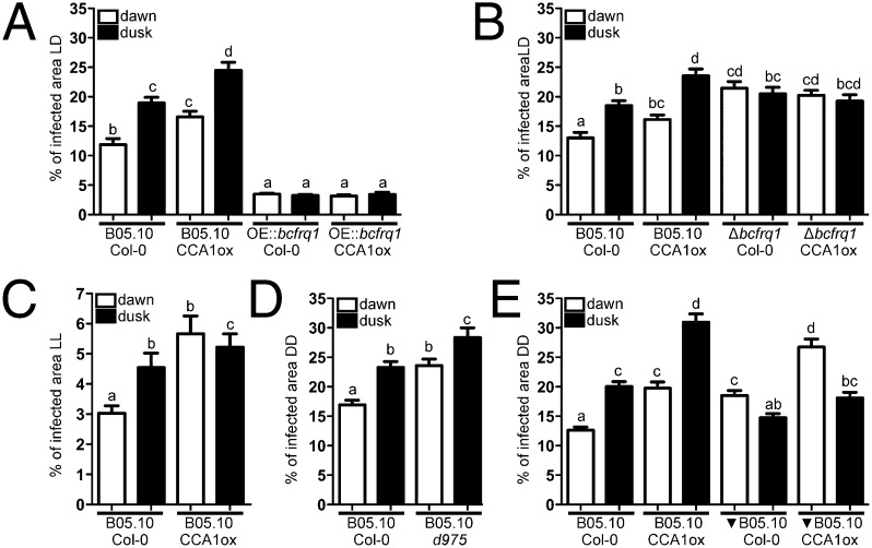
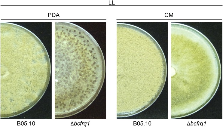
The circadian clock of the plant model Arabidopsis thaliana modulates defense mechanisms impacting plant-pathogen interactions. Nevertheless, the effect of clock regulation on pathogenic traits has not been explored in detail. Moreover, molecular description of clocks in pathogenic fungi--or fungi in general other than the model ascomycete Neurospora crassa--has been neglected, leaving this type of question largely unaddressed. We sought to characterize, therefore, the circadian system of the plant pathogen Botrytis cinerea to assess if such oscillatory machinery can modulate its virulence potential. Herein, we show the existence of a functional clock in B. cinerea, which shares similar components and circuitry with the Neurospora circadian system, although we found that its core negative clock element FREQUENCY (BcFRQ1) serves additional roles, suggesting extracircadian functions for this protein. We observe that the lesions produced by this necrotrophic fungus on Arabidopsis leaves are smaller when the interaction between these two organisms occurs at dawn. Remarkably, this effect does not depend solely on the plant clock, but instead largely relies on the pathogen circadian system. Genetic disruption of the B. cinerea oscillator by mutation, overexpression of BcFRQ1, or by suppression of its rhythmicity by constant light, abrogates circadian regulation of fungal virulence. By conducting experiments with out-of-phase light:dark cycles, we confirm that indeed, it is the fungal clock that plays the main role in defining the outcome of the Arabidopsis-Botrytis interaction, providing to our knowledge the first evidence of a microbial clock modulating pathogenic traits at specific times of the day.

Keywords: Arabidopsis thaliana; Botrytis cinerea; circadian clock; plant–pathogen interaction; virulence.


Conflict of interest statement

The authors declare no conflict of interest.

Figures

Fig. 1.
Fig. 1.
B. cinerea presents a functional circadian oscillator. (A) RT-qPCR showing bcfrq1 mRNA circadian oscillations under FRCs (DD). (B) BcFRQ1-LUC protein levels display circadian rhythms under DD. (C) bcfrq1 mRNA levels oscillate under LD conditions. (D) BcFRQ1-LUC levels reveal an anticipatory behavior under temperature cycles in DD. (E) BcFRQ1-LUC traces confirm entrainment to symmetric temperature cycles of different period length (T16, T20, and T24) under DD. The black diagonal line represents the phase of entrainment. (F) bcfrq1 mRNA levels depend on the bcwcl1 gene. (G) Under LL conditions, bcfrq1 mRNA levels fail to show rhythmic oscillations. For A, C, F, and G, each point, in black, represents mean ± SEM, whereas a trend line is depicted in orange. For B, D, and E, mean values are plotted as a black line, whereas SEM is represented as gray-filled area.
Fig. S1.
Fig. S1.
Characterization of BcFRQ1 from B. cinerea. (A) Phylogenetic reconstruction of 60 FRQ-like fungal proteins. FRQ proteins from B. cinerea (BcFRQ1) and N. crassa (NcFRQ) are indicated in blue. (B) Schematic representation of BcFRQ1 and NcFRQ showing the position of the translational start and stop codons, structural domains, and their respective positions. Previously defined functional domains (blue) include: CC, coiled-coil domain; NLS, nuclear localization signal; FCD1 and FCD2, FRQ-CKI interacting domains; FFD, FRQ-FRH interacting domain; PEST1 and PEST2, [proline (P), glutamate (E), serine (S), and threonine (T) rich sequence] domains. Protein domains were inferred from multiple sequence alignments of the FRQ-like proteins.
Fig. S2.
Fig. S2.
Generation of the BcFRQ1-LUC reporter strain. (A) Replacement strategy scheme showing the expected in-locus insertion of the knock-in cassette. The bcfrq1 gene and its transcriptional orientation (2,958 bp; BC1G_13940, genomic coordinates 37,815–40,772) are represented as a green arrow. The gene replacement cassette used to generate the BcFRQ1-LUC allele is shown bellow. The positions of the regions used for the homologous recombination (orange boxes) are shown. Black arrows show primers used for diagnostic PCRs, indicating their respective relative position and orientation. (B) Diagnostic PCRs showing in-locus integration. (C) Sequence chromatogram showing the junction of bcfrq1 and luc. (D) Primer pairs used in diagnostic PCRs.
Fig. S3.
Fig. S3.
Generation of the OE::bcfrq1 strain. (A) Replacement strategy scheme showing the expected in-locus insertion of the OE::bcfrq1 construct, as well as the used replacement cassette (Bottom), at the bcku70 locus. The latter and its transcriptional orientation (2,063 bp; BC1G_16015, genomic coordinates 15,622–17,684) are represented as a purple arrow. To overexpress bcfrq1, the B. cinerea actin promoter (ActA, Bc1G_08198; 1,000 bp) was used. Genomic regions used for the homologous recombination (orange boxes) are shown. Black arrows show primers used for diagnostic PCRs, indicating their respective relative position and orientation. (B) Diagnostic PCRs showing in-locus integration (a representative mutant is shown). (C) Primer pairs used in diagnostic PCRs.
Fig. S4.
Fig. S4.
bcfrq1 transcript levels become elevated and arrhythmic in the OE::bcfrq1 genetic background. (A) Total and endogenous bcfrq1 transcript levels were determined by RT-qPCR in the OE::bcfrq1 mutant and compared with bcfrq1 transcript levels in the B05.10 strain. Each point indicates mean values ± SEM and a trend line is depicted in orange. (B) Primer pairs used in the RT-qPCR procedure.
Fig. S5.
Fig. S5.
Generation of the Δbcfrq1 strain. (A) Schematic representation of the Δbcfrq1 locus depicting the in-locus insertion of the gene replacement cassette used for generation of the mutant (Bottom). Orange boxes denote the genomic regions used for homologous recombination of the replacement cassette, whereas black arrows show primers used for diagnostic PCRs, indicating their relative position and orientation. (B) Diagnostic PCRs showing in-locus integration. (C) Southern blot hybridization of a representative independent mutant (mutant 1). (D) Primer pairs used in diagnostic PCRs.
Fig. S6.
Fig. S6.
The Δbcfrq1 strain presents a culture media-dependent differentiation phenotype. (A) B05.10 and Δbcfrq1 strains were incubated on PDA and CM plates during 14 d under LD and DD conditions. (B) Hoechst-stained spores of B05.10 and Δbcfrq1 strains visualized by fluorescence microscopy.
Fig. S7.
Fig. S7.
Δbcfrq1 strain presents an altered infection phenotype. (A) Conidia from the B05.10 and microconidia (obtained from PDA plates) of a representative Δbcfrq1 mutant were inoculated on A. thaliana Col-0 plants and kept under LD conditions for 3 d. Representative leaves for each infection condition are shown. (B) Trypan blue staining showing fungal growth on plants at 3 dpi. (Scale bars, 500 μm.) (C) Quantification of the lesion area. Bars represent mean values ± SEM. Significant differences in comparison with the lesion area observed for B05.10 are indicated with asterisks.
Fig. S8.
Fig. S8.
Genetic complementation of the Δbcfrq1 strain allows recovering wild-type phenotype. (A) B05.10, Δbcfrq1 and Δbcfrq1+bcfrq1 strains were incubated on PDA plates for 14 d under LD. (B) Hoechst-stained spores of B05.10 and one representative Δbcfrq1 and Δbcfrq1+bcfrq1 strain visualized by fluorescence microscopy. (C) Lesion spreading of B05.10, Δbcfrq1, and Δbcfrq1+bcfrq1 strains on A. thaliana Col-0 plants incubated under LD conditions for 3 d. (D) Quantification of the lesion area produced by B05.10 and one representative Δbcfrq1 and Δbcfrq1+bcfrq1 mutant at 3 dpi. Bars represent mean values ± SEM. Different letters indicate statistical differences.
Fig. 2.
Fig. 2.
The outcome of the B. cinereaA. thaliana interaction differs with the time of day. (A) Lesion spreading of B05.10 strain on A. thaliana Col-0 plants. Arabidopsis leaves were inoculated at dawn or dusk, and then plants were incubated in LD for 72 h. Representative pictures are shown. (B) Trypan blue staining showing fungal growth after 3 dpi. (Scale bars, 500 μm.) (C) Quantification of lesion spreading of three independent virulence assays. Bars represent mean ± SEM (different letters indicate statistical differences).
Fig. 3.
Fig. 3.
A fungal circadian clock controls the infection process of B. cinerea on A. thaliana. (A) Measurements of lesion spreading of B05.10 and Δbcwcl1 strains. (B) Comparison of lesion spreading between B05.10 and OE::bcfrq1 strains. (C) Lesion spreading of B05.10 and Δbcfrq1 strains. Conidia were inoculated at dawn or dusk on A. thaliana Col-0 and CCA1ox plants, as indicated. Lesions were quantified 3 dpi under DD. Bars represent mean ± SEM (different letters indicate statistical differences).
Fig. S9.
Fig. S9.
B. cinerea has increased ability to infect at night in rhythmic and clock-null Arabidopsis plants. Representative pictures of lesion spreading of B05.10, Δbcwcl1, OE::bcfrq1, and Δbcfrq1 strains on A. thaliana Col-0 and CCA1ox plants, inoculated with a conidial suspension at dawn or dusk under FRCs (DD).
Fig. 4.
Fig. 4.
The B. cinerea circadian clock influences the outcome of the interaction under 12:12 photocycles as well as under constant darkness conditions. (A) Disruption of the clock by bcfrq1 overexpression (OE::bcfrq1) affects temporal variation of virulence in LD. (B) There are no differences in the infection behavior observed between dawn and dusk for the Δbcfrq1 strain in LD. (C) Under LL, the Arabidopsis clock—but not the Botrytis clock—provides a time-of-day effect on susceptibility. (D) The B05.10 strain has the ability to produce bigger lesions at dusk in both Col-0 and d975 arrhythmic mutant plants. (E) The relative magnitude of lesions achieved at dawn or dusk follows the time sensed by the fungus and not the plant. WT B05.10 strain was grown under the same LD cycle or 12 h out of phase (represented as a black inverted triangle) than the plants. Arabidopsis were inoculated at dawn or dusk with B. cinerea grown in the same phase or having opposite times. In all graphs, after macroconidia inoculations, WT (Col-0) and mutant plants were kept under the specified LD, LL, or DD regimes. White and black bars indicate conidia inoculation at dawn and dusk, respectively. Lesion area measurements were performed 3 dpi. Bars represent mean ± SEM (different letters indicate statistical differences).
Fig. S10.
Fig. S10.
The Δbcfrq1 strain develops sclerotia under LL culture conditions. B05.10 and Δbcfrq1 strains were incubated on PDA and CM plates during 21 d under LL conditions.

References

    1. Dunlap JC. Molecular bases for circadian clocks. Cell. 1999;96(2):271–290. - PubMed
    1. Bell-Pedersen D, et al. Circadian rhythms from multiple oscillators: Lessons from diverse organisms. Nat Rev Genet. 2005;6(7):544–556. - PMC - PubMed
    1. Dodd AN, et al. Plant circadian clocks increase photosynthesis, growth, survival, and competitive advantage. Science. 2005;309(5734):630–633. - PubMed
    1. Lowrey PL, Takahashi JS. Genetics of circadian rhythms in mammalian model organisms. Adv Genet. 2011;74:175–230. - PMC - PubMed
    1. Ouyang Y, Andersson CR, Kondo T, Golden SS, Johnson CH. Resonating circadian clocks enhance fitness in cyanobacteria. Proc Natl Acad Sci USA. 1998;95(15):8660–8664. - PMC - PubMed

Publication types