. 2015 Jul 14;112(28):E3679-88.
doi: 10.1073/pnas.1505995112. Epub 2015 Jun 29.

Small GTP-binding protein Ran is regulated by posttranslational lysine acetylation

Affiliations

Small GTP-binding protein Ran is regulated by posttranslational lysine acetylation

Susanne de Boor et al. Proc Natl Acad Sci U S A. .

Abstract

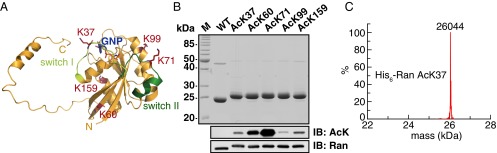
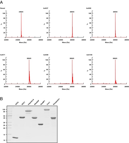
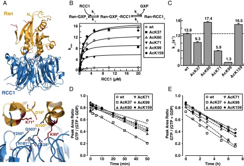
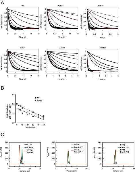
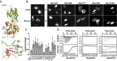
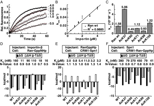
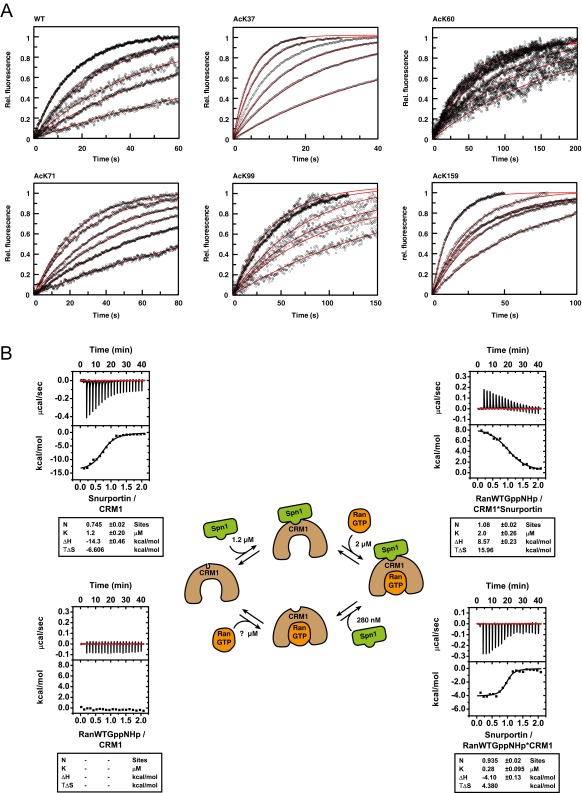
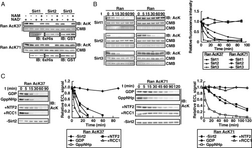
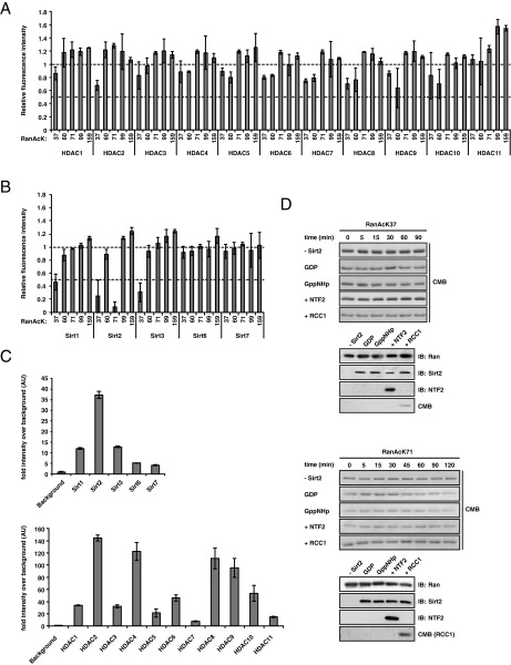
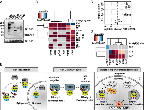
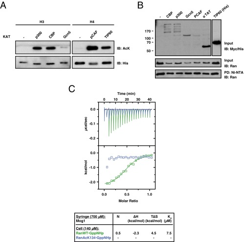
Ran is a small GTP-binding protein of the Ras superfamily regulating fundamental cellular processes: nucleo-cytoplasmic transport, nuclear envelope formation and mitotic spindle assembly. An intracellular Ran•GTP/Ran•GDP gradient created by the distinct subcellular localization of its regulators RCC1 and RanGAP mediates many of its cellular effects. Recent proteomic screens identified five Ran lysine acetylation sites in human and eleven sites in mouse/rat tissues. Some of these sites are located in functionally highly important regions such as switch I and switch II. Here, we show that lysine acetylation interferes with essential aspects of Ran function: nucleotide exchange and hydrolysis, subcellular Ran localization, GTP hydrolysis, and the interaction with import and export receptors. Deacetylation activity of certain sirtuins was detected for two Ran acetylation sites in vitro. Moreover, Ran was acetylated by CBP/p300 and Tip60 in vitro and on transferase overexpression in vivo. Overall, this study addresses many important challenges of the acetylome field, which will be discussed.

Keywords: Ran; genetic code expansion concept; lysine acetylation; nuclear cytosolic transport; nucleus.


Conflict of interest statement

The authors declare no conflict of interest.

Figures

Fig. 1.
Fig. 1.
Incorporation of N-(ε)-acetyl-L-lysine into Ran using the genetic code expansion concept. (A) Ribbon representation of Ran (yellow) and position of the five lysine acetylation sites (red) studied here (PDB ID code 1K5D). K37R in switch I (light green), K60R in β3, K71R in switch II (dark green), K99R in α3 and K159R in α5. (B) Final purity of the recombinantly expressed WT Ran and lysine acetylated proteins shown by SDS/PAGE (Top). Immunoblotting (IB) of Ran proteins using a specific anti–acetyl-lysine (ab21623) antibody (Middle). The antibody differentially recognizes the different acetylation sites in Ran and does not detect RanWT. The immunoblotting using an anti–Ran-antibody shows equal loading. (C) Acetyl-lysine is quantitatively incorporated at position 37 in Ran. The corresponding theoretical molecular mass of the nonacetylated His6-Ran protein is 26,001 Da; the acetyl group has a molecular weight of 42 Da.
Fig. S1.
Fig. S1.
Quantitative incorporation of N-(ε)-acetyl-L-lysine into Ran at five distinct sites (A) and the final purity of the proteins used in this study as shown by SDS/PAGE and Coomassie-brillant blue staining (B). The determined molecular masses do correspond exactly (±1 Da) to the protein mass (Ran: 26001 Da) plus the incorporated acetyl-group (42 Da). RanWT was purified using a GST-tag (cleaved by TEV-protease) and does not carry a hexahistidine tag.
Fig. 2.
Fig. 2.
Acetylation of Ran interferes with RCC1 catalyzed nucleotide exchange and RanGAP-catalyzed and intrinsic nucleotide hydrolysis. (A) Structure of the Ran•RCC1-complex and close up of the binding interface, showing interactions of Ran K71/K99 as described in the text (PDB ID code 1I2M). RCC1 (blue), Ran (yellow), acetylation sites (red). (B) Pseudo–first-order kinetics of nucleotide exchange rates of 500 nM Ran (final concentration) titrated with increasing RCC1 concentrations (0.0195–20 µM). The scheme shows that Ran•GDP with tightly bound nucleotide (GXP: GTP or GDP; subscript: T) binds RCC1 first loosely in a ternary Ran•GXP•RCC1-complex (subscript: L), and in the second step, the nucleotide is released with a dissociation rate k−2 to result in a tight Ran•RCC1 complex. (C) The hyperbolic fit resulted in the rate of nucleotide dissociation from the ternary Ran•GDP•RCC1 complex, k−2. (D) RanGAP-stimulated nucleotide hydrolysis on Ran. GTP hydrolysis rates were examined by HPLC determining the GTP/(GTP + GDP) ratio as a function of time. The acetylation does not alter GAP-catalyzed nucleotide hydrolysis on Ran. (E) Intrinsic nucleotide hydrolysis on Ran and acetylated Ran. The rates were determined as described in D. Ran AcK71 leads to a 1.5-fold increase in the intrinsic GTP hydrolysis rate, whereas the other Ran AcKs are similar to WT Ran.
Fig. S2.
Fig. S2.
Primary kinetics data of the Ran-RCC1 nucleotide exchange experiment as determined by stopped-flow (A), HPLC analysis of RanGAP-stimulated nucleotide hydrolysis (B), and analytical size exclusion chromatography of Ran•NTF2-complexes (C). (A) Traces and fits (red) for the development of the fluorescence intensity over time for various concentrations of RCC1. Ran•mantGDP and the lysine acetylated proteins (final concentration: 500 nM) were titrated with increasing concentrations of RCC1 (final concentration: 0.0195–20 µM). (B) HPLC analysis of RanGAP-stimulated nucleotide hydrolysis on Ran in presence of RanBP1. A 1:1 complex of RanBP1 and RanGTP (each 150 µM) was incubated with 5 nM RanGAP at 23 °C. Samples taken at indicated time points were boiled for 5 min, centrifuged for 10 min at 17.000 × g, and subsequently subjected to HPLC analysis. (C) Analytical size exclusion chromatography of Ran wt/AcK71/K71Q and NTF2; 150 µM protein (RanWT, AcK71, K71Q, and NTF2) or the equimolar mixture was applied to a Superdex S75 10/300 column at a flow-rate of 0.6 mL/min in phosphate buffer (buffer E). Protein was detected by absorption at 280 nm. The RanWT•NTF2-complex elutes earlier than the single proteins. Ran Ack71 and K71Q do not form a complex with NTF2.
Fig. 3.
Fig. 3.
Ran AcK71 abolishes nuclear localization of Ran by blocking NTF2 binding. (A) Ribbon representation of the NTF2-Ran•GDP complex (PDB ID code 1A2K). K71 of Ran forms a salt bridge to D92/D94 in NTF2. Shown are the distances in Angstroms. (B) EGFP fluorescence of the Ran-EGFP K to Q and K to R mutants in HeLa cells. Ran localizes mainly to the nucleus for WT and all mutants except for Ran K71Q and K99R, which are primarily cytosolic. (C) Quantification of subcellular Ran by measurement of the EGFP fluorescence in the nucleus and the cytosol. As shown in B, Ran K71Q and K99R localize to the cytosol, whereas the others are predominantly nuclear. (D) Thermodynamic characterization of the interaction of NTF2 and Ran, Ran AcK71, Ran K71Q (acetylation mimic), and Ran K71R (charge conserving) by ITC. K71Q and AcK71 abolish binding toward NTF2.
Fig. 4.
Fig. 4.
Acetylation of Ran in import and export complex formation. (A) Association kinetics of Ran•mantGppNHp WT (100 nM final) and increasing concentrations of Importin-β (final: 0.5–4 µM) as determined by stopped-flow. The kinetics were fitted single exponentially to result in the observed rate constants, kobs. (B) Determination of the Ran•mantGppNHp-Importin-β association rate constant. The obtained kobs values were plotted against the Importin-β concentration. The linear fit resulted in the association rate constant, kon. (C) Comparison of the association rates for Importin-β-RanWT and the acetylated Ran proteins. Ran AcK37 increases the association rate fivefold. (D) Thermodynamics of the Importin-β (268 μΜ) and Ran•GppNHp (40 µM) interaction as determined by ITC. Ran AcK37, AcK99, and AcK159 increase the affinity toward Importin-β. (E) Thermodynamics of the interaction of Ran•GppNHp (200 µM) titrated onto a Crm1•Spn1-complex (20/40 µM) determined by ITC. Ran AcK71 decreases the Ran•GppNHp affinity to the complex fivefold. (F) Thermodynamic profile of the interaction of 200 µM Spn1 titrated onto a preformed Crm1•Ran•GppNHp-complex (20/40 µM) as determined by ITC. Ran AcK37, AcK99, and AcK159 increase the binding affinity of Spn1 to the preformed complex.
Fig. S3.
Fig. S3.
Influence of Ran acetylation on the association with Importin-β and protein export complex formation. (A) Stopped-flow primary data for the association kinetics of Importin-β and Ran•GppNHp. 100 nM Ran•mantGppNHp and the acetylated Ran proteins were titrated with increasing concentrations of Importin-β (final: 0.5–4 µM). (B) Interplay of Ran•GppNHp, Spn1, and CRM1 in export complex formation as determined by ITC. Spn1 binds CRM1 with an affinity of 1.2 µM (Upper Left). No heat signal was obtained for the direct interaction of Ran•GppNHp and CRM1 (Lower Left). Ran•GppNHp binds with 2 µM to a preformed Crm1•Spn1-complex (Upper Right) and Spn1 binds with 280 nM to a preformed Ran•GppNHp-CRM1 complex. The model in the center shows that CRM1 can be primed to bind Ran•GTP by first binding to the export substrate Spn1 or, alternatively, to bind Spn1 by first binding Ran•GTP.
Fig. 5.
Fig. 5.
Regulation of Ran acetylation by KDACs. (A) Ran AcK37 is deacetylated by Sirt1, -2, and -3, whereas Ran AcK71 is specifically deacetylated only by Sirt2. Three micrograms recombinant Ran was incubated with Sirt1, -2, and -3 (0.6, 0.2, and 0.55 µg) for 2 h at room temperature in the presence or absence of NAD+ and nicotinamide (NAM). Shown are the immunoblots using the anti-AcK antibody after the in vitro deacetylase reaction. Coomassie (CMB) staining is shown as loading control for Ran AcK37, immunoblots using anti-His6- and anti-GST antibodies for the sirtuins. (B) Kinetics of deacetylation of Ran AcK37 and Ran AcK71 by Sirt1, -2, and -3. Twenty-five micrograms recombinant Ran was incubated with Sirt1, -2, and -3 (4.5, 1.5, and 4.4 µg) depending on the individual enzyme activity (Fig. S4B). Shown is the immunoblot using the anti-AcK antibody (IB: AcK; Left) and the quantification of the time courses (Right). Ran AcK71 is only deacetylated by Sirt2; Ran AcK37 is deacetylated by all three sirtuins. (C) Dependence of Sirt2 deacetylation of Ran AcK37 and AcK71 on the nucleotide state and presence of the interactors NTF2 and RCC1. Sixty-five micrograms recombinant Ran was incubated with Sirt2 at 25 °C, and samples taken after the indicated time points. To compensate for the slower deacetylation rate, 3.7 µg Sirt2 was used for Ran AcK71, whereas only 1 µg Sirt2 was used for Ran AcK71. The immunodetection with the anti-AcK antibody and the corresponding quantification of the time course is shown. The deacetylation of Ran AcK37 depends on the nucleotide state; AcK71 is accelerated in the GppNHp-loaded state. Presence of NTF2 decelerates the deacetylation of Ran AcK37, whereas RCC1 accelerates it. For Ran AcK71, presence of NTF2 has no influence on the deacetylation kinetics by Sirt2; RCC1 blocks deacetylation. For loading and input controls of the time courses, please refer to Fig. S4D.
Fig. S4.
Fig. S4.
Screen of acetylated Ran proteins as substrate for classical (HDAC1-11) and sirtuin (Sirt1-7) deacetylases and activity controls of KDACs used in this study. Ran AcK37/60/71/99/159 was used as substrate for classical HDACs (A) or sirtuins (B). For all enzymatic reactions, enzyme concentrations used in the in vitro assays were normalized to yield a comparable activity for all enzymes. (C) The enzymatic activities were determined using a fluor-de-lys substrate for sirtuins (Upper) and classical HDACs (Lower). (D) Loading and input controls for the Sirt2 deacetylation time course experiments with Ran AcK37 (Upper) and Ran AcK71 (Lower). Coomassie staining (CMB) of the time courses are shown as Ran loading controls. As input controls, immunoblots for Ran, Sirt2, and NTF2, as well as Coomassie staining of RCC1, are shown.
Fig. 6.
Fig. 6.
Ran is acetylated by the KATs CBP and Tip60 in vitro and on KAT overexpression in cells. (A) Immunoblotting of the in vitro KAT assay of Ran using recombinant p300, CBP, pCAF, Tip60, and Gcn5. p300 and CBP acetylate Ran (anti-AcK). As loading control Ran was stained with an anti-Ran antibody. (B) Heat map with hierarchical clustering of identified acetylation sites. Increased Ran acetylation was detected for Tip60, CBP, and p300, predominantly at lysines 134, 142, and 37. The mean intensities of two independent in vitro KAT assays are shown. (C) Volcano plot of three independent in vitro KAT-assays with Ran and CBP. The Ran lysines 37, 134, and 142 were identified as the most significant acetylation sites (P < 0.05). (D) Heat map with hierarchical clustering of identified acetylation sites after transfection of KATs and subsequent Ni pulldown of Ran from HEK cells. Increased Ran acetylation at lysine 134 was detected for Tip60, CBP, and p300. Lysine 152 was exclusively acetylated by α-TAT. The mean intensities of two independent in vivo KAT assays are shown. (E) Working model of the regulation of Ran by posttranslational lysine acetylation. (Left) Ran acetylation at K71 abolishes NTF2 binding, thereby preventing nuclear Ran localization. Also, K99R does show cytosolic distribution by an unidentified mechanism. D, GDP. (Center) Ran acetylation at K71 and K99 affects the Ran•GDP/GTP cycle by interfering with RCC1-catalyzed nucleotide exchange and binding. AcK71 increases RCC1 binding and decreases RCC1 activity on Ran (dominant negative); AcK99 decreases RCC1 binding and RCC1 activity on Ran (loss of function). Furthermore, AcK71 decreases the intrinsic GTP hydrolysis rate. (Right) Ran acetylation at K37, K99, and K159 increases the affinity toward Importin-β and Spn1 if complexed with Crm1. Thereby, lysine acetylation might interfere with import substrate release and export substrate binding in the nucleus. T, GTP.
Fig. S5.
Fig. S5.
Controls for KAT assays and interaction of Ran AcK134 and Mog1. (A) The lysine-acetyltransferase (KAT) activities were tested using histone substrates (0.75 µg H3, 7.5 µg H4) and immunoblotting with the anti-AcK antibody. One microliter commercially available p300, pCAF, CBP, Gcn5, or Tip60 and 100 μM acetyl-CoA were used in transferase buffer (50 mM Tris⋅HCl, 50 mM KCl, 5% glycerol, 1 mM DTT, 0.1 mM EDTA, pH 7.3) for 4 h at 25 °C; 250 ng H3 (−, p300, CBP, Gcn5) and 2.5 μg H4 (−, pCAF, Tip60) were loaded for immunoblotting. (B) Coexpression of selected KATs and Ran to study in vivo acetylation. Successful transfection of KATs was verified by immunoblotting against Myc-tag (His-tag for Tip60). (C) Impact of Ran K134 acetylation on the interaction with Mog1 as shown by ITC; 700 µM Mog1 (syringe) was stepwise injected into the cell containing 140 µM GppNHp-loaded RanWT or Ran AcK134.

Similar articles

Cited by

References

    1. Clarke PR, Zhang C. Spatial and temporal coordination of mitosis by Ran GTPase. Nat Rev Mol Cell Biol. 2008;9(6):464–477. - PubMed
    1. Scheffzek K, Klebe C, Fritz-Wolf K, Kabsch W, Wittinghofer A. Crystal structure of the nuclear Ras-related protein Ran in its GDP-bound form. Nature. 1995;374(6520):378–381. - PubMed
    1. Monecke T, et al. Crystal structure of the nuclear export receptor CRM1 in complex with Snurportin1 and RanGTP. Science. 2009;324(5930):1087–1091. - PubMed
    1. Stewart M, Kent HM, McCoy AJ. Structural basis for molecular recognition between nuclear transport factor 2 (NTF2) and the GDP-bound form of the Ras-family GTPase Ran. J Mol Biol. 1998;277(3):635–646. - PubMed
    1. Ren M, Drivas G, D’Eustachio P, Rush MG. Ran/TC4: A small nuclear GTP-binding protein that regulates DNA synthesis. J Cell Biol. 1993;120(2):313–323. - PMC - PubMed

Publication types

LinkOut - more resources