. 2015 Jul 21;112(29):E3816-25.
doi: 10.1073/pnas.1510077112. Epub 2015 Jun 29.

Sex hormone-dependent tRNA halves enhance cell proliferation in breast and prostate cancers

Affiliations

Sex hormone-dependent tRNA halves enhance cell proliferation in breast and prostate cancers

Shozo Honda et al. Proc Natl Acad Sci U S A. .

Abstract

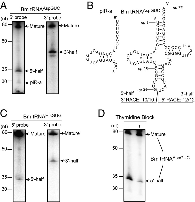
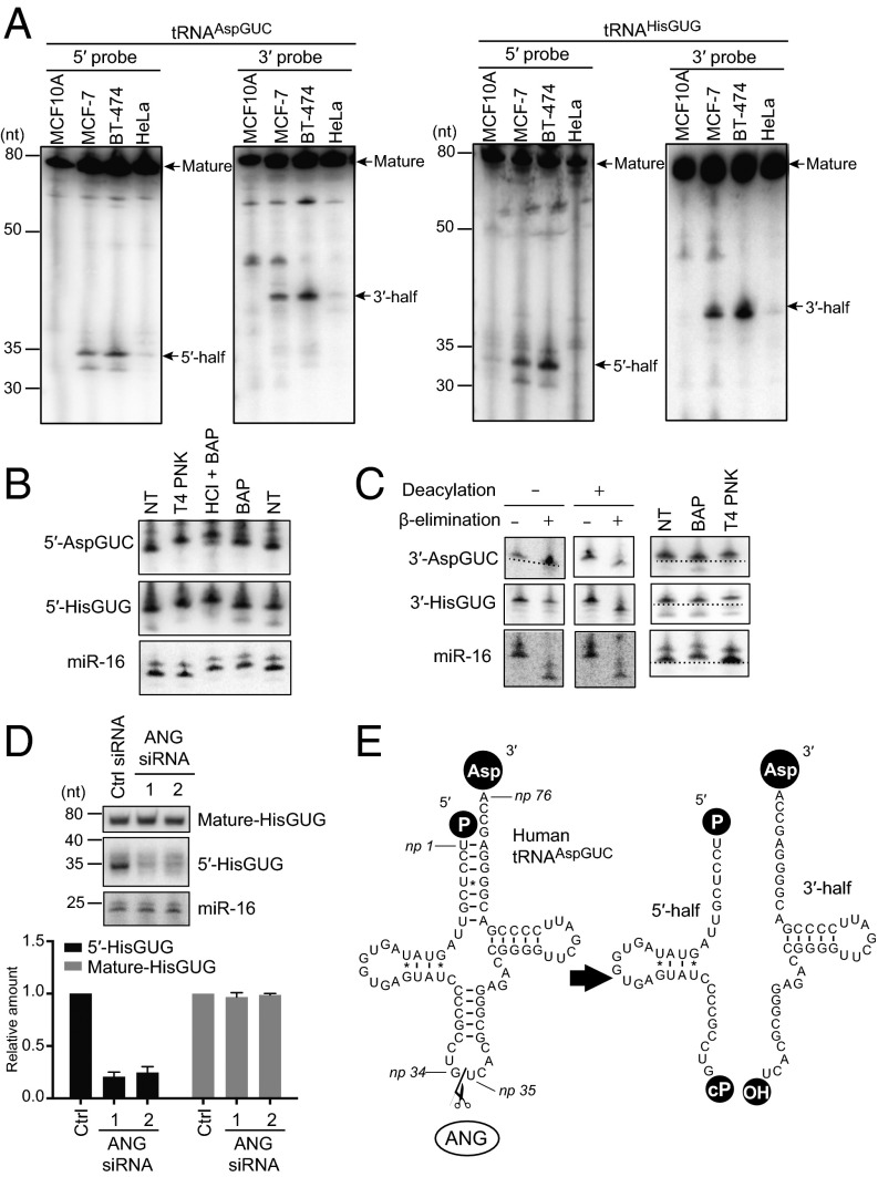
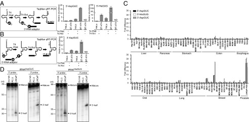
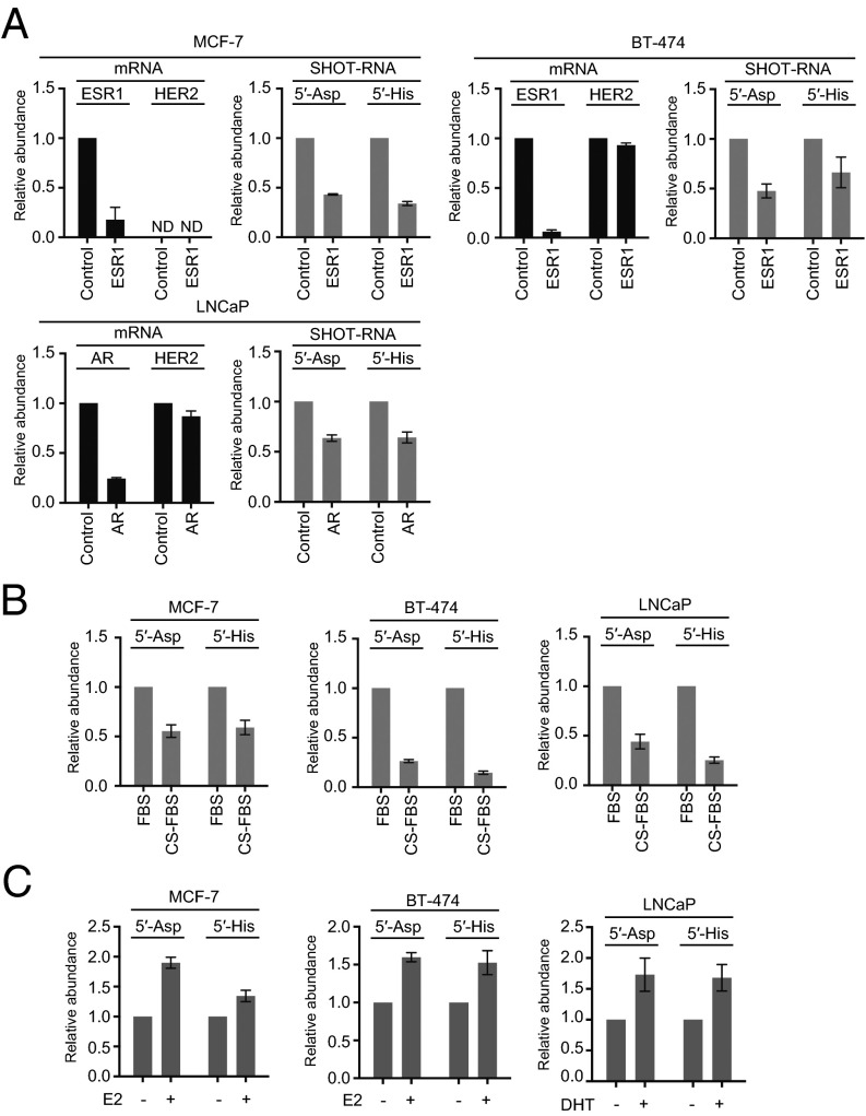
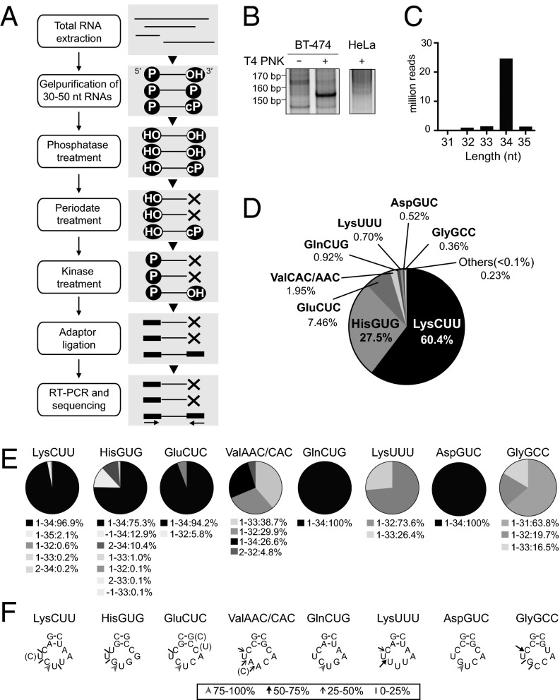
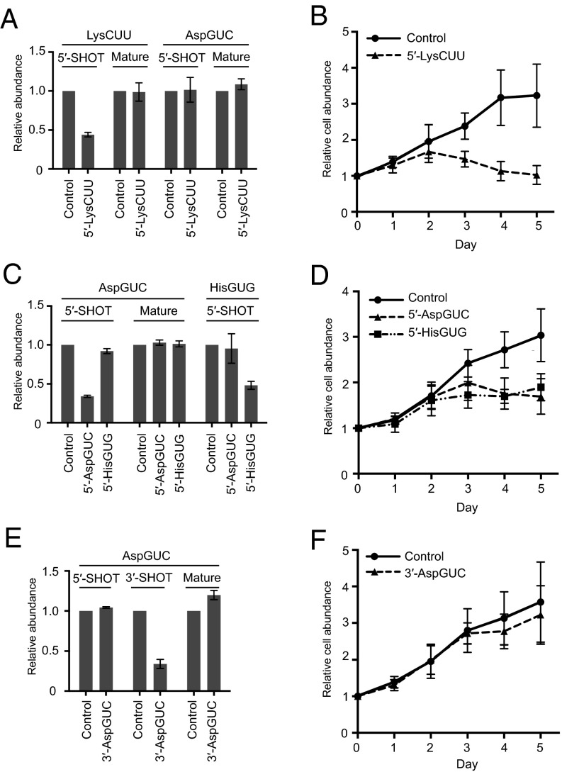
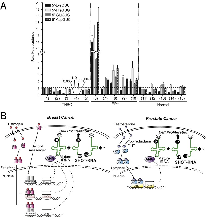
Sex hormones and their receptors play critical roles in the development and progression of the breast and prostate cancers. Here we report that a novel type of transfer RNA (tRNA)-derived small RNA, termed Sex HOrmone-dependent TRNA-derived RNAs (SHOT-RNAs), are specifically and abundantly expressed in estrogen receptor (ER)-positive breast cancer and androgen receptor (AR)-positive prostate cancer cell lines. SHOT-RNAs are not abundantly present in ER(-) breast cancer, AR(-) prostate cancer, or other examined cancer cell lines from other tissues. ER-dependent accumulation of SHOT-RNAs is not limited to a cell culture system, but it also occurs in luminal-type breast cancer patient tissues. SHOT-RNAs are produced from aminoacylated mature tRNAs by angiogenin-mediated anticodon cleavage, which is promoted by sex hormones and their receptors. Resultant 5'- and 3'-SHOT-RNAs, corresponding to 5'- and 3'-tRNA halves, bear a cyclic phosphate (cP) and an amino acid at the 3'-end, respectively. By devising a "cP-RNA-seq" method that is able to exclusively amplify and sequence cP-containing RNAs, we identified the complete repertoire of 5'-SHOT-RNAs. Furthermore, 5'-SHOT-RNA, but not 3'-SHOT-RNA, has significant functional involvement in cell proliferation. These results have unveiled a novel tRNA-engaged pathway in tumorigenesis of hormone-dependent cancers and implicate SHOT-RNAs as potential candidates for biomarkers and therapeutic targets.

Keywords: breast cancer; prostate cancer; sex hormone; tRNA; tRNA half.


Conflict of interest statement

The authors declare no conflict of interest.

Figures

Fig. 1.
Fig. 1.
tRNA halves were highly expressed in BmN4 cells. (A) BmN4 total RNA was subjected to Northern blot using a probe targeting the 5′- (5′-probe) or 3′-part (3′-probe) of Bombyx cytoplasmic tRNAAspGUC. Detected mature tRNA, piRNA-a, and 5′- and 3′-tRNA halves are indicated by arrows. (B) The cloverleaf secondary structure of Bombyx cytoplasmic tRNAAspGUC-V1 (SI Appendix, Fig. S1A) is shown. Nucleotide positions (np) are indicated according to the nucleotide numbering system of tRNAs (21). Sequences of the tRNAAspGUC halves were identified by RACE analyses. All 3′-RACE products (10 of 10) cloned from 5′-tRNA halves had their 3′-terminal positions at np 34, whereas all 5′-RACE products (12 of 12) cloned from 3′-tRNA halves had their 5′-terminal positions at np 35. piRNA-a was found to be derived from np 1–28 of tRNAAspGUC. (C) BmN4 total RNA was subjected to Northern blot targeting Bombyx cytoplasmic tRNAHisGUG. (D) BmN4 cells were subjected to thymidine block treatment and total RNA from the cells was subjected to Northern blot using a 5′-probe for tRNAAspGUC.
Fig. 2.
Fig. 2.
tRNA halves were abundantly present in human breast cancer cells. (A) Total RNA from the indicated cells was subjected to Northern blot using a 5′- or 3′-probe targeting human cytoplasmic tRNAAspGUC and tRNAHisGUG. Detected mature tRNAs and tRNA halves are indicated by arrows. (B) Terminal structure of 5′-tRNA halves in BT-474 cells were analyzed enzymatically. Total RNA was treated with T4 PNK, BAP, or acid followed by BAP treatment (HCl+BAP). NT designates nontreated sample used as a negative control. The treated total RNA was subjected to Northern blots targeting the 5′-tRNAAspGUC half, 5′-tRNAHisGUG half, and microRNA-16 (miR-16). miR-16 was investigated as a control RNA containing 5′-phosphate and 3′-hydroxyl terminal structures, which are thought to react with BAP but not with T4 PNK. Indeed, BAP treatment up-shifted the miR-16 band, whereas T4 PNK did not. (C) To analyze the terminal structure of 3′-tRNA halves, BT-474 total RNA was subjected to deacylation treatment, sodium periodate oxidation followed by β-elimination, BAP treatment, or T4 PNK treatment. The 3′-tRNAAspGUC half, 3′-tRNAHisGUG half, and miR-16 in the treated total RNA were analyzed by Northern blots. Because miR-16 contains 5′-phosphate and 3′-hydroxyl ends, β-elimination shortened miR-16 regardless of deacylation treatment. BAP treatment, but not T4 PNK treatment, up-shifted the miR-16 band by removing its 5′-phosphate. The PAGE for 3′-AspGUC detection in the deacylation minus lane leaned (entire picture of the PAGE is shown in SI Appendix, Fig. S5), and a dotted line indicates the extent of the leaning. (D) BT-474 cells were transfected with control siRNA or the two different siRNAs targeting ANG. (Upper) Total RNA was extracted and subjected to Northern blots for detection of mature tRNAHisGUG, 5′-tRNAHisGUG, and miR-16 (negative control). (Lower) The Northern blot bands were quantified and shown as relative abundance; amounts in control cells were set as 1. (E) Schematic description of the production of tRNA halves in breast cancer cells.
Fig. 3.
Fig. 3.
Screening of cancer cell lines for the expressions of tRNA halves. (A, Left) A schematic representation of 5′-tRNA half quantification. (Right) The 5′-tRNAAspGUC half and 5′-tRNAHisGUG half in BT-474 and HeLa total RNA were quantified using a method with or without T4 PNK or T4 RNA ligase (T4 Rnl) treatment. (B, Left) A schematic representation of 3′-tRNA half quantification. (Right) The 3′-tRNAAspGUC half was quantified by using the method described for BT-474 and HeLa total RNA. (C) Expression levels of the 5′-tRNAAspGUC half, 5′-tRNAHisGUG, and 3′-tRNAAspGUC half in the indicated cancer cell lines were examined. Expression levels in BT-20 cells were set as 1, and fold-changes are indicated. Averages of three independent experiments with SD values are shown. (D) Total RNA from the indicated prostate cancer cell lines was subjected to Northern blot for detection of tRNA halves derived from tRNAAspGUC and tRNAHisGUG. Detected mature tRNAs and tRNA halves are indicated by arrows.
Fig. 4.
Fig. 4.
Dependency of tRNA halves expression on sex hormones and their receptors. (A) MCF-7 and BT-474 cells were transfected with control siRNA or siRNA targeting the ESR1 gene encoding ERα, whereas LNCaP-FGC cells were treated with control siRNA or siRNA targeting the AR. After 72 h of transfection, expression levels of ESR1, AR, and HER2 (negative control) mRNAs and of the 5′-tRNAAspGUC half and 5′-tRNAHisGUG half were quantified. Expression levels of control siRNA-treated cells were set as 1. Each dataset represents the average of three independent experiments with bars showing the SD. (B) The indicated cells were cultured in medium containing normal FBS or hormone-free CS-FBS. After culturing for 120 h, the 5′-tRNAAspGUC half and 5′-tRNAHisGUG half were quantified. (C) The indicated cells were cultured in medium containing 17-β estradiol (E2) or DHT. After culturing for 48 h, the 5′-tRNAAspGUC half and 5′-tRNAHisGUG half were quantified.
Fig. 5.
Fig. 5.
Identification of 5′-SHOT-RNAs by cP-RNA-seq. (A) A flowchart of the cP-RNA-seq procedure. (B) Total RNA extracted from BT-474 and HeLa cells was applied to cP-RNA-seq method. The method amplified ∼153-bp cDNA products (5′-adapter, 55 bp; 3′-adapter, 63 bp; and therefore inserted sequences, ∼35 bp) from BT-474 cells. (C) Read-length distribution of the tRNA-mapped reads. (D) Pie charts showing the percentage of reads derived from respective cytoplasmic tRNA species. (E) Pie charts showing the mature tRNA regions from which the sequenced 5′-SHOT-RNAs were derived. (F) Based on the 3′-terminal sequences of 5′-SHOT-RNAs, the ANG cleavage sites in the anticodon loops were predicted. Arrowheads, thick arrows, thin arrows, and short strokes designates the 3′-terminal positions of each 5′-SHOT-RNA occupying 75–100%, 50–75%, 25–50%, and 10–25% of the reads, respectively.
Fig. 6.
Fig. 6.
Knockdown of 5′-SHOT-RNA inhibited cell proliferation. (A, C, and E) LNCaP-FGC cells were transfected with control siRNA or siRNA targeting the indicated SHOT-RNAs. After 72 h of transfection, expression levels of SHOT-RNAs were quantified by the method described in Fig. 3. Mature tRNA levels were quantified by FL-PCR (49). Expression levels from control siRNA-treated cells were set as 1. Each dataset represents the average of three independent experiments with bars showing the SD. (B, D, and F) The relative abundance of LNCaP-FGC cells at 1, 2, 3, 4, and 5 d after transfection of the indicated siRNAs were examined using AlamarBlue assay. Cell abundance on the day of transfection was set at 1. Each dataset represents the average of three independent experiments with bars showing the SD.
Fig. 7.
Fig. 7.
SHOT-RNA expressions in breast cancer patient tissues. (A) Total RNA was extracted from FFPE tissues obtained from five ER+ breast cancer patients (ER+), five triple-negative breast cancer patients (TNBC), and five normal breast tissues (Normal), and subjected to TaqMan qRT-PCR quantification (shown in Fig. 3) of the indicated SHOT-RNAs. Expression levels of sample 1 (TNBC) were set as 1. Each dataset represents the average of three independent experiments with bars showing the SD. (B) A proposed model for SHOT-RNA involvement in sex hormone-dependent breast and prostate cancers.

Comment in

References

    1. Chan PP, Lowe TM. GtRNAdb: A database of transfer RNA genes detected in genomic sequence. Nucleic Acids Res. 2009;37(Database issue):D93–D97. - PMC - PubMed
    1. Telonis AG, Loher P, Kirino Y, Rigoutsos I. Nuclear and mitochondrial tRNA-lookalikes in the human genome. Front Genet. 2014;5:344. - PMC - PubMed
    1. Dittmar KA, Goodenbour JM, Pan T. Tissue-specific differences in human transfer RNA expression. PLoS Genet. 2006;2(12):e221. - PMC - PubMed
    1. Gingold H, et al. A dual program for translation regulation in cellular proliferation and differentiation. Cell. 2014;158(6):1281–1292. - PubMed
    1. Ishimura R, et al. RNA function. Ribosome stalling induced by mutation of a CNS-specific tRNA causes neurodegeneration. Science. 2014;345(6195):455–459. - PMC - PubMed

Publication types

MeSH terms

Associated data