. 2015 Jun;2(6):623-35.
doi: 10.1002/acn3.198. Epub 2015 May 1.

De novo mutations in KIF1A cause progressive encephalopathy and brain atrophy

Affiliations

De novo mutations in KIF1A cause progressive encephalopathy and brain atrophy

Sahar Esmaeeli Nieh et al. Ann Clin Transl Neurol. 2015 Jun.

Abstract

Objective: To determine the cause and course of a novel syndrome with progressive encephalopathy and brain atrophy in children.

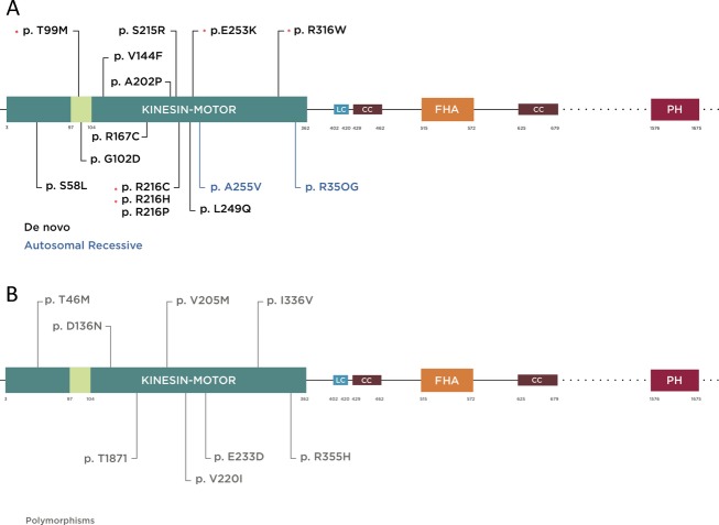
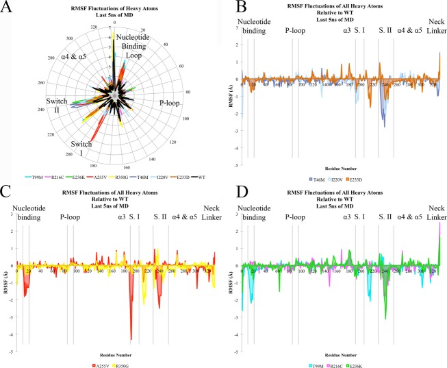
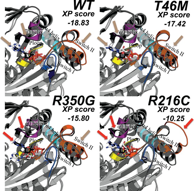
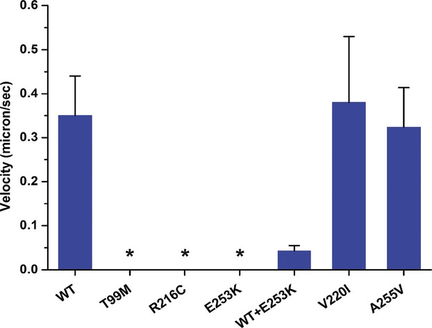
Methods: Clinical whole-exome sequencing was performed for global developmental delay and intellectual disability; some patients also had spastic paraparesis and evidence of clinical regression. Six patients were identified with de novo missense mutations in the kinesin gene KIF1A. The predicted functional disruption of these mutations was assessed in silico to compare the calculated conformational flexibility and estimated efficiency of ATP binding to kinesin motor domains of wild-type (WT) versus mutant alleles. Additionally, an in vitro microtubule gliding assay was performed to assess the effects of de novo dominant, inherited recessive, and polymorphic variants on KIF1A motor function.

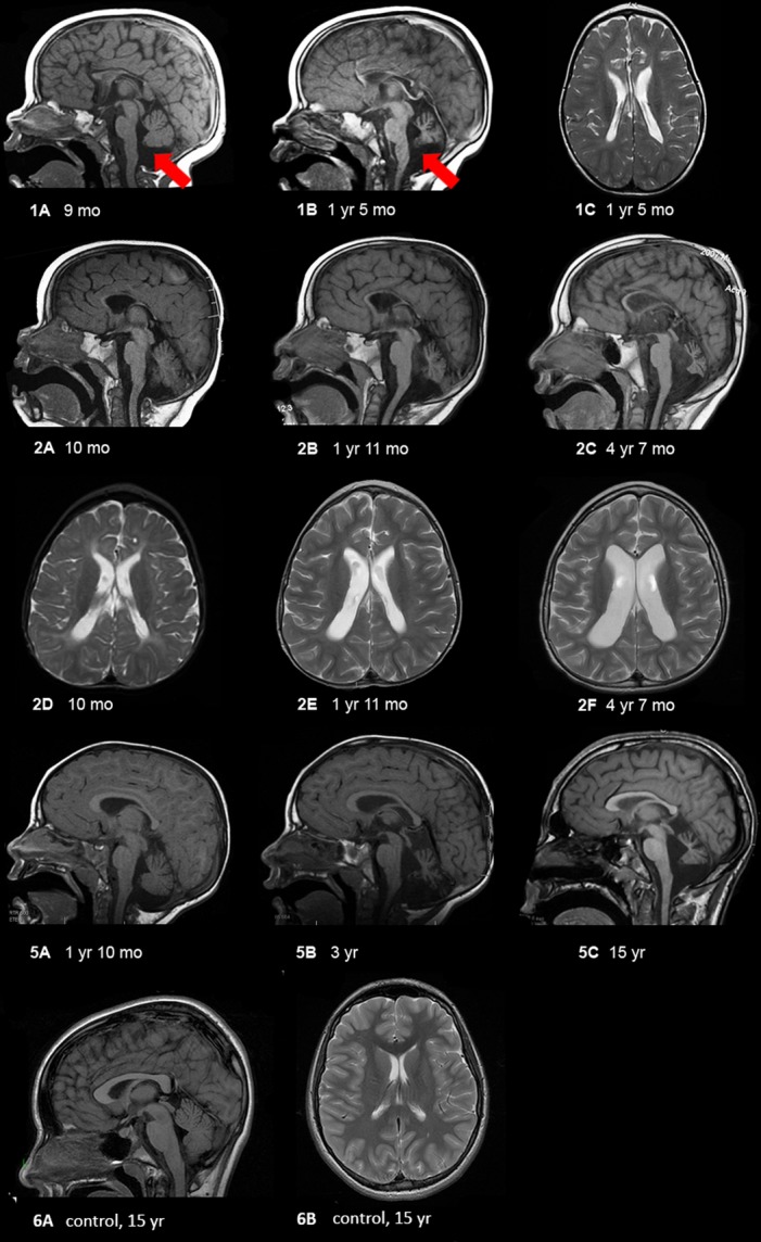
Results: All six subjects had severe developmental delay, hypotonia, and varying degrees of hyperreflexia and spastic paraparesis. Microcephaly, cortical visual impairment, optic neuropathy, peripheral neuropathy, ataxia, epilepsy, and movement disorders were also observed. All six patients had a degenerative neurologic course with progressive cerebral and cerebellar atrophy seen on sequential magnetic resonance imaging scans. Computational modeling of mutant protein structures when compared to WT kinesin showed substantial differences in conformational flexibility and ATP-binding efficiency. The de novo KIF1A mutants were nonmotile in the microtubule gliding assay.

Interpretation: De novo mutations in KIF1A cause a degenerative neurologic syndrome with brain atrophy. Computational and in vitro assays differentiate the severity of dominant de novo heterozygous versus inherited recessive KIF1A mutations. The profound effect de novo mutations have on axonal transport is likely related to the cause of progressive neurologic impairment in these patients.


Figures

Figure 1
Figure 1
Schematic diagram of KIF1A protein illustrating functional domains and mutations reported to date (more information in the main text). Kinesin motor domain 3–362 aa, low complexity domains (LC); 402–420, 684–697, 753–769, 981–994, 1415–1435, 1533–1540 aa, (only 402–420 is depicted for limited space), coiled coil domain (CC) 429–462, 625–672 aa, forkhead-associated domain (FHA) 515–572 aa. The regions extending beyond this figure are represented by dotted lines. (A) Mutations are color coded according to inheritance pattern and clinical severity, with de novo in black and recessive in blue. De novo mutations identified specifically in this study are shown by asterisk. T99M has been previously reported and occurred twice with the same nucleotide and predicted amino acid change in our cohort. It is located within the ATP-binding cassette of the kinesin motor domain. (B) Mutations reported as rare polymorphic variants in a control cohort based on data from the Exome Variant Server (http://evs.gs.washington.edu/EVS/) are shown in gray.
Figure 2
Figure 2
MRI findings in selected patients showing progressive cerebellar and cerebral atrophy. (1) Patient 1 at 9 months (1A) and 17 months (1B and C) showing progressive loss of white matter, extremely thin corpus callosum, and cerebellar atrophy. The red arrows delineate vermian atrophy. (2) Patient 2 at 10 months (2A and D), 23 months (2B and E), and 4 years and 7 months (2C and F) showing progressive atrophy of the entire brain, with early cerebral atrophy (mainly white matter) followed by very severe cerebellar (nuclei > vermis > hemispheres, and anterior vermis earlier than posterior vermis) and optic nerve atrophy (not shown). Midbrain and pontine atrophy occur simultaneously with cerebellar atrophy. (5) Patient 5 at 22 months (5A), 36 months (5B), and 15 years (5C) of age showing mildly diminished white matter with thinning of the corpus callosum, moderate ex vacuo enlargement of the 4th ventricle and severe cerebellar atrophy including the cerebellar hemispheres and deep nuclei, rostra > caudal vermis, and superior cerebellar peduncles. For comparison, we have shown a mid-sagittal T1-weighted image showing a large corpus callosum and intact cerebellar vermis and a axial T2-weighted image showing no volume loss with normal sized ventricles and no increased extra-axial space.
Figure 3
Figure 3
Motor domain molecular dynamic simulation. (A–D) RMSF (root mean square fluctuation) of KIF1A motor-ATP complex over the final 5 nsec of simulation time based on heavy atoms of each residue. (A) Radial representation of RMSF values (Å) for all mutants and wild-type (WT) simulation. (B–D) Depiction of the extent of flexibility relative to the WT simulation, in which a negative number corresponds to a higher degree of flexibility. (B) corresponds to the autosomal dominant de novo mutations, (C) to the recessive mutants, and (D) to rare polymorphic variants from the exome variant server. The color-coding legend for the different mutations is represented at the bottom of each panel.
Figure 4
Figure 4
Best-scoring poses (after running molecular dynamic simulations with the program Glide) for wild-type (WT) and three mutant kinesin motor domains. Distinct regions of the kinesin protein include the P-loop (yellow), Switch I (blue), Switch II (orange), the α0 helix (purple), and the α4-α5 helices (cyan). The location of ATP in the crystal structure (PDB ID 4HNA) is displayed in white as sticks and the docked ATP from the molecular dynamic simulations is displayed with black sticks. Colored arrows that indicate a high degree of overlap between the crystal and computed structures are labeled beige in the WT nonpathogenic (T46M) variant while a lower degree of overlap was observed (red arrows) for recessive (R350G) and dominant (R216C) mutations.
Figure 5
Figure 5
Microtubule gliding velocities of KIF1a motor domain as (described in the gliding assay methods). For absolute values and sample size see Table S2. No motility was observed for T99M, R216C, and E253K mutants during the assay (marked with asterisks).

References

    1. Need AC, Shashi V, Hitomi Y, et al. Clinical application of exome sequencing in undiagnosed genetic conditions. J Med Genet. 2012;49:353–361. - PMC - PubMed
    1. Yang Y, Muzny DM, Reid JG, et al. Clinical whole-exome sequencing for the diagnosis of mendelian disorders. N Engl J Med. 2013;369:1502–1511. - PMC - PubMed
    1. Rauch A, Wieczorek D, Graf E, et al. Range of genetic mutations associated with severe non-syndromic sporadic intellectual disability: an exome sequencing study. Lancet. 2012;380:1674–1682. - PubMed
    1. Riviere JB, Ramalingam S, Lavastre V, et al. KIF1A, an axonal transporter of synaptic vesicles, is mutated in hereditary sensory and autonomic neuropathy type 2. Am J Hum Genet. 2011;89:219–230. - PMC - PubMed
    1. Erlich Y, Edvardson S, Hodges E, et al. Exome sequencing and disease-network analysis of a single family implicate a mutation in KIF1A in hereditary spastic paraparesis. Genome Res. 2011;21:658–664. - PMC - PubMed

LinkOut - more resources