. 2015 Sep;156(9):3370-80.
doi: 10.1210/en.2015-1121. Epub 2015 Jun 30.

miR-122 Regulates LH Receptor Expression by Activating Sterol Response Element Binding Protein in Rat Ovaries

Affiliations

miR-122 Regulates LH Receptor Expression by Activating Sterol Response Element Binding Protein in Rat Ovaries

Bindu Menon et al. Endocrinology. 2015 Sep.

Abstract

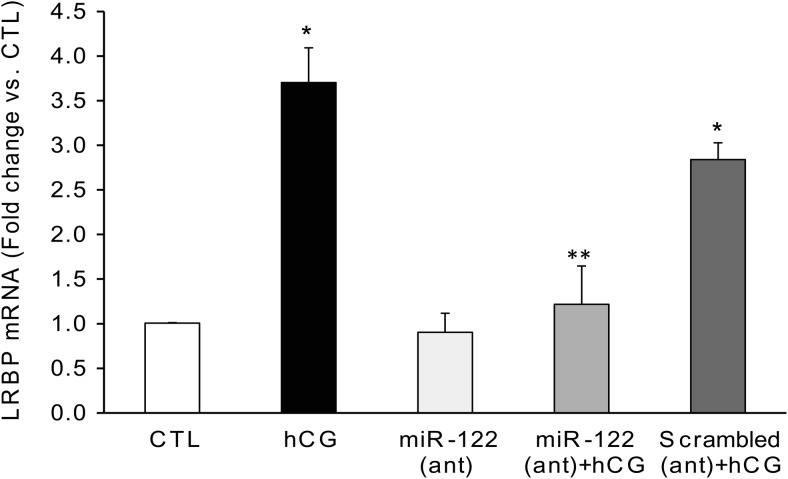
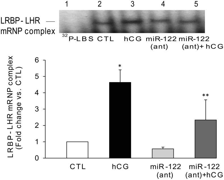
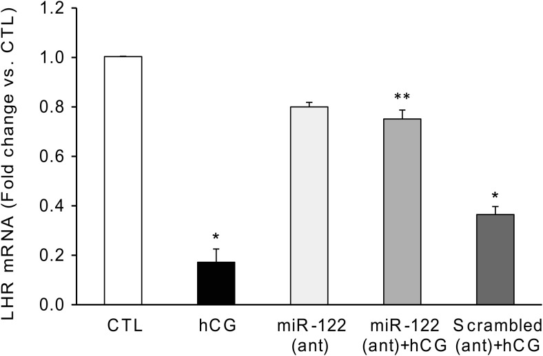
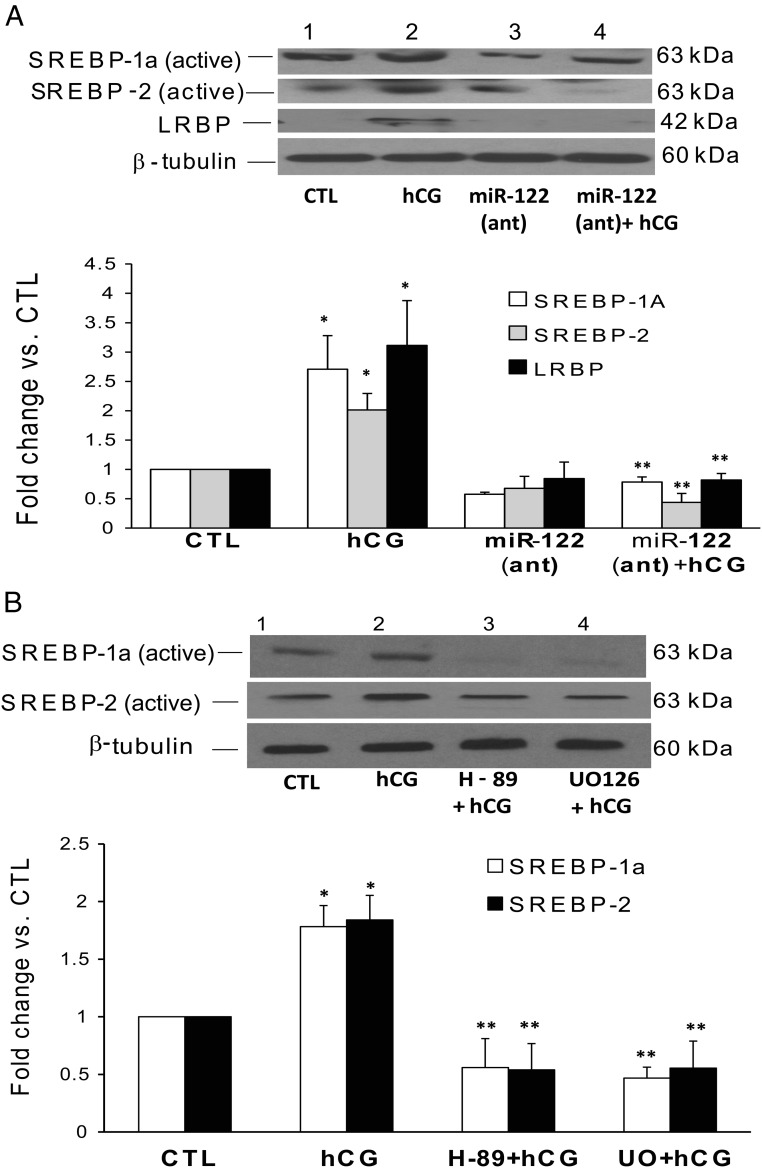
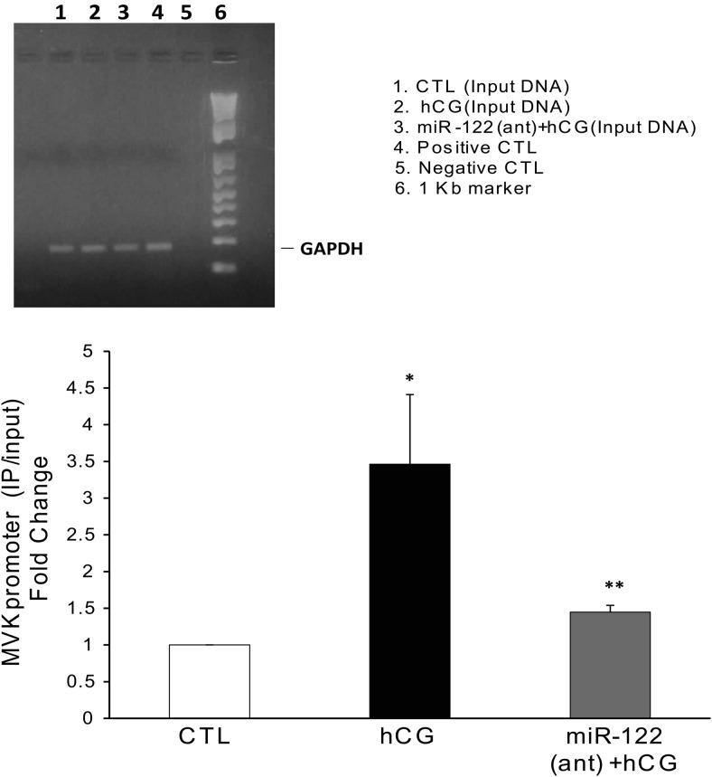
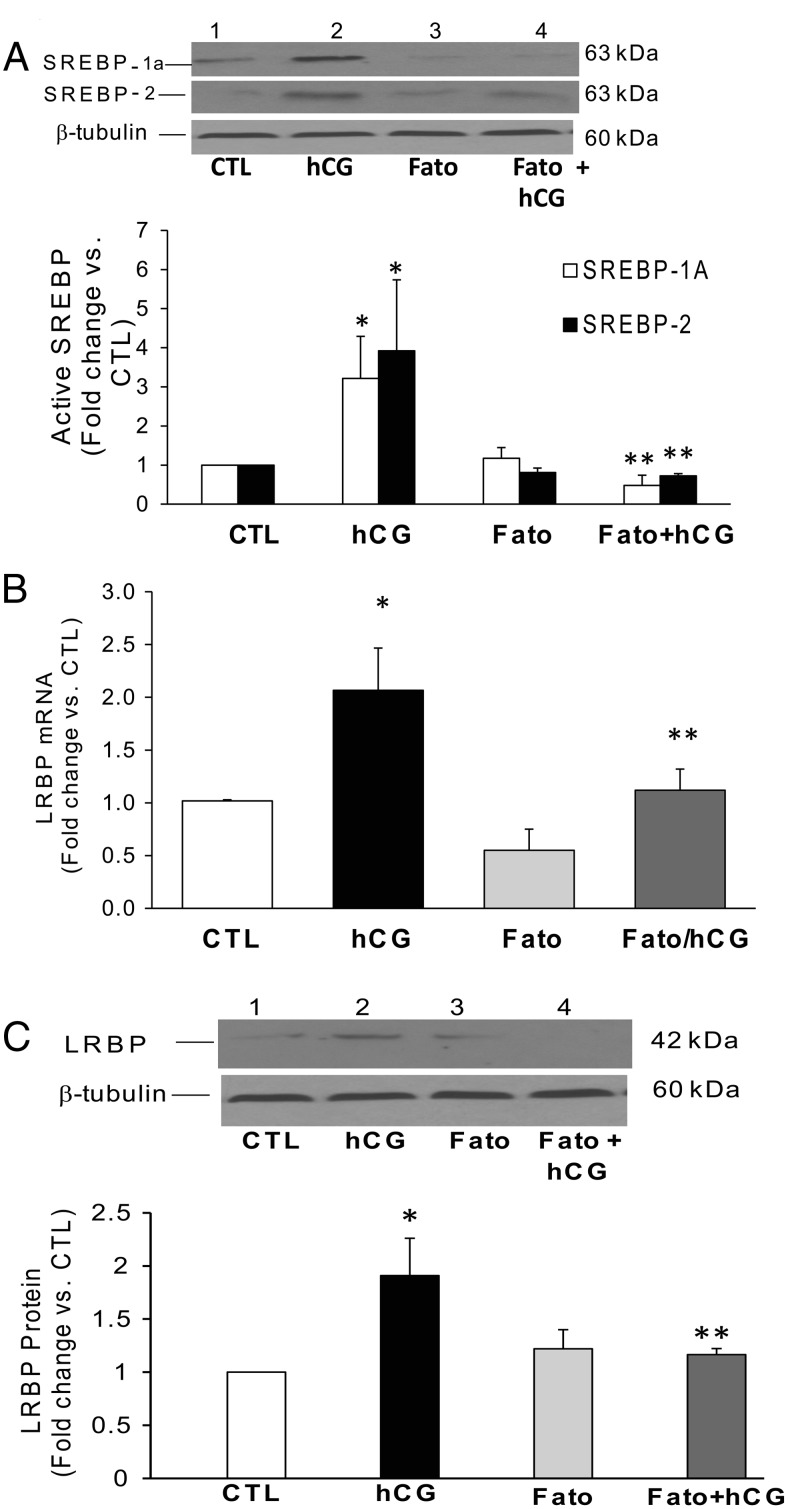
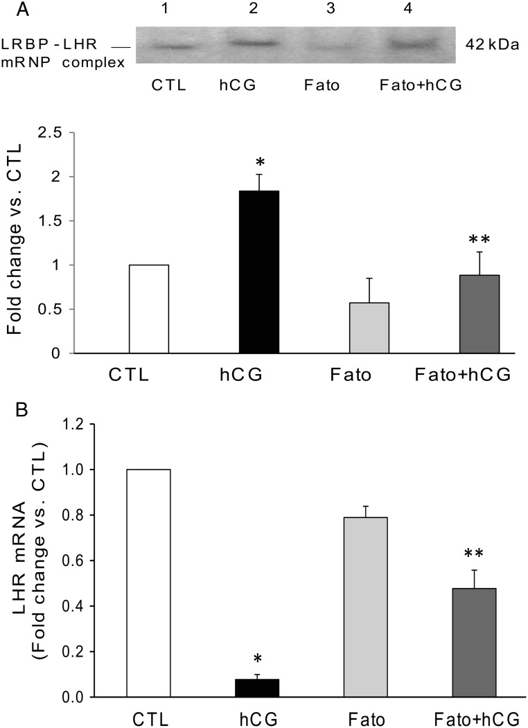
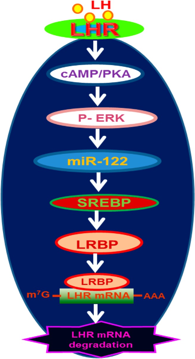
LH/human chorionic gonadotropin receptor (LHR) undergoes down-regulation during preovulatory LH surge or in response to exposure to a supraphysiological concentration of its ligands through a posttranscriptional mechanism involving an RNA binding protein designated as LHR mRNA binding protein (LRBP). miR-122, a short noncoding RNA, has been shown to mediate the up-regulation of LRBP. In the present study, we show that inhibition of miR-122 using a locked nucleic acid (LNA)-conjugated antagomir suppressed human chorionic gonadotropin (hCG)-induced up-regulation of LRBP as well as its association with LHR mRNA, as analyzed by RNA EMSA. Most importantly, inhibition of miR-122 resulted in the abolishment of hCG-mediated LHR mRNA down-regulation. We also show that the transcription factor, sterol regulatory element binding protein (SREBP) (SREBP-1a and SREBP-2 isoforms), is an intermediate in miR-122-mediated LHR mRNA regulation. HCG-stimulated increase in the activation of both SREBP-1a and SREBP-2 was inhibited by pretreatment with the miR-122 antagomir. The inhibition of cAMP/protein kinase A (PKA) and ERK pathways, upstream activators of miR-122, abolished SREBP activation after hCG treatment. SREBP-mediated regulation of LRBP expression is mediated by recruitment of LRBP promoter element to SREBP-1a, because chromatin immunoprecipitation assay revealed that association of LRBP promoter to SREBP was increased by hCG treatment. Pretreatment with miR-122 antagomir suppressed this response. Inhibition of SREBP activation by pretreating the rats with a chemical compound, fatostatin abrogated hCG-induced up-regulation of LRBP mRNA and protein. Fatostatin also inhibited LHR-LRBP mRNA-protein complex formation and LHR down-regulation. These results conclusively show that miR-122 plays a regulatory role in LH/hCG-induced LHR mRNA down-regulation by increasing LRBP expression through the activation of SREBP pathway.


Figures

Figure 1.
Figure 1.
Inhibition of miR-122 abrogated hCG-induced increases in LRBP gene expression. LNA-conjugated miR-122 antagomir (10 mg/kg b.w.) or scrambled LNA CTL was injected into the bursa of the ovaries of superovulated rats on day 4. Animals were allowed to recover and treated with hCG (50 IU) on day 5. Ovaries were collected 4 hours later and were processed for total RNA isolation. Total RNAs were reverse transcribed, and the resulting cDNAs were subjected to real-time PCR quantitation using predesigned primers and probes for LRBP as described in Materials and Methods. The graphs represent ratio of LRBP levels normalized to 18S rRNA and are expressed as percent change vs CTL. Error bars represent mean ± SE. *, P < .05 vs CTL and **, P < .05 vs hCG; n = 4.
Figure 2.
Figure 2.
Inhibition of miR-122 using a specific antagomir inhibits hCG-induced increase in LRBP binding activity. LNA-conjugated miR-122 antagomir was injected into the bursa of the ovaries of superovulated rats on day 4, followed by hCG on day 5. Ovaries were collected 4 hours later and S100 fractions isolated as described previously (23). Electrophoretic gel mobility shift analysis was performed using S100 fractions isolated from ovaries from different treatment groups, containing equal amounts of total protein, after incubating with [32P]-labeled LBS (1.5 × 10⁵ c.p.m). The autoradiogram shown is representative of 3 independent experiments (biological replicates). The graph represents quantitative analysis of the bands in the autoradiogram. Error bars represent mean ± SE. *, P < .05 vs CTL and **, P < .05 vs hCG; n = 3.
Figure 3.
Figure 3.
miR-122 antagomir significantly reversed hCG-induced LHR mRNA down-regulation. LNA-conjugated miR-122 antagomir or scrambled LNA CTL was injected into the bursa of the ovaries of superovulated rats on day 4, followed by hCG on day 5. Ovaries were collected 12 hours later and were processed for total RNA isolation. Total RNAs were reverse transcribed, and the resulting cDNAs were subjected to real-time PCR quantitation using specific primers and probes for LHR. The graph represents changes in mRNA levels normalized to 18S rRNA and shown as fold change vs CTL. Error bars represent mean ± SE. *, P < .05 vs CTL and **, P < .05 vs hCG; n = 6.
Figure 4.
Figure 4.
Inhibition of miR-122, PKA, and ERK1/2 abrogates hCG-induced increases in SREBP activation in the superovulated rat ovaries. Superovulated rats were injected either with (A) miR-122 antagomir on day 4 or with H-89 (100 mg/kg b.w.) or U0126 (10 mg/kg b.w.) (B) 1 hour before hCG injection on day 5, and ovaries were collected 4 hours later. S10 fractions were prepared using RIPA buffer. Equal amounts of protein from the CTL or hCG-treated S10 fractions were subjected to Western blot analysis using SREBP-1a antibody. The blots were stripped and reprobed with SREBP-2, LRBP, and finally with tubulin antibody. The blots shown are representative of 3 independent experiments. The graphs represent SREBP levels normalized to tubulin and are shown as percent change vs CTL. Error bars represent mean ± SE. *, P < .05 vs CTL and **, P < .05 vs hCG.
Figure 5.
Figure 5.
HCG-induced increases in the binding of SREBP to the LRBP promoter was abrogated on pretreatment with miR-122 antagomir. LNA-conjugated miR-122 antagomir was injected into the bursa of the ovaries of superovulated rats on day 4, followed by hCG on day 5. Ovaries were collected 4 hours later and were processed for ChIP assay as described in detail in Materials and Methods. Upper panel, Tissues were cross-linked, and ChIP assay was performed with mouse IgG (negative CTL) or Pol II antibody (positive CTL), as described in Materials and Methods. The purified DNA samples from ChIP assay (positive and negative CTLs) as well as input DNA were amplified by PCR using rat primer sequences for the GAPDH gene promoter, and the PCR products were analyzed using 1.2% agarose gel electrophoresis. Lower panel, The immunoprecipitated DNA, along with input DNA, was subjected to real-time PCR quantitation using specific primers and probes for MVK promoter and GAPDH. The graph represents MVK promoter levels in the immunoprecipitated samples normalized to input DNA and shown as fold change vs CTL. Error bars represent mean ± SE. *, P < .05 vs CTL and **, P < .05 vs hCG; n = 3.
Figure 6.
Figure 6.
Inhibition of hCG-induced increases in SREBP activation and LRBP expression using fatostatin (Fato) pretreatment. Superovulated rats were injected with fatostatin (30 mg/kg b.w.) 4 hours before hCG injection on day 5, and ovaries were collected 4 hours later. S10 fractions were prepared using RIPA buffer. Equal amounts of protein from the S10 fractions were subjected to Western blot analysis using (A) SREBP-1a antibody followed by stripping and reprobing with SREBP-2 and tubulin antibodies or with (C) LRBP antibody followed by stripping and reprobing with tubulin. The blots shown are representative of 3 independent experiments. The graphs represent active SREBP (68 kDa) levels normalized to tubulin and are shown as percent change vs CTL. Error bars represent mean ± SE. *, P < .05 vs CTL and **, P < .05 vs hCG. B. Separate set of ovaries were processed for total RNA isolation, which were then reverse transcribed, and the resulting cDNAs subjected to real-time PCR quantitation using specific primers and probes for LRBP. The graph represents changes in mRNA levels normalized to 18S rRNA and shown as fold change vs CTL. Error bars represent mean ± SE. *, P < .05 vs CTL and **, P < .05 vs hCG; n = 3.
Figure 7.
Figure 7.
Fatostatin (Fato) pretreatment results in the reversal of hCG-induced increases in LHR mRNA binding activity of LRBP and LHR down-regulation. Superovulated rats were injected with fatostatin (30 mg/kg b.w.) 4 hours before hCG injection on day 5, and ovaries were collected 6 hours later. Ovaries were processed for total RNA isolation or REMSA. A, Gel mobility shift analysis was performed using S100 fractions isolated from ovaries from different treatment groups, containing equal amounts of total protein, after incubating with [32P]-labeled LBS (1.5 × 10⁵ c.p.m). The autoradiogram shown is representative of 3 independent experiments. B, Total RNAs were reverse transcribed, and the resulting cDNAs were subjected to real-time PCR quantitation using specific primers and probes for LHR. The graph represents changes in mRNA levels normalized to 18S rRNA and shown as fold change vs CTL. Error bars represent mean ± SE. *, P < .05 vs CTL; **, P < .05 vs hCG; n = 4.
Figure 8.
Figure 8.
Schematic model depicting the proposed signaling pathway in LH/hCG-induced miR-122-mediated LHR mRNA down-regulation. Binding of ligand to LH receptor induces activation of cAMP/PKA signaling followed by ERK1/2. This leads to an increase in the expression of miR-122, which causes the activation of SREBPs. Activated SREBPs increase the gene expression of LRBP, and the increase in translated protein is expressed as an increase in its LHR mRNA binding activity, ultimately leading to the degradation of LHR mRNA.

References

    1. McFarland KC, Sprengel R, Phillips HS, et al. Lutropin-choriogonadotropin receptor: an unusual member of the G protein-coupled receptor family. Science. 1989;245(4917):494–499. - PubMed
    1. Loosfelt H, Misrahi M, Atger M, et al. Cloning and sequencing of porcine LH-hCG receptor cDNA: variants lacking transmembrane domain. Science. 1989;245(4917):525–528. - PubMed
    1. LaPolt PS, Oikawa M, Jia XC, Dargan C, Hsueh AJ. Gonadotropin-induced up- and down-regulation of rat ovarian LH receptor message levels during follicular growth, ovulation and luteinization. Endocrinology. 1990;126(6):3277–3279. - PubMed
    1. Segaloff DL, Wang HY, Richards JS. Hormonal regulation of luteinizing hormone/chorionic gonadotropin receptor mRNA in rat ovarian cells during follicular development and luteinization. Mol Endocrinol. 1990;4(12):1856–1865. - PubMed
    1. Hoffman YM, Peegel H, Sprock MJ, Zhang QY, Menon KMJ. Evidence that human chorionic gonadotropin/luteinizing hormone receptor down-regulation involves decreased levels of receptor messenger ribonucleic acid. Endocrinology. 1991;128(1):388–393. - PubMed

Publication types

MeSH terms