. 2015 Jun 30;10(6):e0130811.
doi: 10.1371/journal.pone.0130811. eCollection 2015.

HLA-B27-Homodimer-Specific Antibody Modulates the Expansion of Pro-Inflammatory T-Cells in HLA-B27 Transgenic Rats

Affiliations

HLA-B27-Homodimer-Specific Antibody Modulates the Expansion of Pro-Inflammatory T-Cells in HLA-B27 Transgenic Rats

Osiris Marroquin Belaunzaran et al. PLoS One. .

Abstract

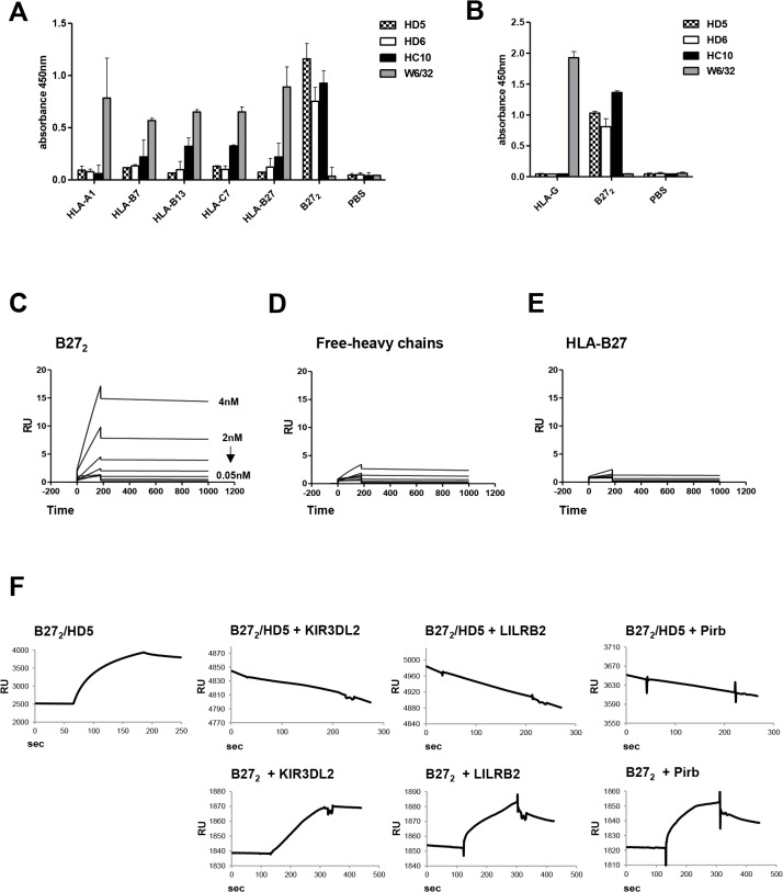
Objectives: HLA-B27 is a common genetic risk factor for the development of Spondyloarthritides (SpA). HLA-B27 can misfold to form cell-surface heavy chain homodimers (B272) and induce pro-inflammatory responses that may lead to SpA pathogenesis. The presence of B272 can be detected on leukocytes of HLA-B27+ Ankylosing spondylitis (AS) patients and HLA-B27 transgenic rats. We characterized a novel B272-specific monoclonal antibody to study its therapeutic use in HLA-B27 associated disorders.

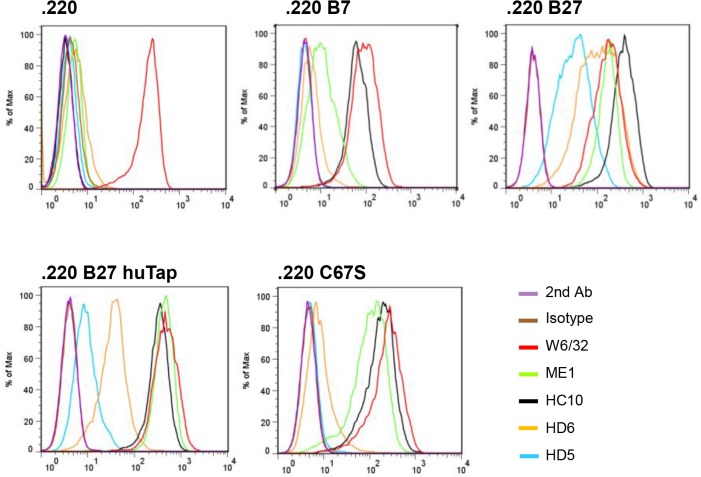
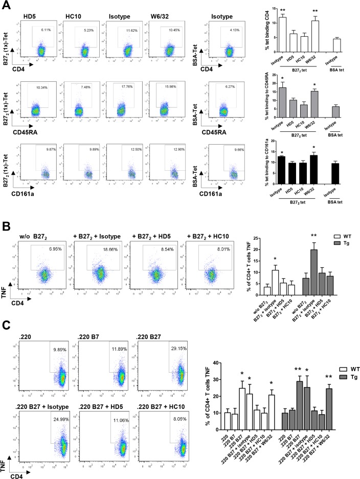
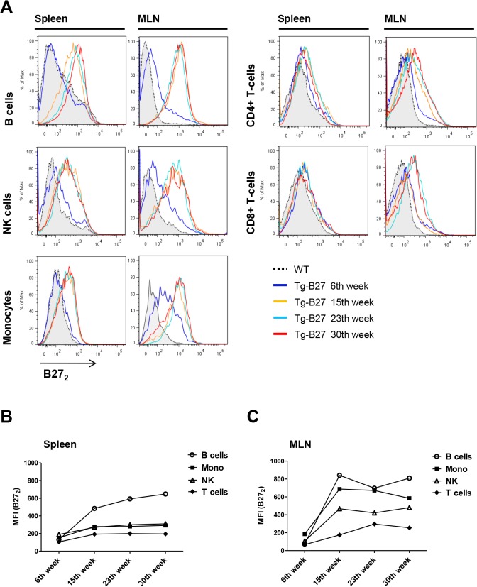
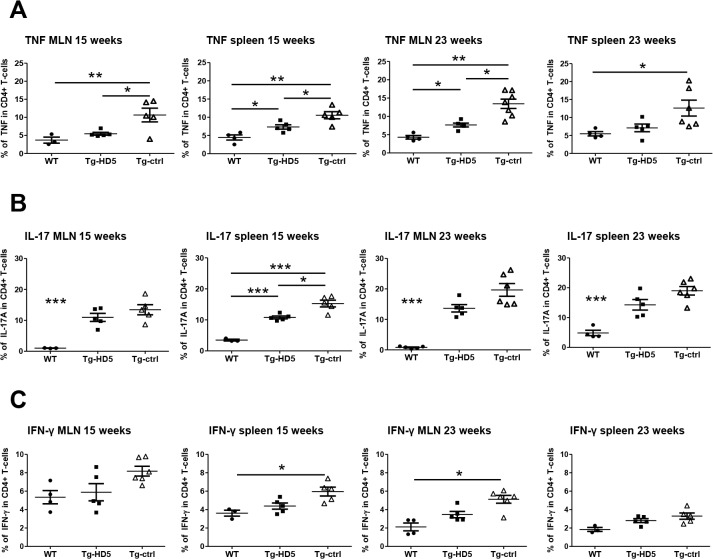
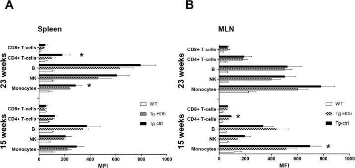
Methods: The monoclonal HD5 antibody was selected from a phage library to target cell-surface B272 homodimers and characterized for affinity, specificity and ligand binding. The immune modulating effect of HD5 was tested in HLA-B27 transgenic rats. Onset and progression of disease profiles were monitored during therapy. Cell-surface B272 and expansion of pro-inflammatory cells from blood, spleen and draining lymph nodes were assessed by flow cytometry.

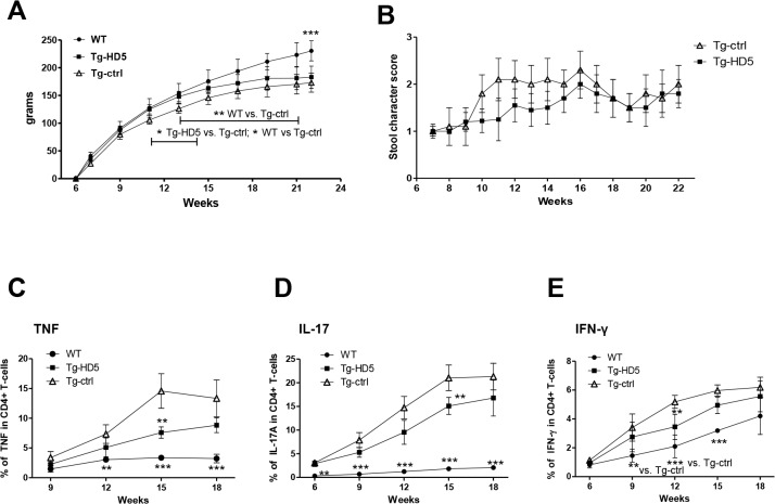
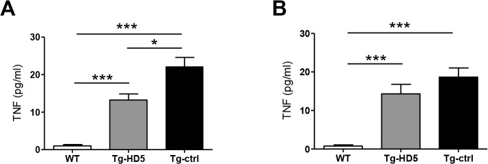
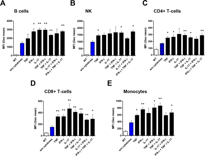
Results: HD5 bound B272 with high specificity and affinity (Kd = 0.32 nM). HD5 blocked cell-surface interaction of B272 with immune regulatory receptors KIR3DL2, LILRB2 and Pirb. In addition, HD5 modulated the production of TNF from CD4+ T-cells by limiting B272 interactions in vitro. In an HLA-B27 transgenic rat model repetitive dosing of HD5 reduced the expansion of pro-inflammatory CD4+ T-cells, and decreased the levels of soluble TNF and number of cell-surface B272 molecules.

Conclusion: HD5 predominantly inhibits early TNF production and expansion of pro-inflammatory CD4+ T-cells in HLA-B27 transgenic rats. Monoclonal antibodies targeting cell-surface B272 propose a new concept for the modulation of inflammatory responses in HLA-B27 related disorders.


Conflict of interest statement

Competing Interests: The authors have declared that no competing interests exist.

Figures

Fig 1
Fig 1. HD5 mAb is specific to B272, binds to recombinant B272, and blocks interaction with immunoregulatory cell receptors.
(A) ELISA results showed the specificity of HD5 and HD6 to B272 homodimers when challenged for different recombinant HLA class I complexes (-A1,-B7,-B13,-C7,-B27, and-B272). Control HC10 and W6/32 antibodies were used as positive and negative control, respectively. (B) Direct ELISA against HLA-G and B272 homodimers using HD5, HD6, HC10 and W6/32 antibodies showed specificity of HD5 and HD6 to immobilized B272 homodimers but not to HLA-G homodimers. (C-E) Recombinant B272, B27-free-heavy chain and HLA-B27 heterotrimers were immobilized into chips for kinetic characterization by SPR. C) HD5 and ligand (B272) have a Kd of 0.32 nM. Immobilized free-heavy chains (D) or HLA–B27 heterotrimers (E) did not interact with HD5 and Kd values were not fitted. (F) Blocking competition experiments in SPR were performed by immobilizing B272 to a streptavidin chip followed by injection of HD5 to form B272-HD5 complexes. Injections of LILRB2, KIR3DL2 and Pirb were assessed and binding events recorded.
Fig 2
Fig 2. HD5 binds specifically to cell-surface B272 homodimers expressed in human .220 B cell lines.
Representative flow cytometry analysis of HD5 and control antibodies (IgG1 Isotype, W6/32, ME1, HC10, and HD6) binding to LBL721.220 cells either untransfected (.220), or transfected with HLA–B7, B27.C67S (HLA-B27 with a mutation of Cys-67 to serine that abrogates cell-surface B272 expression), B27.hutpn (HLA-B27 together with transfection of human tapasin, that reduces B272 expression and increases HLA-B27 heterotrimer formation), or HLA-B27 (HLA-B27 transfected cells expressing high levels of cell-surface B272). RU = responsive units. ELISA values are expressed as mean±SEM.
Fig 3
Fig 3. HD5 mAb blocks interaction of B272 to leukocytes and modulates activation of CD4+ T-cells by B272.
(A) B272 (1x)-tetramers were shown to bind to CD4+ T-cells, B-cells (CD45RA,-lineage) and NK (CD161a,-lineage) populations. Pre-incubation of HD5 or HC10 with B272(1x)-tetramers partially blocked interactions with cells when compared to control antibodies (W6/32 and isotype). (B) Tg and WT CD4+ T-cells produce TNF after incubation with B272 (1x)-tetramers. Pre-incubation of HD5 or HC10 with B272 (1x)-tetramers inhibited the interaction to CD4+ T-cells and the production of TNF. (C) Tg and WT CD4+ T-cells produce TNF after incubation with .220 B27cells expressing B272 dimers, but not with .220 and .220 B7 cells. Pre-incubation of HD5 or HC10 with .220 B27 cells blocked the interaction of B272 to CD4+ T-cells and the production of TNF. Representative flow cytometry plots belong to leukocytes populations of Tg rats. Tests were performed in triplicates. Tet = tetramer. Values are expressed as mean±SEM. *p<0.05, **p<0.01, as determined by one-way ANOVA followed by Bonferroni post-hoc analysis.
Fig 4
Fig 4. Detection of cell-surface B272 in leukocyte populations of HLA-B27 transgenic rats at different ages.
(A) Representative flow cytometry analysis of cell-surface B272 expression in leukocytes populations of Tg rats at different ages (6, 15, 23 and 30 weeks) from spleen and MLNs. WT leukocytes represent the control population where B272 is absent. (B) MFI values plotted of positive B272 stains from splenocytes. (C) MFI values plotted of positive B272 stains from MLNs. Detection of cell-surface B272 homodimers was performed using HD6-biotinylated and detected by streptavidin-APC. HD6 had been previously assessed as an antibody capable of recognizing cell-surface B272 in human [12] and rat [41] leukocyte populations. Antibody panels: CD4+ T-cells (+CD3, +CD4), CD8+ T-cells (+CD3, +CD8), NK (+CD161a,—lineage), B cells (+CD45RA,-lineage) and Monocytes (+CD172a,—RP-1).
Fig 5
Fig 5. Increased body weight and decreased expansion of pro-inflammatory CD4+ T-cells from blood in Tg-HD5 rats.
(A) Body weight gain from the 6th week of age where the first antibody injections were performed. (B) Stool score from Tg-HD5 and Tg-ctrl groups. (C-E) In vitro stimulated cells obtained at different time points from the blood of Tg-HD5, Tg-ctrl and WT-littermates were assessed by ICS for the presence of pro-inflammatory cells expressing TNF (C), IL-17 (D) and IFN-γ (E). Results are expressed as percentages of CD4+ T gated cells. Values are expressed as mean±SEM. *p<0.05, **p<0.01, ***p<0.005 as determined by one-way ANOVA followed by Bonferroni post-hoc analysis.
Fig 6
Fig 6. Reduced expansion of pro-inflammatory CD4+ T-cells from spleen and MLN in Tg-HD5 rats.
In vitro stimulated cells obtained from Tg-HD5, Tg-ctrl and WT-littermates were assessed by ICS for the presence of pro-inflammatory cells expressing TNF (A), IL-17 (B) and IFN-γ (C). MLN and spleens cells were obtained at week 15 (n = 5) and at week 23 (n = 5). Results are plotted as the percentage of CD4+ T-cells gated positive for TNF, IL-17 and IFN-γ. Values are expressed as mean±SEM. *p<0.05, **p<0.01, ***p<0.005 as determined by one-way ANOVA followed by Bonferroni post-hoc analysis.
Fig 7
Fig 7. Reduced amount of soluble TNF in Tg-HD5 rats.
Serum obtained from the blood after euthanasia was assessed for the presence of soluble TNF. (A-B) TNF serum ELISA from WT, Tg-HD5 and Tg-ctrl (n = 5 per group) at 15 weeks (A) and at 23 weeks (B).
Fig 8
Fig 8. Pro-inflammatory cytokines increase cell-surface B272 homodimers.
(A-E) Cell cultures of leukocyte populations were analyzed by flow cytometry for presence of cell-surface B272 homodimers after stimulation with cytokines. HD6-biotinylated and streptavidin APC were used for detection. Mean fluorescence intensity (MFI) values were plotted from data analysis. Statistical analysis was plotted against “without cytokines” data values. Values are expressed as mean±SEM. *p<0.05, **p<0.01, ***p<0.005 as determined by one-way ANOVA followed by Bonferroni post-hoc analysis.
Fig 9
Fig 9. Reduced accumulation of cell-surface B272 molecules in Tg-HD5 rats.
Analysis of cell-surface B272 from spleen and MLN was performed using HD6-biotinylated and detected by streptavidin-APC in WT-littermates and HLA-B27 rat groups at 15 and 23 weeks. (A-B) MFI values plotted of positive B272 stains from flow cytometry analyzed splenocytes (n = 5) (A) and MLN (n = 5) (B) subpopulations. Values are expressed as mean±SEM. *p<0.05, ***p<0.005 as determined by one-way ANOVA followed by Bonferroni post-hoc analysis.

Similar articles

Cited by

References

    1. Khan MA. Update on spondyloarthropathies. Ann Intern Med. 2002;136(12):896–907. . - PubMed
    1. Dougados M, Baeten D. Spondyloarthritis. Lancet. 2011;377(9783):2127–37. 10.1016/S0140-6736(11)60071-8 . - DOI - PubMed
    1. Brewerton DA, Caffrey M, Nicholls A, Walters D, Oates JK, James DC. Reiter's disease and HL-A 27. Lancet. 1973;302(7836):996–8. . - PubMed
    1. Bird LA, Peh CA, Kollnberger S, Elliott T, McMichael AJ, Bowness P. Lymphoblastoid cells express HLA-B27 homodimers both intracellularly and at the cell surface following endosomal recycling. Eur J Immunol. 2003;33(3):748–59. 10.1002/eji.200323678 . - DOI - PubMed
    1. Santos SG, Lynch S, Campbell EC, Antoniou AN, Powis SJ. Induction of HLA-B27 heavy chain homodimer formation after activation in dendritic cells. Arthritis Res Ther. 2008;10(4):R100 10.1186/ar2492 - DOI - PMC - PubMed

Publication types