. 2015 Jun 30;10(6):e0131113.
doi: 10.1371/journal.pone.0131113. eCollection 2015.

Hippo Stabilises Its Adaptor Salvador by Antagonising the HECT Ubiquitin Ligase Herc4

Affiliations

Hippo Stabilises Its Adaptor Salvador by Antagonising the HECT Ubiquitin Ligase Herc4

Birgit L Aerne et al. PLoS One. .

Abstract

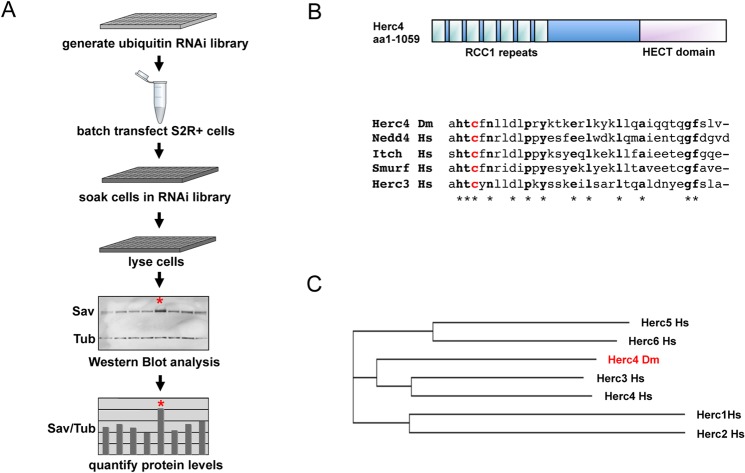
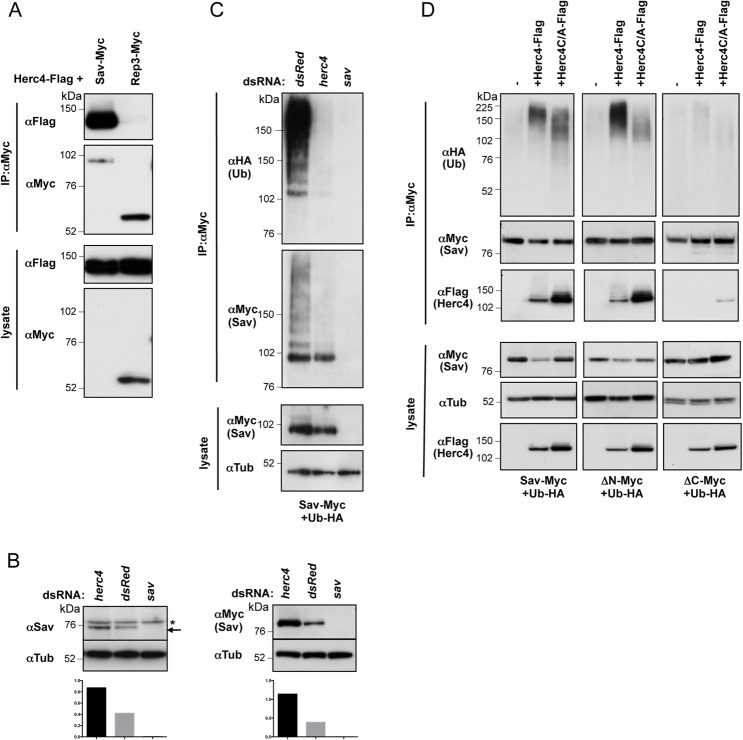
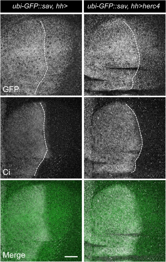
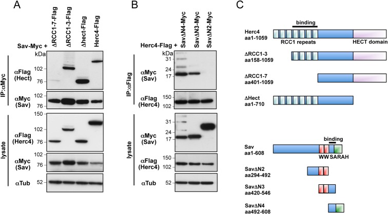
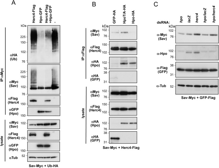
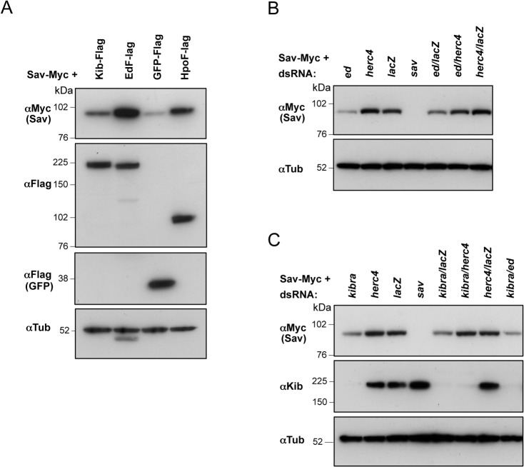
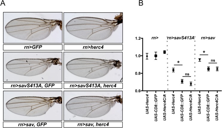
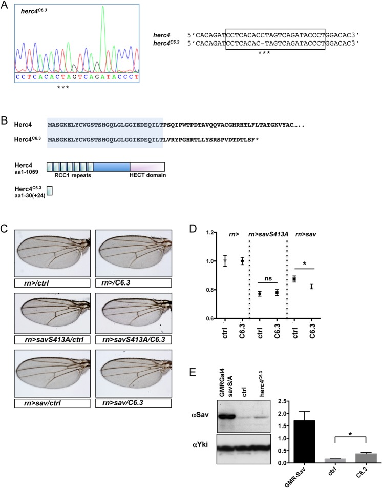
Signalling through the Hippo (Hpo) pathway involves a kinase cascade, which leads to the phosphorylation and inactivation of the pro-growth transcriptional co-activator Yorkie (Yki). Despite the identification of a large number of pathway members and modulators, our understanding of the molecular events that lead to activation of Hpo and the downstream kinase Warts (Wts) remain incomplete. Recently, targeted degradation of several Hpo pathway components has been demonstrated as a means of regulating pathway activity. In particular, the stability of scaffold protein Salvador (Sav), which is believed to promote Hpo/Wts association, is crucially dependent on its binding partner Hpo. In a cell-based RNAi screen for ubiquitin regulators involved in Sav stability, we identify the HECT domain protein Herc4 (HECT and RLD domain containing E3 ligase) as a Sav E3 ligase. Herc4 expression promotes Sav ubiquitylation and degradation, while Herc4 depletion stabilises Sav. Interestingly, Hpo reduces Sav/Herc4 interaction in a kinase-dependent manner. This suggests the existence of a positive feedback loop, where Hpo stabilises its own positive regulator by antagonising Herc4-mediated degradation of Sav.


Conflict of interest statement

Competing Interests: The authors have declared that no competing interests exist.

Figures

Fig 1
Fig 1. Sav is ubiquitylated predominantly in the C-term half of the protein.
(A) Sav is stabilised and shows increased ubiquitylation in the presence of proteasome inhibitors (MG132 and LLnL). Myc-tagged Sav was expressed in S2 cells with either Ub-HA or ß-Galactosidase (ß-Gal) in the presence of proteasome inhibitors (MG132 and LLnL, M/L) or DMSO control. Sav ubiquitylation assay shows incorporation of HA-tagged ubiquitin. (B) Left panel: Sav ubiquitylation occurs in the C-terminal half of the protein. Sav deletions (∆C and ∆N) were tested for ubiquitylation as described above. Right panel: schematic representation of Sav protein structure and distribution of Lysine residues. Sav contains eighteen Lysine residues (K), which could serve as potential ubiquitylation sites: ten are located near the N-terminal end of the protein, whereas the remaining eight are located in the C-terminus.
Fig 2
Fig 2. Hpo stabilises Sav by inhibiting Sav ubiquitylation.
(A) Sav is stabilised when co-expressed with Hpo. Baseline Sav expression is increased upon proteasome inhibitor treatment (M/L). (B) hpo depletion by RNAi destabilises Sav protein levels. Top panel: endogenous Sav levels (arrow) in non-transfected S2 cells. Asterisk indicates a non-specific band detected by the anti-Sav antibody. Bottom panel: levels of transfected Myc-tagged Sav. Sav depletion by RNAi is used as a control. (C) Hpo inhibits ubiquitylation of Sav. Sav, Sav∆N, and Sav∆C were analysed for ubiquitylation in the presence or absence of co-expressed Hpo. Hpo prevents ubiquitylation of full-length Sav and Sav∆N, while Sav∆C is not ubiquitylated. (A-C) S2 cells were transfected with the indicated plasmids or treated with the indicated dsRNAs prior to lysis. Lysates were processed for western blot analysis or used in co-IP.
Fig 3
Fig 3. Binding of active Hpo to Sav is required to inhibit Sav ubiquitylation.
(A) Schematic representation of Hpo constructs used. Protein domains and point mutations in the Hpo T-loop inactivating Hpo kinase (HpoT195A) and PPxY motif (Y591G) are indicated. (B) Sav ubiquitylation assays with co-expressed Hpo truncations (Hpo∆N and ∆C) or Hpo kinase dead (HpoT195A). While full-length Hpo prevents Sav ubiquitylation, truncations lacking the kinase domain or C-terminus, or a kinase-dead Hpo have no effect. (C) Hpo C-terminal deletions (Hpo∆C2 and Hpo∆C3) and a Hpo C-terminal deletion mutated for the PPxY motif (Hpo∆C3/Y591G) affect Sav ubiquitylation proportionally to their ability to bind Sav (compare upper and lower panels).
Fig 4
Fig 4. Ubiquitin ligase RNAi screen identifies Herc4 as a Sav E3 ligase.
(A) E3 RNAi screen work-plan (B) Diagram depicting the Herc4 protein domain structure and alignment of HECT domains. Herc4 contains seven RCC1 repeats and a HECT E3 ligase domain. Protein alignment shows the last 36 aa of Drosophila melanogaster Herc4 aligned with the corresponding sequences of human Nedd4, Itch, Smurf, and Herc3. The conserved catalytic cysteine is shown in red. (C) Phylogenetic tree of human Herc1-6 and Drosophila melanogaster Herc4.
Fig 5
Fig 5. Herc4 controls Sav stability and ubiquitylation.
(A) Sav binds to Herc4. Myc-tagged Sav was immunoprecipitated using a Myc antibody and bound Herc4-Flag protein detected using Flag antibody. Myc-tagged Drosophila Rep3 was used as control. (B) herc4 RNAi stabilises Sav protein levels. Left panel: endogenous Sav levels (arrow) in non-transfected S2 cells. Asterisk indicates a non-specific band detected by the anti-Sav antibody. Left bottom panel: quantification presented as ratio of Sav/Tub levels. Right panel: levels of transfected Myc-tagged Sav. Right bottom panel: quantification presented as ratio of Sav-Myc/Tub levels (C) herc4 RNAi reduces Sav ubiquitylation. Sav ubiquitylation assay of Myc-tagged Sav expressed in S2 cells, treated with dsRed RNAi, herc4 RNAi or sav RNAi. Ubiquitylation assay was performed in the presence of 5mM NEM to block deubiquitylating enzyme activity (D) Overexpression of Herc4 results in destabilisation and increased ubiquitylation of Sav, which is dependent on the active site cysteine. Sav ubiquitylation assay of Myc-tagged Sav, Sav∆N and Sav∆C co-expressing either Flag-Herc4 or Flag-Herc4C/A.
Fig 6
Fig 6. Herc4 overexpression reduces Sav protein levels in vivo.
GFP-tagged Sav was expressed under the control of the ubiquitin promoter (ubi-GFP::Sav) and analysed in 3rd instar larval wing discs expressing hh-GAL4 alone (left panel) or UAS-herc4 in the posterior compartment (hh>herc4). White dotted lines denote the border between the anterior and posterior compartments of the wing. Anti-Ci staining marks the anterior compartment. Scale bar = 100μm.
Fig 7
Fig 7. Mapping of Herc4/Sav binding.
(A) Mapping of Herc4 interaction domain with Sav. Co-IPs of Sav with Herc4 truncations lacking the N-terminal RCC1 domains (∆RCC1-3, ∆RCC1-7) or the HECT domain (∆HECT). The RCC4-7 repeats are required for binding of Herc4 to Sav. (B) Mapping of the Sav interaction domain with Herc4. C-terminal fragments of Sav were used in co-immunoprecipitation experiments with Herc4. (C) Schematic representation of Herc4 and Sav truncations used in A and B.
Fig 8
Fig 8. Hpo competes with Herc4 for Sav binding and antagonises Herc4-mediated ubiquitylation of Sav.
(A) Hpo reduces Herc4-induced ubiquitylation of Sav. Sav ubiquitylation assay was performed using Myc-tagged Sav co-expressed with either Hpo, Herc4 or Hpo and Herc4 combined. Ubiquitylation assay was performed in the presence of 5mM NEM to block deubiquitylating enzyme activity. (B) Binding of Sav to Herc4 is prevented by active Hpo kinase (compare left with right lane), while kinase-dead Hpo has no effect (compare left and middle lanes). Herc4/Sav co-IP experiments were performed in the presence of a control protein (GFP), wild-type or kinase dead version of Hpo (HpoT195A). (C) herc4 RNAi rescues the destabilizing effect of hpo RNAi on Sav protein levels. S2 cells expressing Myc-tagged Sav (and GFP-Flag as a control) were treated with the indicated combinations of dsRNA.
Fig 9
Fig 9. herc4 depletion prevents the decrease in Sav protein levels elicited by loss of apical Hpo pathway regulators.
(A) Co-expression of Ed and Kibra stabilizes Myc-tagged Sav. Hpo co-transfection is used as a positive control. (B) herc4 RNAi rescues ed RNAi-induced reduction in Sav protein levels. (C) herc4 RNAi rescues kibra RNAi-induced reduction in Sav protein levels. (A-C) S2 cells were transfected with Sav-Myc and the indicated plasmids, and treated with the indicated dsRNAs before lysis and western blot analysis.
Fig 10
Fig 10. Growth regulatory function of Herc4.
(A) Overexpression of Herc4 leads to a partial rescue of the Sav overexpression phenotype. Expression of sav (rn>sav) and savS413A (rn>savS413A) transgenes under the control of the rotund (rn) promoter causes tissue undergrowth and results in smaller wings. Both transgenes were combined with either UAS-herc4, UAS-herc4C/A or UAS-CD8-GFP to address the effect of Herc4. (B) Quantification of wing sizes. Shown are fold changes of average wing areas for the indicated genotypes compared to the control (rn>UAS-CD8-GFP), which was set as 1. Statistical significance assessed by Student’s t-test. * p < 0.0001. N.S. p > 0.05. n = 10–21. Error bars denote standard deviations.
Fig 11
Fig 11. Generation and in vivo analysis of herc4 C6.3 mutant.
(A) Sequence analysis of herc4 CRISPR mutation. Sequence chromatogram of the sgRNA target sequence (boxed in the sequence alignment on the right) showing the loss of cytosine 92 (C92) in the herc4 C6.3 strain (marked by asterisk). (B) Diagram and sequence of the predicted protein resulting from frame-shift mutation. The loss of C92 results in a frame-shift mutation and a truncated protein of 30aa plus 24aa of out-of-frame sequence. (C) herc4 C6.3 enhances the Sav overexpression phenotype in the wing. Both sav (rn>sav) and savS413A (rn>savS413A) transgenes were combined with heterozygosity for either herc4 C6.3 or herc4 ctrl and wings sizes were analysed. (D) Quantification of wing sizes. Shown are fold changes of average wing areas for the indicated genotypes compared to the control (rn/ctrl), which was set as 1. Statistical significance assessed by Student’s t-test. * p < 0.0001. N.S. p > 0.05. n = 10–21. Error bars denote standard deviations. (E) Levels of Sav protein in fly heads of herc4 C6.3, herc4 ctrl and GMR>savS413A were analysed by Western blot. Bottom panel: quantification presented as ratio of Sav/Yki levels from three independent repeats. * p = 0.0385 with a Student’s t-test.

References

    1. Yu FX, Guan KL (2013) The Hippo pathway: regulators and regulations. Genes Dev 27: 355–371. 10.1101/gad.210773.112 - DOI - PMC - PubMed
    1. Lin JI, Poon CL, Harvey KF (2013) The Hippo size control pathway—ever expanding. Sci Signal 6: pe4. - PubMed
    1. Justice RW, Zilian O, Woods DF, Noll M, Bryant PJ (1995) The Drosophila tumor suppressor gene warts encodes a homolog of human myotonic dystrophy kinase and is required for the control of cell shape and proliferation. Genes Dev 9: 534–546. - PubMed
    1. Xu T, Wang W, Zhang S, Stewart RA, Yu W (1995) Identifying tumor suppressors in genetic mosaics: the Drosophila lats gene encodes a putative protein kinase. Development 121: 1053–1063. - PubMed
    1. Tapon N, Harvey KF, Bell DW, Wahrer DC, Schiripo TA, Haber D, et al. (2002) Salvador Promotes both cell cycle exit and apoptosis in Drosophila and is mutated in human cancer cell lines. Cell 110: 467–478. - PubMed

Publication types

MeSH terms

LinkOut - more resources