Review
. 2015 Oct;72(20):3883-96.
doi: 10.1007/s00018-015-1975-2. Epub 2015 Jul 1.

Mesenchymal-epithelial interactions during digestive tract development and epithelial stem cell regeneration

Affiliations
Review

Mesenchymal-epithelial interactions during digestive tract development and epithelial stem cell regeneration

Ludovic Le Guen et al. Cell Mol Life Sci. 2015 Oct.

Abstract

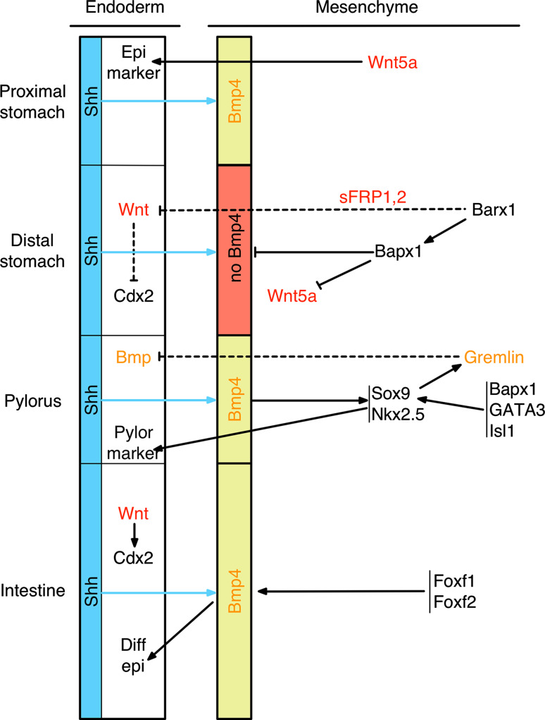
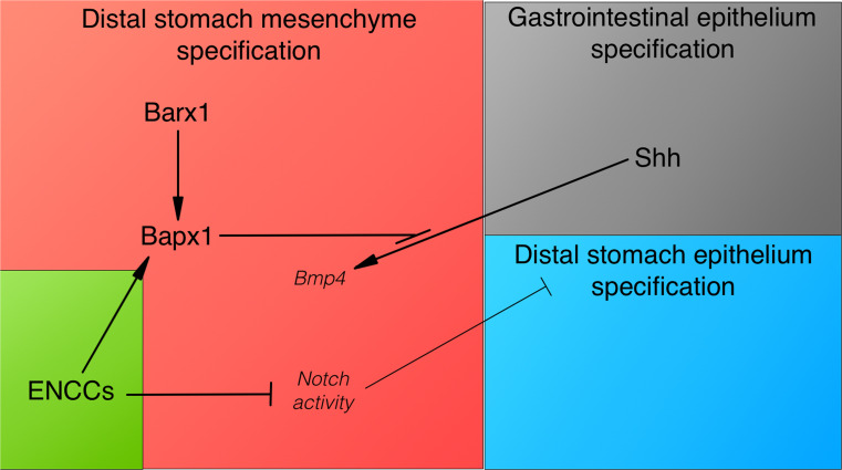
The gastrointestinal tract develops from a simple and uniform tube into a complex organ with specific differentiation patterns along the anterior-posterior and dorso-ventral axes of asymmetry. It is derived from all three germ layers and their cross-talk is important for the regulated development of fetal and adult gastrointestinal structures and organs. Signals from the adjacent mesoderm are essential for the morphogenesis of the overlying epithelium. These mesenchymal-epithelial interactions govern the development and regionalization of the different gastrointestinal epithelia and involve most of the key morphogens and signaling pathways, such as the Hedgehog, BMPs, Notch, WNT, HOX, SOX and FOXF cascades. Moreover, the mechanisms underlying mesenchyme differentiation into smooth muscle cells influence the regionalization of the gastrointestinal epithelium through interactions with the enteric nervous system. In the neonatal and adult gastrointestinal tract, mesenchymal-epithelial interactions are essential for the maintenance of the epithelial regionalization and digestive epithelial homeostasis. Disruption of these interactions is also associated with bowel dysfunction potentially leading to epithelial tumor development. In this review, we will discuss various aspects of the mesenchymal-epithelial interactions observed during digestive epithelium development and differentiation and also during epithelial stem cell regeneration.

Keywords: Anterior-posterior axis; BMP pathway; Colorectal cancer; Enteric nervous system; Epithelial cell; FOXF; Gastrointestinal tract; Hedgehog; Homeotic HOX genes; Mesenchymal–epithelial interactions; Metaplasia; Myofibroblast; NKX2.5; Notch pathway; Regeneration; SOX9; Smooth muscle cell; Stem cell.


Figures

Fig. 1
Fig. 1
Molecular control of the mesenchymal–epithelial interactions in the developing GI tract in vertebrates. Throughout the AP axis, epithelial Shh induces Bmp4 expression in the adjacent mesenchyme, with the exception of the distal stomach where Bapx1 represses Bmp4 and Wnt5a expression. In the distal stomach, Barx1, which is upstream of Bapx1, regulates the mesenchymal expression of the WNT antagonists sFRP1 and sFRP2 that inhibit WNT activity and Cdx expression in the gastric epithelium. In the small intestine, Foxf1 and Foxf2 activate Bmp4, leading to BMP activity in both mesoderm and endoderm. In the pyloric sphincter, Bmp4 activates the expression of Nkx2.5 and Sox9 that induce the pyloric epithelial phenotype through modulation of mesenchymal–epithelial signaling. In addition, Bapx1, Gata3 and Isl1 regulate Sox9 expression in the pyloric structure. SOX9 controls Gremlin expression in the pyloric sphincter mesenchyme. Gremlin, a diffusible factor, modulates the activation of the endodermal BMP pathway to induce the specific pyloric epithelium differentiation
Fig. 2
Fig. 2
Smooth muscle cell differentiation in the small intestine of chicken embryos. Smooth muscle cells (SMC) were detected by immunostaining with an antibody against the SMC-specific marker αSMA. a Non-organized and undifferentiated mesenchymal cells in the small intestine of a 6-day/old (E6) chick embryo. b αSMA-positive cells are present in the smooth muscle layer in the small intestine of an E9 chick embryo. a, b During early embryonic development, the visceral endoderm is uniform with stratified layers of cells. c αSMA-positive cells in the circular smooth muscle layer of the small intestine of an E13 chick embryo. The ongoing intestinal epithelial cytodifferentiation leads to the formation of an epithelial monolayer. d αSMA-positive cells in the longitudinal and circular smooth muscle layers of the small intestine of an E15 chick embryo. e αSMA-positive cells in the longitudinal, circular and submucosal smooth muscle layers in the small intestine of an E16 chick embryo. f αSMA-positive cells (myofibroblasts) are detected also in the lamina propria of the small intestine in a E18 chick embryo. df Intestinal epithelial cytodifferentiation is marked by mesodermal growth into the lumen and villi formation, characterized by specific long and thin villi in the small intestine. Abbreviations: mes mesenchyme, csm circular smooth muscle, lsm longitudinal smooth muscle, myof myofibroblast, αSMA alpha smooth muscle actin, mm muscularis mucosae
Fig. 3
Fig. 3
Model of vENCC role in reciprocal epithelial–mesenchymal interactions during distal stomach development. The schematic shows the molecular pathways and their potential interactions during stomach patterning in vertebrates. Shh from the gastrointestinal epithelium induces Bmp4 expression in the adjacent mesenchyme, with the exception of the distal stomach, where Bapx1 prevents its expression. Bapx1 expression in the distal stomach mesenchyme requires Barx1. vENCCs are required for Bapx1 expression in the distal stomach mesenchyme, independently of Barx1. Importantly, inhibition of Notch activity in the distal stomach mesenchyme is essential for conferring stomach identity to the mesenchymal and epithelial layers. Indeed, ectopic activation of the Notch pathway in the stomach leads to a mixed stomach–intestinal phenotype. In conclusion, in the vertebrate stomach, mesenchymal–epithelial interactions involve also vENCCs required for stomach patterning during development

References

    1. Buller NV, Rosekrans SL, Westerlund J, van den Brink GR. Hedgehog signaling and maintenance of homeostasis in the intestinal epithelium. Physiology (Bethesda) 2012;27(3):148–155. doi: 10.1152/physiol.00003.2012. - DOI - PubMed
    1. de Santa Barbara P, van den Brink GR, Roberts DJ. Development and differentiation of the intestinal epithelium. Cell Mol Life Sci. 2003;60(7):1322–1332. doi: 10.1007/s00018-003-2289-3. - DOI - PMC - PubMed
    1. Smith DM, Grasty RC, Theodosiou NA, Tabin CJ, Nascone-Yoder NM. Evolutionary relationships between the amphibian, avian, and mammalian stomachs. Evol Dev. 2000;2(6):348–359. doi: 10.1046/j.1525-142x.2000.00076.x. - DOI - PubMed
    1. Narita N, Bielinska M, Wilson DB. Wild-type endoderm abrogates the ventral developmental defects associated with GATA-4 deficiency in the mouse. Dev Biol. 1997;189(2):270–274. doi: 10.1006/dbio.1997.8684. - DOI - PubMed
    1. de Santa Barbara P, Roberts DJ. Tail gut endoderm and gut/genitourinary/tail development: a new tissue-specific role for Hoxa13. Development. 2002;129(3):551–561. - PMC - PubMed

Publication types

MeSH terms

Substances