Review
. 2015 Aug;16(8):486-98.
doi: 10.1038/nrm4012. Epub 2015 Jul 1.

Forcing cells into shape: the mechanics of actomyosin contractility

Affiliations
Review

Forcing cells into shape: the mechanics of actomyosin contractility

Michael Murrell et al. Nat Rev Mol Cell Biol. 2015 Aug.

Abstract

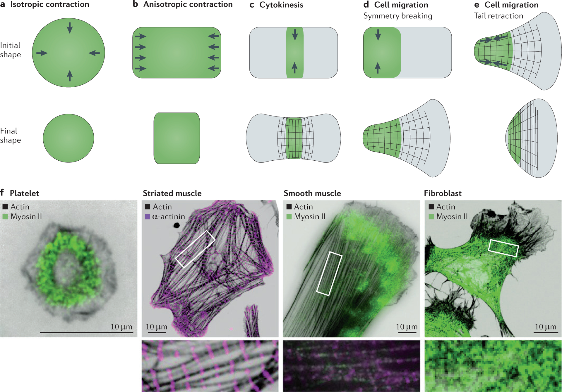
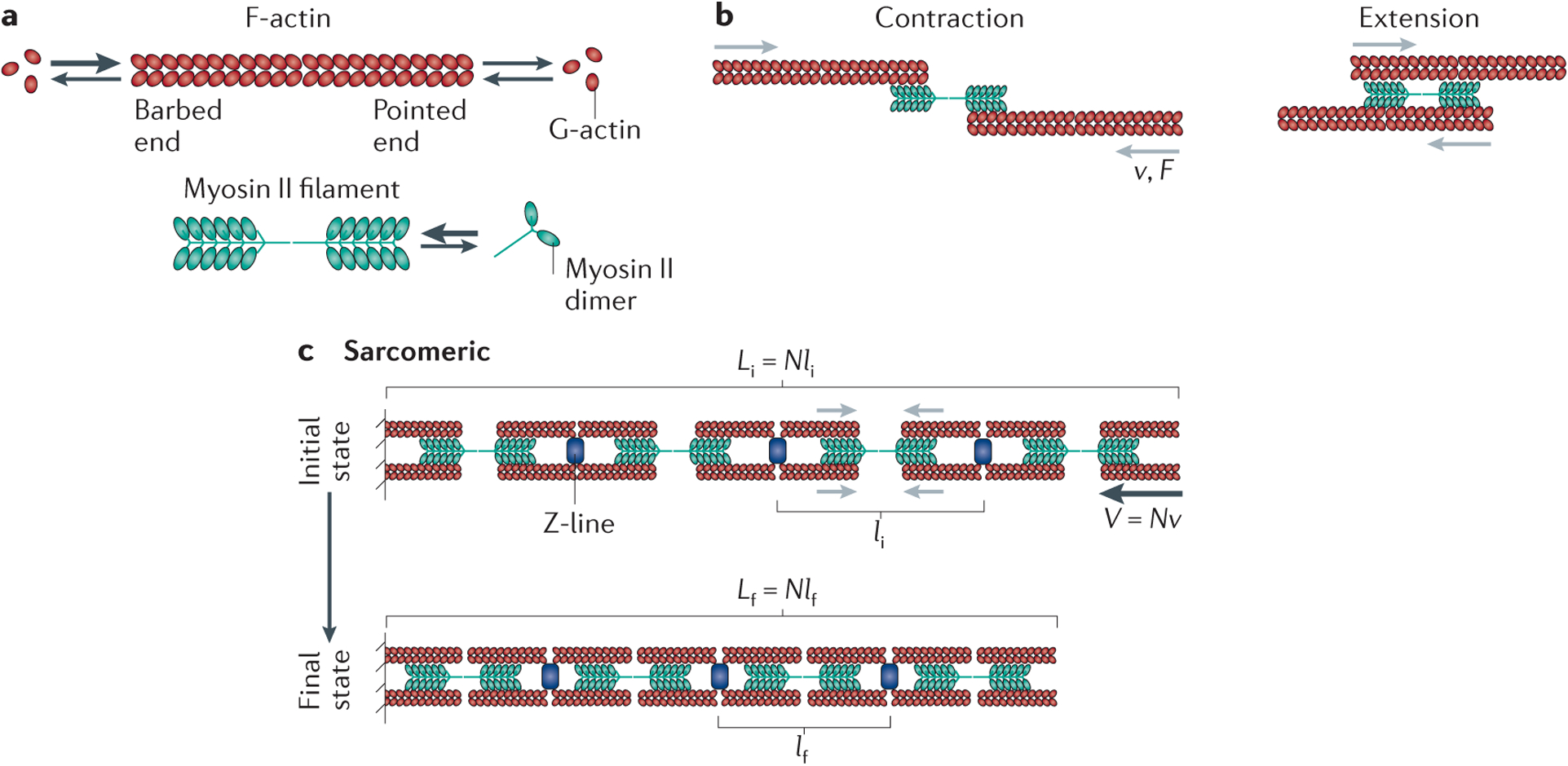
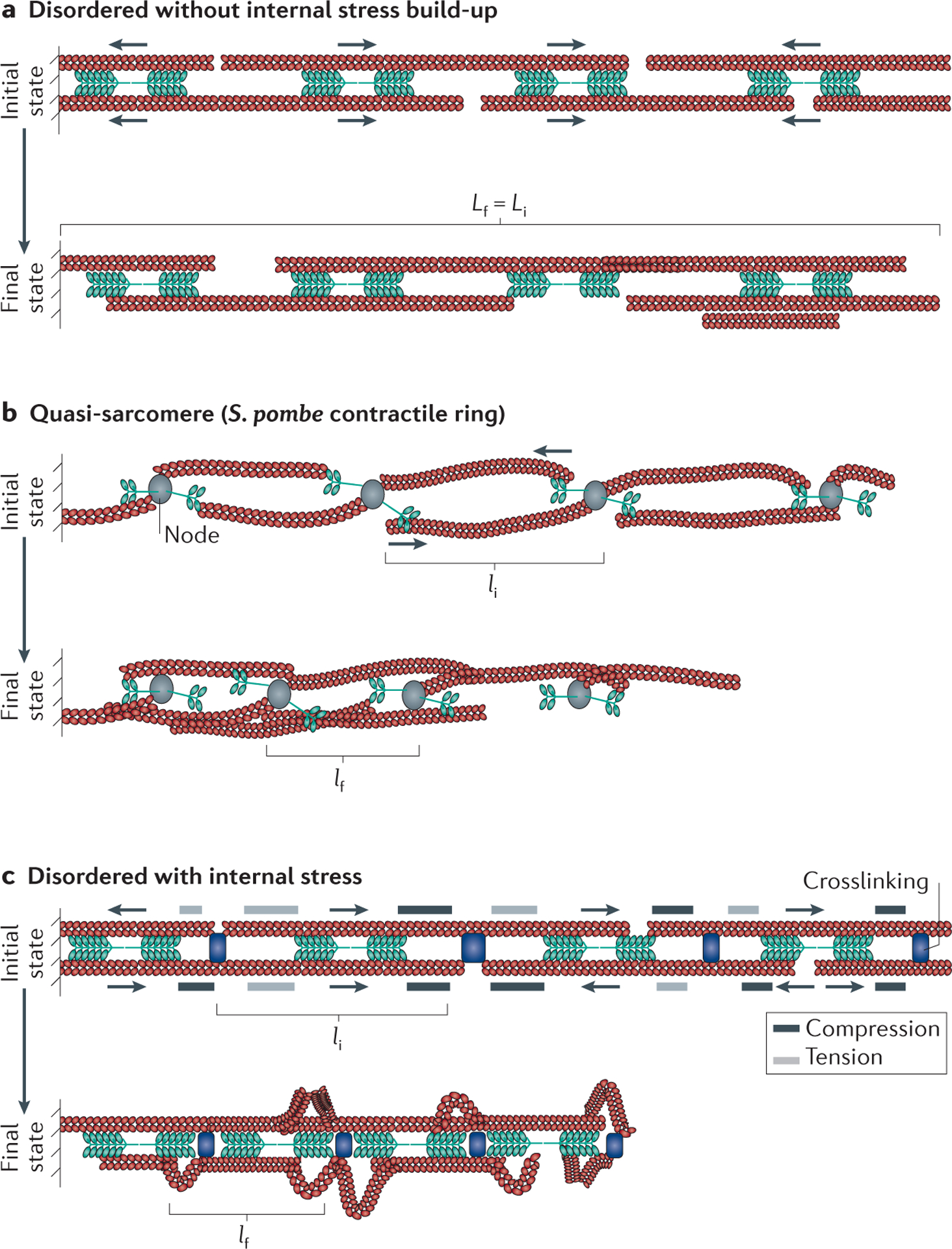
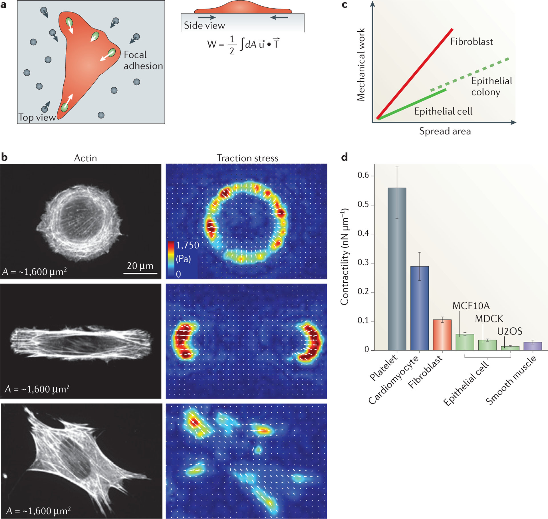
Actomyosin-mediated contractility is a highly conserved mechanism for generating mechanical stress in animal cells and underlies muscle contraction, cell migration, cell division and tissue morphogenesis. Whereas actomyosin-mediated contractility in striated muscle is well understood, the regulation of such contractility in non-muscle and smooth muscle cells is less certain. Our increased understanding of the mechanics of actomyosin arrays that lack sarcomeric organization has revealed novel modes of regulation and force transmission. This work also provides an example of how diverse mechanical behaviours at cellular scales can arise from common molecular components, underscoring the need for experiments and theories to bridge the molecular to cellular length scales.


Conflict of interest statement

Competing interests statement

The authors declare no competing interests.

Figures

Figure 1 |
Figure 1 |. Types of contractile deformations generated by cells and tissues.
a | Isotropic contraction, which is performed by platelets, is uniform around the cell perimeter and induces a uniform change in shape and in the force generated. b | Anisotropic contraction, which is performed by striated and smooth muscle cells, induces contraction and force generation along one axis. ce | In cytokinesis and cell migration, contractile stresses are spatially localized in a particular region of the cell to generate large deformations in cytokinesis (panel c), during symmetry breaking in migrating cells (panel d) and during tail retraction in migrating cells (panel e). f | Immunofluorescence images of several adherent cell types stained for actin, myosin II and α-actinin, including human platelets, striated muscle from a rat heart, smooth muscle from a human airway and mouse NIH 3T3 fibroblasts. Insets are a magnification of the corresponding boxed region and highlight the actomyosin organization within the cell. Myosin II was visualized using an antibody against phosphorylated myosin light chain, α-actinin was visualized via direct antibody staining and actin was visualized via phalloidin staining. Image of striated muscle cell courtesy of B. Hissa, University of Chicago, Illinois, USA, and image of smooth muscle cell courtesy of Y. Beckham, University of Chicago, Illinois, USA.
Figure 2 |
Figure 2 |. Contractility in sarcomeres.
a | Filamentous (F)-actin has a barbed end and a pointed end (indicated by the ‘open’ and ‘closed’ direction, respectively, of the depicted actin chevrons that make up the polymer), and it can associate with globular (G)-actin from a pool of monomers or add G-actin back to this pool, as indicated by the arrows. Higher association rates of monomeric actin to the barbed end are indicated by a larger arrow. Bipolar myosin filaments with a central bare zone that lacks motor heads are assembled from myosin II dimers. b | Myosin II filaments drive the translocation of F-actin filaments towards their barbed ends with a characteristic force (F) and gliding velocity (v) relationship. This can result in the contraction (left) or extension (right) of two bound actin filaments, depending on the location of myosin II with respect to the middle of these filaments. c | Actomyosin organization within sarcomeres. Here myosin filaments are segregated towards the F-actin pointed ends, and F-actin barbed ends are localized at Z-bands, which contain numerous regulatory proteins, including α-actinin crosslinkers. The initial and final contractile unit length are indicated by li and lf, respectively, in the initial (i) and final (f) states. Black arrows in the initial state indicate the direction of F-actin translocation. The contractile unit size is set by the sarcomere geometry, with the bundle shortening velocity (V) equal to the number of contractile units (N) times the myosin gliding velocity (v) such that V = Nv. The reduction in sarcomere length between the initial state and the final state arises from increased overlap between the F-actin and the myosin bare zone. The entire bundle length L is determined by the number N of contractile units multiplied by their length (l). Thus, the initial and final bundle lengths are given by Li = Nli and Lf = Nlf respectively.
Figure 3 |
Figure 3 |. Contractility in disordered actomyosin bundles.
Throughout the figure the initial (i) and final (f) configurations are indicated, and the initial and final contractile unit length is indicated by li and lf, respectively. Actin chevrons indicate the direction of the actin; the actin barbed end and actin pointed end are depicted by the ‘open’ and ‘closed’ direction of the chevrons, respectively. a | In a bundle with disordered actomyosin orientations, myosin II activity results in the internal sorting of F-actin polarity but does not lead to an overall reduction in the average bundle length (the initial and final bundle lengths are equal Lf = Li). b | Quasi-sarcomeric organizations arise in some cytoskeletal assemblies, such as the Schizosaccharomyces pombe contractile ring, in which myosin II and formins are localized to nodes. Formins cluster F-actin barbed ends, thus localizing myosin II activity from neighbouring nodes towards F-actin pointed ends. This is effectively a sarcomere-like geometry and results in contractility over time. c | In a disordered actomyosin bundle similar to that shown in part a but with added crosslinking, myosin II activity generates internal compressive and tensile stresses, which cause the compression or extension of F-actin portions, respectively, depending on the relative position of motors and crosslinks with respect to F-actin barbed ends. If sufficiently large, these internal stresses deform and buckle portions of F-actin, which relieves compressive stress and enables bundle shortening. In this model, the average contractile unit size is the average distance between F-actin buckling events.
Figure 4 |
Figure 4 |. Inherent contractility of adherent cells.
a | Traction force microscopy measures the distribution and magnitude of traction stresses of adherent cells (indicated in red) exerted through focal adhesions (green ovals) by probing the deformation of the underlying compliant matrix (grey) as measured by fiduciary markers (grey circles). Using this technique, we can calculate the strain in the substrate (u; grey arrows), the traction stresses applied by the cell (T; white arrows) and the amount of work performed to deform the substrate (W). The strain and stress are both vector fields, meaning that at each position these quantities have both a direction and magnitude. The total work is determined by integrating the dot product of the strain and stress vectors over the entire area (dA). b | The traction stress direction and magnitude for NIH 3T3 fibroblast cells of similar areas (~1,600 μm2) plated on a circular (top), oblong (middle) and unpatterned (bottom) surface are shown. The cell area is approximately constant in each of the three conditions, resulting in a similar amount of mechanical work performed on the environment by each of these three cells. Different cell geometries, however, result in different distributions of stresses (measured in Pa, as indicated) on the surface. c | Numerous experimental groups have found that the strain energy is proportional to the spread area for a wide range of cell types and for multicellular islands. The magnitude of this ratio (that is, slope of the line) is a measure of the characteristic contractility, and thus is cell type dependent. Multicellular islands of epithelial cells scale similarly to those for single cells when the area of the entire island is considered. d | The ratio of strain energy to spread area shows a characteristic contractility value for different muscle and non-muscle cells.

Similar articles

Cited by

References

    1. Munjal A & Lecuit T Actomyosin networks and tissue morphogenesis. Development 141, 1789–1793 (2014). - PubMed
    1. Gardel ML, Schneider IC, Aratyn-Schaus Y & Waterman CM Mechanical integration of actin and adhesion dynamics in cell migration. Annu. Rev. Cell Dev. Biol 26, 315–333 (2010). - PMC - PubMed
    1. Vicente-Manzanares M, Ma X, Adelstein RS & Horwitz AR Non-muscle myosin II takes centre stage in cell adhesion and migration. Nat. Rev. Mol. Cell Biol 10, 778–790 (2009). - PMC - PubMed
    1. Salbreux G, Charras G & Paluch E Actin cortex mechanics and cellular morphogenesis. Trends Cell Biol 10, 536–545 (2012). - PubMed
    1. Green RA, Paluch E & Oegema K Cytokinesis in animal cells. Annu. Rev. Cell Dev. Biol 28, 29–58 (2012). - PubMed

Publication types