. 2015 Nov:50:115-124.
doi: 10.1016/j.bbi.2015.06.022. Epub 2015 Jun 27.

Indoleamine 2,3-dioxygenase-dependent neurotoxic kynurenine metabolism mediates inflammation-induced deficit in recognition memory

Affiliations

Indoleamine 2,3-dioxygenase-dependent neurotoxic kynurenine metabolism mediates inflammation-induced deficit in recognition memory

Jillian M Heisler et al. Brain Behav Immun. 2015 Nov.

Abstract

Cognitive dysfunction in depression is a prevalent and debilitating symptom that is poorly treated by the currently available pharmacotherapies. Research over the past decade has provided evidence for proinflammatory involvement in the neurobiology of depressive disorders and symptoms associated with these disorders, including aspects of memory dysfunction. Recent clinical studies implicate inflammation-related changes in kynurenine metabolism as a potential pathogenic factor in the development of a range of depressive symptoms, including deficits in cognition and memory. Additionally, preclinical work has demonstrated a number of mood-related depressive-like behaviors to be dependent on indoleamine 2,3-dioxygenase-1 (IDO1), the inflammation-induced rate-limiting enzyme of the kynurenine pathway. Here, we demonstrate in a mouse model, that peripheral administration of endotoxin induced a deficit in recognition memory. Mice deficient in IDO were protected from cognitive impairment. Furthermore, endotoxin-induced inflammation increased kynurenine metabolism within the perirhinal/entorhinal cortices, brain regions which have been implicated in recognition memory. A single peripheral injection of kynurenine, the metabolic product of IDO1, was sufficient to induce a deficit in recognition memory in both control and IDO null mice. Finally, kynurenine monooxygenase (KMO) deficient mice were also protected from inflammation-induced deficits on novel object recognition. These data implicate IDO-dependent neurotoxic kynurenine metabolism as a pathogenic factor for cognitive dysfunction in inflammation-induced depressive disorders and a potential novel target for the treatment of these disorders.

Keywords: Behavior; Indoleamine 2,3-dioxygenase; Kynurenine; Kynurenine monooxygenase; Mouse; Neuroinflammation; Neuropsychiatric symptom; Novel object recognition; Perirhinal cortex; Recognition memory.


Conflict of interest statement

Conflict of Interest

None of the authors have any conflicts of interest to disclose

Figures

Figure 1
Figure 1
The kynurenine pathway of tryptophan metabolism. In the brain, kynurenine metabolism can begin with the catabolism of tryptophan to N-formylkynurenine via either tryptophan 2,3-dioxygenase (TDO) or indoleamine 2,3-dioxygenase (IDO), expressed in both neurons and glia. Kynurenine is metabolized either via kynurenine aminotransferases (KATs) to kynurenic acid (KYNA) in astrocytes, or via kynurenine monooxygenase (KMO), kynureninase, and 3-hydroxyanthranilic acid oxygenase (3-HAO) to quinolinic acid (QUIN). KYNA and QUIN act at the NMDA receptor as an antagonist and agonist, respectively.
Figure 2
Figure 2
Indoleamine 2,3-dioxygenase 1 mediates a deficit in Novel Object Recognition following peripheral immune challenge. (A) Schematic representation of Novel Object Recognition protocol. (B) LPS administration induces a significant reduction in preference for the novel object in WT mice, whereas IDO −/− mice are protected (Genotype x Treatment Interaction: F(1,36)=13.00, p=0.0009). The total number of contacts with the objects (C) and the total time spent exploring the objects (D) is not different between any of the experimental groups. Data represent mean ± SEM. *p<0.05, **p<0.01. n=7–12 mice/group.
Figure 2
Figure 2
Indoleamine 2,3-dioxygenase 1 mediates a deficit in Novel Object Recognition following peripheral immune challenge. (A) Schematic representation of Novel Object Recognition protocol. (B) LPS administration induces a significant reduction in preference for the novel object in WT mice, whereas IDO −/− mice are protected (Genotype x Treatment Interaction: F(1,36)=13.00, p=0.0009). The total number of contacts with the objects (C) and the total time spent exploring the objects (D) is not different between any of the experimental groups. Data represent mean ± SEM. *p<0.05, **p<0.01. n=7–12 mice/group.
Figure 3
Figure 3
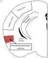
Peripheral LPS administration increases the mRNA expression of proinflammatory cytokines, markers of glial activation in the perirhinal/entorhinal cortex. (A) Representative area of dissection of perirhinal/entorhinal cortices. Two 1mm sections taken from 1.56mm to 3.56mm caudal to Bregma. (B & C) LPS significantly increases expression of both microglia and astrocyte markers (Iba1: LPS effect: F(2,37)=15.47, p<0.0001; GFAP: LPS effect: F(2.37)=15.28, p<0.0001). LPS additionally significantly increases mRNA expression of proinflammatory cytokines (D)(IL-1β: LPS effect: F(2,37)=17.67, p<0.0001; (E) TNF-α (LPS effect: F(2,35)=56.32, p<0.0001). There was also a significant main effect of LPS on (F) IL-6 mRNA expression (LPS effect: F(2,37)=5.724, p=0.0068), however there were no significant between group differences. Data represent mean ± SEM. n=7–12 mice/group.
Figure 3
Figure 3
Peripheral LPS administration increases the mRNA expression of proinflammatory cytokines, markers of glial activation in the perirhinal/entorhinal cortex. (A) Representative area of dissection of perirhinal/entorhinal cortices. Two 1mm sections taken from 1.56mm to 3.56mm caudal to Bregma. (B & C) LPS significantly increases expression of both microglia and astrocyte markers (Iba1: LPS effect: F(2,37)=15.47, p<0.0001; GFAP: LPS effect: F(2.37)=15.28, p<0.0001). LPS additionally significantly increases mRNA expression of proinflammatory cytokines (D)(IL-1β: LPS effect: F(2,37)=17.67, p<0.0001; (E) TNF-α (LPS effect: F(2,35)=56.32, p<0.0001). There was also a significant main effect of LPS on (F) IL-6 mRNA expression (LPS effect: F(2,37)=5.724, p=0.0068), however there were no significant between group differences. Data represent mean ± SEM. n=7–12 mice/group.
Figure 4
Figure 4
Indoleamine 2,3-dioxygenase 1 and hydroxyanthranilic acid oxygenase mRNA expression is increased following peripheral LPS challenge. (A) There was a significant effect of LPS and genotype on IDO1 expression (Genotype x Treatment Interaction: F(2,37)=10.82; p=0.0002). LPS significantly increased mRNA expression of IDO1 in WT mice 48h post-LPS (p<0.0001). (B) There was a significant effect of LPS on KATII expression (LPS effect: F(2,37)=6.09; p=0.005). There was no effect of LPS on mRNA expression of KMO (C) or KYNU (D). LPS significantly increased the mRNA expression of HAAO in both WT and IDO −/− mice (LPS effect: F(2,37)=18.63; p<0.0001). Data represent mean ± SEM. *p<0.001, n=7–12 mice/group.
Figure 4
Figure 4
Indoleamine 2,3-dioxygenase 1 and hydroxyanthranilic acid oxygenase mRNA expression is increased following peripheral LPS challenge. (A) There was a significant effect of LPS and genotype on IDO1 expression (Genotype x Treatment Interaction: F(2,37)=10.82; p=0.0002). LPS significantly increased mRNA expression of IDO1 in WT mice 48h post-LPS (p<0.0001). (B) There was a significant effect of LPS on KATII expression (LPS effect: F(2,37)=6.09; p=0.005). There was no effect of LPS on mRNA expression of KMO (C) or KYNU (D). LPS significantly increased the mRNA expression of HAAO in both WT and IDO −/− mice (LPS effect: F(2,37)=18.63; p<0.0001). Data represent mean ± SEM. *p<0.001, n=7–12 mice/group.
Figure 5
Figure 5
Direct administration of L-kynurenine induces a deficit in Novel Object Recognition. A significant increase in plasma and brain concentration of kynurenine is observed 30 min post administration of L-kynurenine (A–B). Peripheral administration of kynurenine 30 min prior to training results in a significant reduction in novel object preference during testing, 24 hours post administration in both (C) WT and (D) IDO KO mice. There was no effect on overall exploratory activity (not shown). Data represent mean ± SEM. *p<0.05, ****p<0.0001. n=5–6 mice/group.
Figure 5
Figure 5
Direct administration of L-kynurenine induces a deficit in Novel Object Recognition. A significant increase in plasma and brain concentration of kynurenine is observed 30 min post administration of L-kynurenine (A–B). Peripheral administration of kynurenine 30 min prior to training results in a significant reduction in novel object preference during testing, 24 hours post administration in both (C) WT and (D) IDO KO mice. There was no effect on overall exploratory activity (not shown). Data represent mean ± SEM. *p<0.05, ****p<0.0001. n=5–6 mice/group.
Figure 6
Figure 6
Kynurenine 3-monooxygenase mediates a deficit in Novel Object Recognition following peripheral immune challenge. LPS administration induces a significant reduction in preference for the novel object in WT mice, whereas KMO−/− mice are protected (Genotype x Treatment Interaction: F(1,29)=8.23, p=0.01). Data represent mean ± SEM. *p<0.05. n=7–10 mice/group.

References

    1. Andre C, O’Connor JC, Kelley KW, Lestage J, Dantzer R, Castanon N. Spatio-temporal differences in the profile of murine brain expression of proinflammatory cytokines and indoleamine 2,3-dioxygenase in response to peripheral lipopolysaccharide administration. J Neuroimmunol. 2008;200(1–2):90–99. - PMC - PubMed
    1. Capuron L, Miller AH. Cytokines and psychopathology: lessons from interferon-alpha. Biol Psychiatry. 2004;56(11):819–824. - PubMed
    1. Capuron L, Ravaud A, Neveu PJ, Miller AH, Maes M, Dantzer R. Association between decreased serum tryptophan concentrations and depressive symptoms in cancer patients undergoing cytokine therapy. Mol Psychiatry. 2002;7(5):468–473. - PubMed
    1. Chiarugi A, Carpenedo R, Moroni F. Kynurenine disposition in blood and brain of mice: effects of selective inhibitors of kynurenine hydroxylase and of kynureninase. J Neurochem. 1996;67(2):692–698. - PubMed
    1. Collingridge GL, Volianskis A, Bannister N, France G, Hanna L, Mercier M, Tidball P, Fang G, Irvine MW, Costa BM, Monaghan DT, Bortolotto ZA, Molnar E, Lodge D, Jane DE. The NMDA receptor as a target for cognitive enhancement. Neuropharmacology. 2013;64:13–26. - PMC - PubMed

Publication types

MeSH terms