

A histochemical study of the Nras/let-60 activity in filarial nematodes
- PMID: 26130134
- PMCID: PMC4493820
- DOI: 10.1186/s13071-015-0947-6
A histochemical study of the Nras/let-60 activity in filarial nematodes
Abstract
Background: Control and elimination of filarial pathogens is a central focus of major global health efforts directed at parasitic diseases of developing countries. Accomplishment of these goals would be markedly enhanced by the enhanced destruction of the adult stage of filariae. The identification of new, more quantitative biomarkers that correlate with mortality or chemotherapeutic damage to adult filariae, would greatly facilitate, for example, the development of new macrofilaricides.
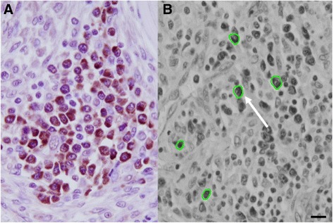
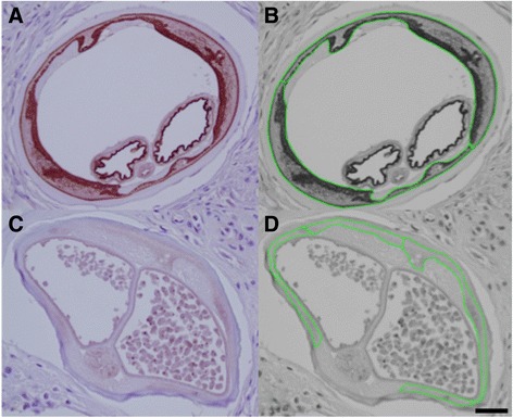
Methods: An immunocytochemical approach using an antibody against human Nras was used to identify and detect changes in the nematode homolog let-60 that is associated with cell growth and maintenance. Single Onchocerca volvulus nodules were removed from each of 13 patients treated with ivermectin (as part of a community-wide mass drug administration programme), and from each of 13 untreated individuals; these 26 nodules were stained with the anti-Nras antibody. The localization and degree of positivity of Nras/let-60 staining were assessed subjectively and compared between the two groups; the positivity of staining was also quantified, using image analysis, in a subgroup of these nodules. In addition, the specific morphological association between Nras/let-60 and the Wolbachia endosymbiont present in these parasites was also observed in 4 additional filarial species using an anti-Wolbachia surface protein (WSP) antibody under light and confocal microscopy.
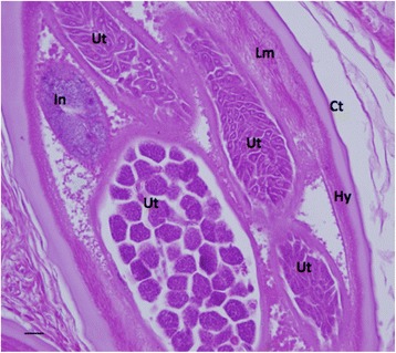
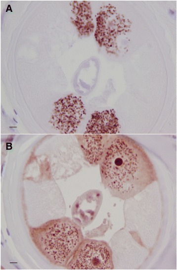
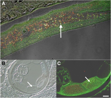
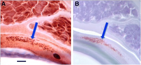
Results: Nras/let-60 is present in many structures within the adult female worms. A statistically significant decrease in the general staining intensity of Nras/let-60 was observed in adult female O. volvulus treated with ivermectin when compared with parasites from untreated patients. Nras/let-60 staining was frequently observed to be co-localized with WSP in O.volvulus, Brugia malayi, Litomosoides sigmodontis and Dirofilaria immitis. Nras/let60 is also present in Onchocerca ochengi.
Conclusion: Nras/let-60, as detected by immunocytochemical staining, is decreased in ivermectin-treated adult female O. volvulus relative to untreated control specimens, suggesting a suppressive effect of ivermectin on the overall biochemical activity of these parasites. Co-localization of Nras/let-60 and WSP suggests the possibility that the endosymbiont utilizes this nematode protein as part of a mutualistic relationship. Nras/let60 appears to be a useful biomarker for assessing the health of filariae.
Figures


Similar articles
-
Macrofilaricidal activity of tetracycline against the filarial nematode Onchocerca ochengi: elimination of Wolbachia precedes worm death and suggests a dependent relationship.Proc Biol Sci. 2000 Jun 7;267(1448):1063-9. doi: 10.1098/rspb.2000.1110. Proc Biol Sci. 2000. PMID: 10885510 Free PMC article.
-
Lipid profiling of the filarial nematodes Onchocerca volvulus, Onchocerca ochengi and Litomosoides sigmodontis reveals the accumulation of nematode-specific ether phospholipids in the host.Int J Parasitol. 2017 Dec;47(14):903-912. doi: 10.1016/j.ijpara.2017.06.001. Epub 2017 Jul 23. Int J Parasitol. 2017. PMID: 28743489 Free PMC article.
-
Localization of a filarial phosphate permease that is up-regulated in response to depletion of essential Wolbachia endobacteria.Exp Parasitol. 2014 Mar;138:30-9. doi: 10.1016/j.exppara.2014.01.006. Epub 2014 Jan 28. Exp Parasitol. 2014. PMID: 24480589
-
Antibiotics for the treatment of onchocerciasis and other filarial infections.Curr Opin Investig Drugs. 2002 Apr;3(4):533-7. Curr Opin Investig Drugs. 2002. PMID: 12090719 Review.
-
Wolbachia in filarial nematodes: evolutionary aspects and implications for the pathogenesis and treatment of filarial diseases.Vet Parasitol. 2001 Jul 12;98(1-3):215-38. doi: 10.1016/s0304-4017(01)00432-0. Vet Parasitol. 2001. PMID: 11516587 Review.
References
Publication types
MeSH terms
Substances
LinkOut - more resources
Full Text Sources
Other Literature Sources
Molecular Biology Databases
Miscellaneous
