. 2015 Jul-Aug;22(4):302-9.
doi: 10.1111/xen.12174. Epub 2015 Jun 29.

Pig kidney graft survival in a baboon for 136 days: longest life-supporting organ graft survival to date

Affiliations

Pig kidney graft survival in a baboon for 136 days: longest life-supporting organ graft survival to date

Hayato Iwase et al. Xenotransplantation. 2015 Jul-Aug.

Abstract

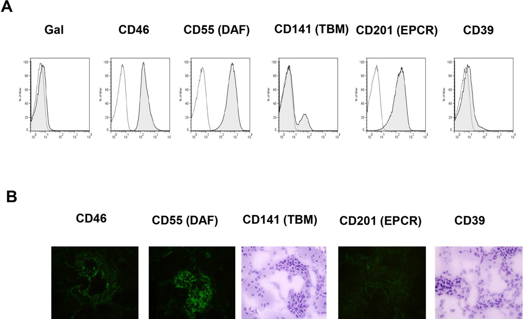
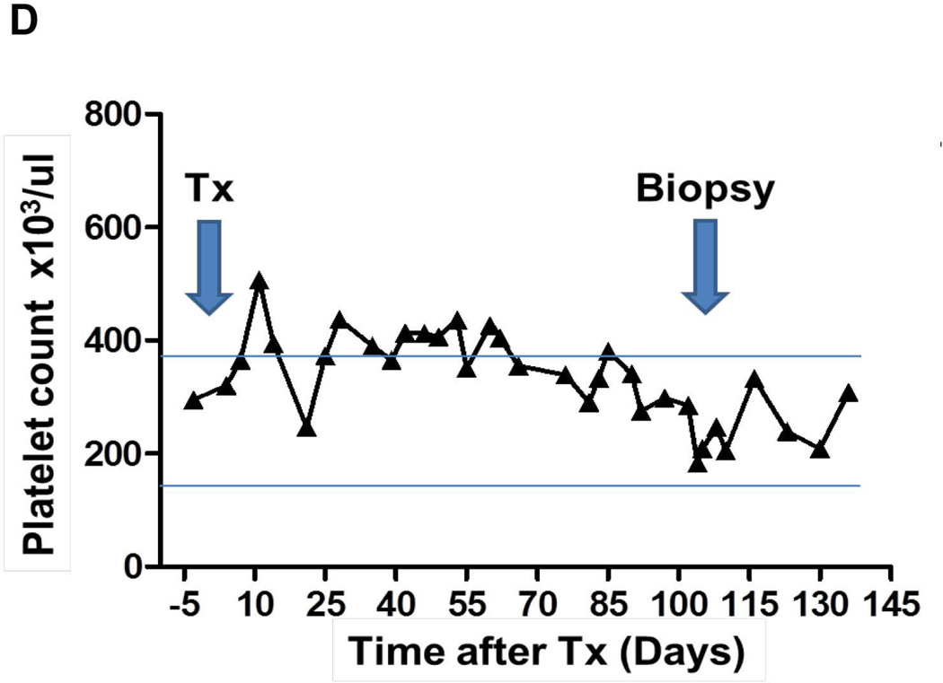
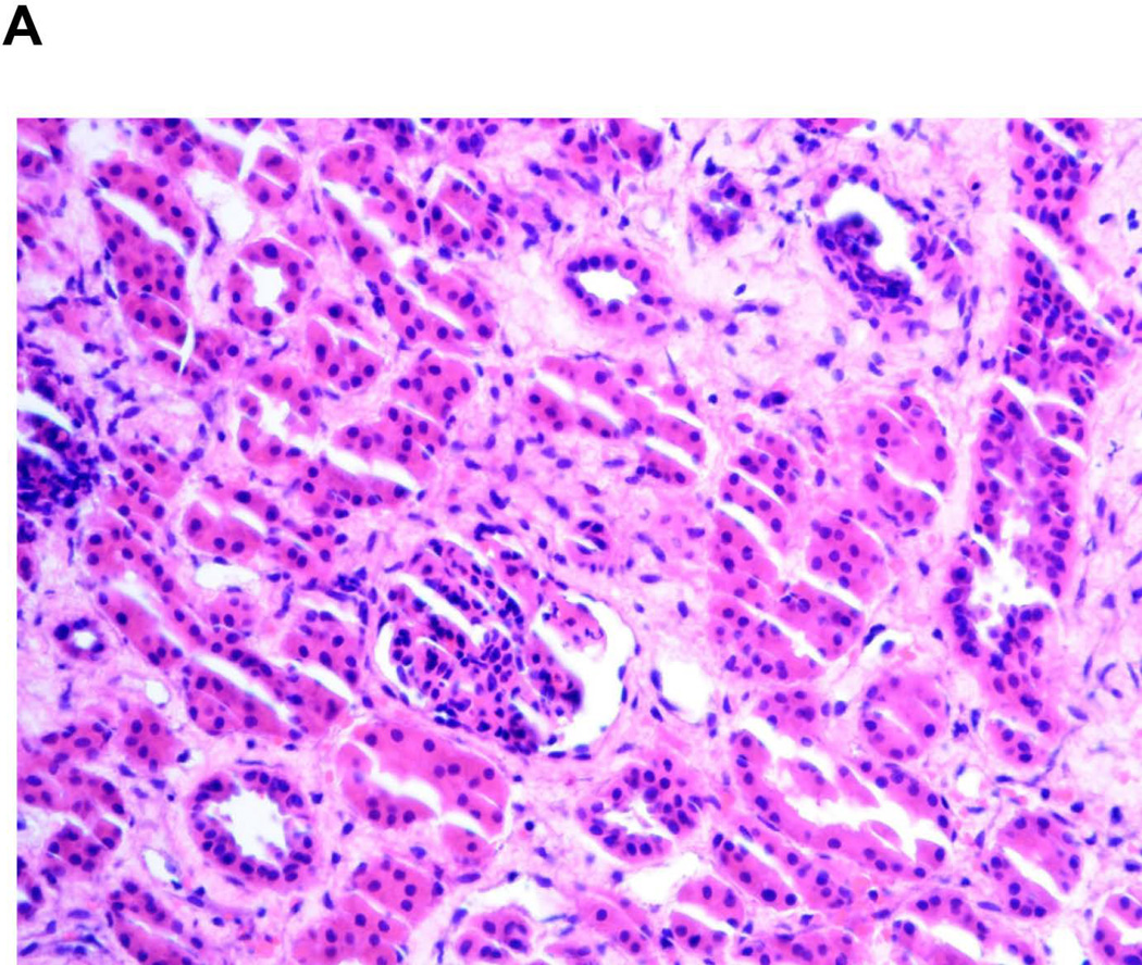
The longest survival of a non-human primate with a life-supporting kidney graft to date has been 90 days, although graft survival > 30 days has been unusual. A baboon received a kidney graft from an α-1,3-galactosyltransferase gene-knockout pig transgenic for two human complement-regulatory proteins and three human coagulation-regulatory proteins (although only one was expressed in the kidney). Immunosuppressive therapy was with ATG+anti-CD20mAb (induction) and anti-CD40mAb+rapamycin+corticosteroids (maintenance). Anti-TNF-α and anti-IL-6R were administered. The baboon survived 136 days with a generally stable serum creatinine (0.6 to 1.6 mg/dl) until termination. No features of a consumptive coagulopathy (e.g., thrombocytopenia, decreased fibrinogen) or of a protein-losing nephropathy were observed. There was no evidence of an elicited anti-pig antibody response. Death was from septic shock (Myroides spp). Histology of a biopsy on day 103 was normal, but by day 136, the kidney showed features of glomerular enlargement, thrombi, and mesangial expansion. The combination of (i) a graft from a specific genetically engineered pig, (ii) an effective immunosuppressive regimen, and (iii) anti-inflammatory agents prevented immune injury and a protein-losing nephropathy, and delayed coagulation dysfunction. This outcome encourages us that clinical renal xenotransplantation may become a reality.

Keywords: anti-IL-6R antagonist; costimulation blockade; genetically engineered; kidney; pig; xenotransplantation.


Conflict of interest statement

DISCLOSURE OF CONFLICT OF INTEREST

David Ayares and Carol Phelps are employees of Revivicor, Inc. No other author has a conflict of interest.

Figures

Figure 1
Figure 1
(A) Expression of human transgenes on donor pig PBMCs by flow cytometry. Gal expression was absent, expression of CD46, CD55, and EPCR was high, whereas expression of TBM and CD39 was low. (B) Expression of human transgenes on donor kidney by immunofluorescence or immunohistochemistry. (Left to right) CD46, CD55, TBM (not detectable), EPCR, CD39 (not detectable). Studies on CD46, CD55, and EPCR were by immunofluorescence but, in view of a high background activity, immunohistochemistry was used for TBM and CD39. This may possibly influence the results. Although there appears to be a discrepancy between expression of CD55 and EPCR, their expression should be equimolar as a single transcript is produced to join these genes. The difference in intensity in these images may be related to specific antibody affinity rather than significant differences in expression.
Figure 2
Figure 2
Monitoring of recipient baboon throughout the course of the experiment.(A) serum creatinine;(B) urinary protein; (C) serum albumin; (D) platelet counts; (E) fibrinogen; (F) D-dimer. The horizontal lines indicate either the upper and lower limits (A, C, D) or lower limit (E), or upper limit (F), of the equivalent parameters measured in healthy humans.
Figure 2
Figure 2
Monitoring of recipient baboon throughout the course of the experiment.(A) serum creatinine;(B) urinary protein; (C) serum albumin; (D) platelet counts; (E) fibrinogen; (F) D-dimer. The horizontal lines indicate either the upper and lower limits (A, C, D) or lower limit (E), or upper limit (F), of the equivalent parameters measured in healthy humans.
Figure 3
Figure 3
(A) Microscopic appearance of the pig kidney graft on biopsy on day 103 (magnification x20). Although there has been some artifact distortion of the tissue in the preparation of the section, the histology is basically normal. (B) Immunohistochemistry staining of the pig kidney graft on biopsy on day 103, demonstrating minimal C3 (left), moderate IgG (center), but no IgM (right) deposition, suggesting an elicited antibody response. (C) Macroscopic appearance of the pig kidney graft at necropsy on day 136. (D) Macroscopic appearqnce of a cross-section of the kidney graft at necropsy on day 136. (E) Low-power (left, magnification x4) and high-power (right, x40) microscopic appearance of the pig kidney graft at necropsy on day 136 from an area with macroscopic hemorrhage. The sections show widespread focal hemorrhage and features of thrombotic microangiopathy (thrombosis of primarily small vessels with focally extensive infarction and tissue destruction); cell infiltrates were minimal. The thrombotic microangiopathy is characterized in part by multiple occluding thrombi within glomerular capillary loops and associated general mesangial expansion.
Figure 3
Figure 3
(A) Microscopic appearance of the pig kidney graft on biopsy on day 103 (magnification x20). Although there has been some artifact distortion of the tissue in the preparation of the section, the histology is basically normal. (B) Immunohistochemistry staining of the pig kidney graft on biopsy on day 103, demonstrating minimal C3 (left), moderate IgG (center), but no IgM (right) deposition, suggesting an elicited antibody response. (C) Macroscopic appearance of the pig kidney graft at necropsy on day 136. (D) Macroscopic appearqnce of a cross-section of the kidney graft at necropsy on day 136. (E) Low-power (left, magnification x4) and high-power (right, x40) microscopic appearance of the pig kidney graft at necropsy on day 136 from an area with macroscopic hemorrhage. The sections show widespread focal hemorrhage and features of thrombotic microangiopathy (thrombosis of primarily small vessels with focally extensive infarction and tissue destruction); cell infiltrates were minimal. The thrombotic microangiopathy is characterized in part by multiple occluding thrombi within glomerular capillary loops and associated general mesangial expansion.
Figure 4
Figure 4
(A) Pre-transplant anti-pig nonGal IgM and IgG levels (determined by flow cytometry) in the recipient baboon (open circle and square) and in 9 other baboons (closed circles and squares) from the Division of Animal Resources, Oklahoma University Health Sciences Center, specific pathogen-free colony received in Pittsburgh during the past 2 years. (B) Anti-pig nonGal IgM (left) and IgG (right) levels in the recipient baboon pre-transplantation (solid line), 60 days post-transplant (dotted line), and 136 days post-transplant (dashed line). (Isotype control - shaded).

References

    1. Lambrigts D, Sachs DH, Cooper DK. Discordant organ xenotransplantation in primates: world experience and current status. Transplantation. 1998;66:547–561. - PubMed
    1. Cooper DK, Satyananda V, Ekser B, Van der Windt DJ, Hara H, Ezzelarab M, et al. Progress in pig-to-nonhuman primate transplantation models (1998–2013): a comprehensive review of the literature. Xenotransplantation. 2014;21:397–419. - PMC - PubMed
    1. Baldan N, Rigotti P, Calabrese F, Cadrobbi R, Dedja A, Iacopetti I, et al. Ureteral stenosis in HDAF pig-to-primate renal xenotransplantation: a phenomenon related to immunological events? Am J Transplant. 2004;4:475–481. - PubMed
    1. Ayares D, Vaught T, Ball S, Kuravi K, Morrill B, Monahan J, et al. Genetic engineering of source pigs for xenotransplantation: progress and prospects. Xenotransplantation. 2013;20:361. (Abstract 408)
    1. Zhou H, Iwase H, Wolf RF, Ekser B, Ezzelarab M, Hara H, et al. Are there advantages in the use of specific pathogen-free baboons in pig organ xenotransplantation models? Xenotransplantation. 2014;21:287–290. - PMC - PubMed

Publication types

MeSH terms

Substances