

Quantitation of the Regional Lymph Node Metastatic Burden and Prognosis in Malignant Mammary Tumors of Dogs
- PMID: 26130166
- PMCID: PMC4858035
- DOI: 10.1111/jvim.13576
Quantitation of the Regional Lymph Node Metastatic Burden and Prognosis in Malignant Mammary Tumors of Dogs
Abstract
Background: As in women, regional lymph node status impacts survival in dogs with malignant mammary tumors. However, few studies have evaluated regional lymph node metastases in dogs with malignant mammary gland tumors.
Objectives: To estimate overall survival based on the assessments of the lymph node status and the morphologic and morphometric features in female dogs with malignant mammary gland tumors.
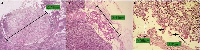
Materials and methods: In total, 178 lymph nodes from 97 female dogs were assessed and reviewed, and after confirmation by immunohistochemistry (IHC), 161 lymph nodes were selected for analysis of metastases. Animals were considered metastasis-free (negative lymph nodes) only after IHC analysis for cytokeratin AE1/AE3. The number of positive lymph nodes, the number of metastatic foci, the maximum diameter and the area of metastasis were analyzed, and estimates of overall survival were made.
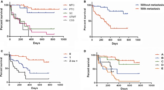
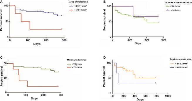
Results: Dogs with metastasis had lower mean survival than those with metastasis-free regional lymph nodes, showing a direct relationship between the number of affected lymph nodes and shorter survival. However, histologic analysis of the lymph nodes identified lower survival rates in animals with macrometastases and isolated tumor cells, areas of metastasis >20.11 mm², and metastatic diameters >7.32 mm.
Conclusion: The identification of ≥1 lymph nodes positive for metastasis and morphometric characterization of lymphatic metastases indicate the prognostic relevance of lymph nodes status in dogs with mammary tumors.
Keywords: Canine; Lymph node; Mammary tumors; Metastasis; Survival.
Copyright © 2015 by the American College of Veterinary Internal Medicine.
Figures
Similar articles
-
Prognostic Value of Occult Isolated Tumour Cells within Regional Lymph Nodes of Dogs with Malignant Mammary Tumours.J Comp Pathol. 2018 Jan;158:32-38. doi: 10.1016/j.jcpa.2017.11.001. Epub 2017 Dec 6. J Comp Pathol. 2018. PMID: 29422313
-
Prognostic value of regional lymph node status in canine mammary carcinomas.Vet Comp Oncol. 2011 Dec;9(4):296-303. doi: 10.1111/j.1476-5829.2011.00268.x. Epub 2011 Apr 21. Vet Comp Oncol. 2011. PMID: 22077411
-
Prognostic factors associated with survival two years after surgery in dogs with malignant mammary tumors: 79 cases (1998-2002).J Am Vet Med Assoc. 2005 Nov 15;227(10):1625-9. doi: 10.2460/javma.2005.227.1625. J Am Vet Med Assoc. 2005. PMID: 16313041
-
Canine mammary gland tumors.Vet Clin North Am Small Anim Pract. 2003 May;33(3):573-96. doi: 10.1016/s0195-5616(03)00020-2. Vet Clin North Am Small Anim Pract. 2003. PMID: 12852237 Review.
-
Identification and characterization of cancer stem cells in canine mammary tumors.Acta Vet Scand. 2016 Dec 19;58(1):86. doi: 10.1186/s13028-016-0268-6. Acta Vet Scand. 2016. PMID: 27993142 Free PMC article. Review.
Cited by
-
Ultrasonography for lymph nodes metastasis identification in bitches with mammary neoplasms.Sci Rep. 2018 Dec 7;8(1):17708. doi: 10.1038/s41598-018-34806-9. Sci Rep. 2018. PMID: 30532025 Free PMC article.
-
Clinical Trials in Veterinary Medicine: A New Era Brings New Challenges.J Vet Intern Med. 2017 Jul;31(4):970-978. doi: 10.1111/jvim.14744. Epub 2017 May 30. J Vet Intern Med. 2017. PMID: 28557000 Free PMC article.
-
The Number and Size of Invasion Areas in Mixed-Type Carcinoma in Female Dogs Are Associated with Regional Metastases.Vet Sci. 2025 Mar 31;12(4):318. doi: 10.3390/vetsci12040318. Vet Sci. 2025. PMID: 40284820 Free PMC article.
-
Pleomorphic lobular carcinoma in a cat: Clinical, histopathological, and immunohistochemical characterization.Braz J Vet Med. 2025 May 30;47:e001725. doi: 10.29374/2527-2179.bjvm001725. eCollection 2025. Braz J Vet Med. 2025. PMID: 40458377 Free PMC article.
-
Ultrasonographic Algorithm for the Assessment of Sentinel Lymph Nodes That Drain the Mammary Carcinomas in Female Dogs.Animals (Basel). 2020 Dec 10;10(12):2366. doi: 10.3390/ani10122366. Animals (Basel). 2020. PMID: 33321917 Free PMC article.
References
-
- Carter CL, Allen C, Henson DE. Relation of tumor size, lymph node status, and survival in 24,740 breast cancer cases. Cancer 1989;63:181–187. - PubMed
-
- Veronesi U, Paganelli G, Viale G, et al. A randomized comparison of sentinel‐node biopsy with routine axillary dissection in breast cancer. N Engl J Med 2003;349:546–553. - PubMed
-
- Lyman GH, Temin S, Edge SB, et al. Sentinel lymph node biopsy for patients with early‐stage breast cancer: American Society of Clinical Oncology clinical practice guideline update. J Clin Oncol 2014;32:1365–1383. - PubMed
-
- Kuehn T, Bembenek A, Decker T, et al. A concept for the clinical implementation of sentinel lymph node biopsy in patients with breast carcinoma with special regard to quality assurance. Cancer 2005;103:451–461. - PubMed
-
- Fitzgibbons PL, Page DL, Weaver D, et al. Prognostic factors in breast cancer. College of American Pathologists Consensus Statement 1999. Arch Pathol Lab Med 2000;124:966–978. - PubMed
Publication types
MeSH terms
LinkOut - more resources
Full Text Sources
Other Literature Sources
Research Materials
Miscellaneous
