

Amyloid PET in European and North American cohorts; and exploring age as a limit to clinical use of amyloid imaging
- PMID: 26130168
- PMCID: PMC4521094
- DOI: 10.1007/s00259-015-3115-5
Amyloid PET in European and North American cohorts; and exploring age as a limit to clinical use of amyloid imaging
Abstract
Purpose: Several radiotracers that bind to fibrillar amyloid-beta in the brain have been developed and used in various patient cohorts. This study aimed to investigate the comparability of two amyloid positron emission tomography (PET) tracers as well as examine how age affects the discriminative properties of amyloid PET imaging.
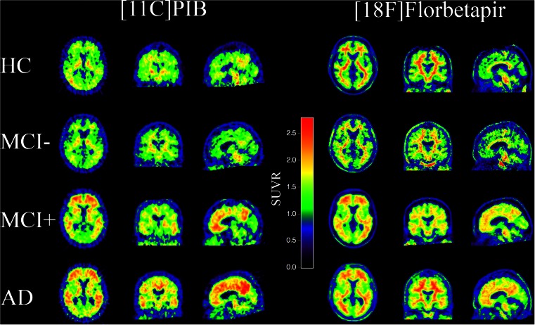
Methods: Fifty-one healthy controls (HCs), 72 patients with mild cognitive impairment (MCI) and 90 patients with Alzheimer's disease (AD) from a European cohort were scanned with [11C]Pittsburgh compound-B (PIB) and compared with an age-, sex- and disease severity-matched population of 51 HC, 72 MCI and 84 AD patients from a North American cohort who were scanned with [18F]Florbetapir. An additional North American population of 246 HC, 342 MCI and 138 AD patients with a Florbetapir scan was split by age (55-75 vs 76-93 y) into groups matched for gender and disease severity. PET template-based analyses were used to quantify regional tracer uptake.
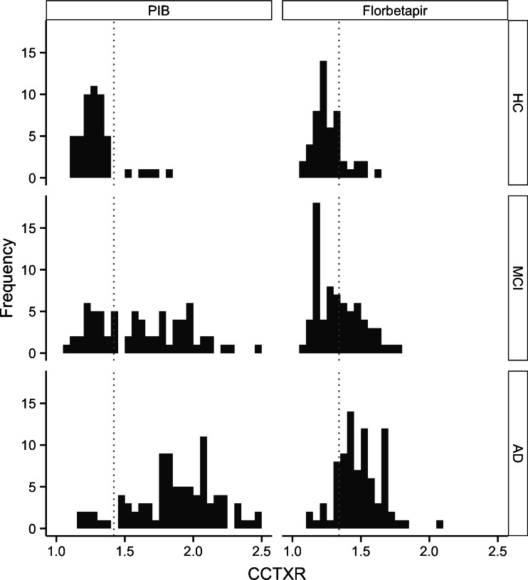
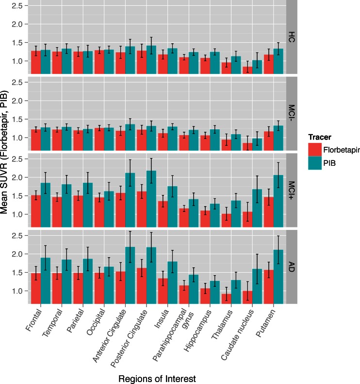
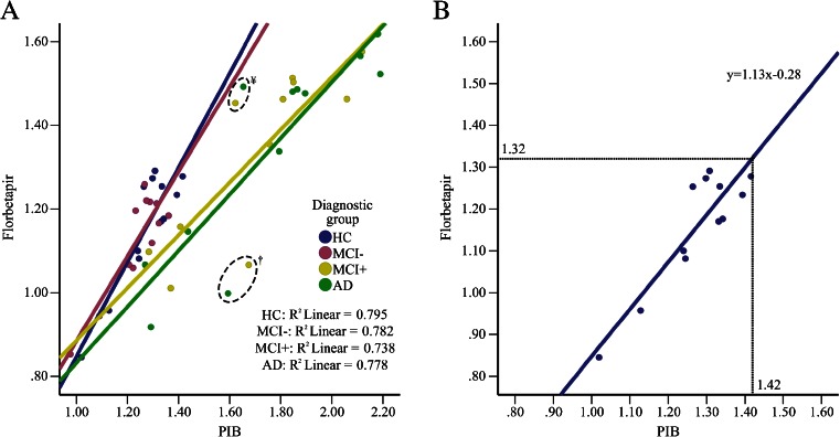
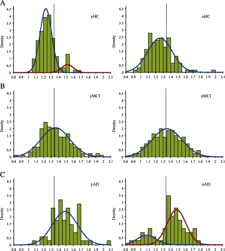
Results: The mean regional uptake patterns were similar and strong correlations were found between the two tracers across the regions of interest in HC (ρ = 0.671, p = 0.02), amyloid-positive MCI (ρ = 0.902, p < 0.001) and AD patients (ρ = 0.853, p < 0.001). The application of the Florbetapir cut-off point resulted in a higher proportion of amyloid-positive HC and a lower proportion of amyloid-positive AD patients in the older group (28 and 30 %, respectively) than in the younger group (19 and 20 %, respectively).
Conclusions: These results illustrate the comparability of Florbetapir and PIB in unrelated but matched patient populations. The role of amyloid PET imaging becomes increasingly important with increasing age in the diagnostic assessment of clinically impaired patients.
Figures
Similar articles
-
Relationships between flortaucipir PET tau binding and amyloid burden, clinical diagnosis, age and cognition.Brain. 2017 Mar 1;140(3):748-763. doi: 10.1093/brain/aww334. Brain. 2017. PMID: 28077397 Free PMC article.
-
Imaging characteristic of dual-phase (18)F-florbetapir (AV-45/Amyvid) PET for the concomitant detection of perfusion deficits and beta-amyloid deposition in Alzheimer's disease and mild cognitive impairment.Eur J Nucl Med Mol Imaging. 2016 Jul;43(7):1304-14. doi: 10.1007/s00259-016-3359-8. Epub 2016 Mar 22. Eur J Nucl Med Mol Imaging. 2016. PMID: 27003417
-
Comparison of imaging biomarkers for Alzheimer's disease: amyloid imaging with [18F]florbetapir positron emission tomography and magnetic resonance imaging voxel-based analysis for entorhinal cortex atrophy.Int J Geriatr Psychiatry. 2015 May;30(5):505-13. doi: 10.1002/gps.4173. Epub 2014 Jul 7. Int J Geriatr Psychiatry. 2015. PMID: 25043833
-
Technical Considerations in Brain Amyloid PET Imaging with 18F-Florbetapir.J Nucl Med Technol. 2015 Sep;43(3):175-84. doi: 10.2967/jnmt.115.156679. Epub 2015 Aug 13. J Nucl Med Technol. 2015. PMID: 26271806 Review.
-
Florbetapir (18F), a PET imaging agent that binds to amyloid plaques for the potential detection of Alzheimer's disease.IDrugs. 2010 Dec;13(12):890-9. IDrugs. 2010. PMID: 21154149 Review.
Cited by
-
Recent publications from the Alzheimer's Disease Neuroimaging Initiative: Reviewing progress toward improved AD clinical trials.Alzheimers Dement. 2017 Apr;13(4):e1-e85. doi: 10.1016/j.jalz.2016.11.007. Epub 2017 Mar 22. Alzheimers Dement. 2017. PMID: 28342697 Free PMC article. Review.
-
Low-dose CT for the spatial normalization of PET images: A validation procedure for amyloid-PET semi-quantification.Neuroimage Clin. 2018 Jul 19;20:153-160. doi: 10.1016/j.nicl.2018.07.013. eCollection 2018. Neuroimage Clin. 2018. PMID: 30094164 Free PMC article.
-
Impact of amyloid-beta changes on cognitive outcomes in Alzheimer's disease: analysis of clinical trials using a quantitative systems pharmacology model.Alzheimers Res Ther. 2018 Feb 2;10(1):14. doi: 10.1186/s13195-018-0343-5. Alzheimers Res Ther. 2018. PMID: 29394903 Free PMC article.
-
Role of Imaging Genetics in Alzheimer's Disease: A Systematic Review and Current Update.CNS Neurol Disord Drug Targets. 2024;23(9):1143-1156. doi: 10.2174/0118715273264879231027070642. CNS Neurol Disord Drug Targets. 2024. PMID: 38243986
-
APOE genotype and sex modulate Alzheimer's disease pathology in aged EFAD transgenic mice.Front Aging Neurosci. 2023 Oct 31;15:1279343. doi: 10.3389/fnagi.2023.1279343. eCollection 2023. Front Aging Neurosci. 2023. PMID: 38020764 Free PMC article.
References
Publication types
MeSH terms
Substances
Grants and funding
LinkOut - more resources
Full Text Sources
Other Literature Sources
Medical
