. 2015 Sep 3;43(15):7349-59.
doi: 10.1093/nar/gkv644. Epub 2015 Jun 30.

Two separable functions of Ctp1 in the early steps of meiotic DNA double-strand break repair

Affiliations

Two separable functions of Ctp1 in the early steps of meiotic DNA double-strand break repair

Lijuan Ma et al. Nucleic Acids Res. .

Abstract

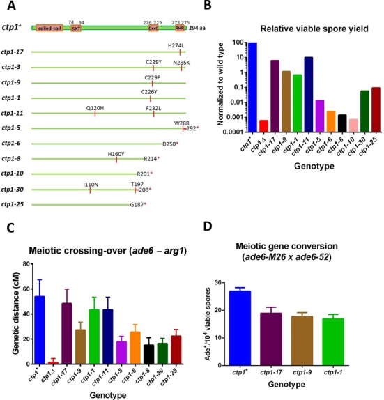
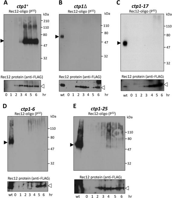
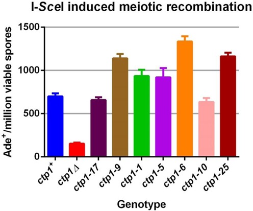
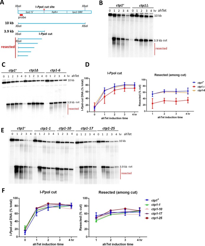
Meiotic programmed DNA double-strand break (DSB) repair is essential for crossing-over and viable gamete formation and requires removal of Spo11-oligonucleotide complexes from 5' ends (clipping) and their resection to generate invasive 3'-end single-stranded DNA (resection). Ctp1 (Com1, Sae2, CtIP homolog) acting with the Mre11-Rad50-Nbs1 (MRN) complex is required in both steps. We isolated multiple S. pombe ctp1 mutants deficient in clipping but proficient in resection during meiosis. Remarkably, all of the mutations clustered in or near the conserved CxxC or RHR motif in the C-terminal portion. The mutants tested, like ctp1Δ, were clipping-deficient by both genetic and physical assays-. But, unlike ctp1Δ, these mutants were recombination-proficient for Rec12 (Spo11 homolog)-independent break-repair and resection-proficient by physical assay. We conclude that the intracellular Ctp1 C-terminal portion is essential for clipping, while the N-terminal portion is sufficient for DSB end-resection. This conclusion agrees with purified human CtIP resection and endonuclease activities being independent. Our mutants provide intracellular evidence for separable functions of Ctp1. Some mutations truncate Ctp1 in the same region as one of the CtIP mutations linked to the Seckel and Jawad severe developmental syndromes, suggesting that these syndromes are caused by a lack of clipping at DSB ends that require repair.


Figures

Figure 1.
Figure 1.
Sequences and phenotypes of new ctp1 mutants. (A) Schematic representation of each of the ctp1 mutations studied here, indicated by the amino acid change at the codon numbered. * indicates non-sense mutation; red wavy line indicates region of amino acids altered by a frameshift mutation and the ensuing out-of-frame non-sense codon. Data in Figures 1–4 are from strains with these alleles integrated at the endogenous chromosomal locus. (B) Relative viable spore yields of ctp1 mutants. Data are from Supplementary Table S2, ‘chr’ column. (C) Meiotic crossing-over between ade6 and arg1 in ctp1 mutants. Data are from Supplementary Table S2, ‘chr’ column. Error bars are the SD of observed frequencies converted to cM using Haldane's equation. (D) Meiotic recombination between ade6-M26 and ade5–52 in ctp1 mutants. Data are from Supplementary Table S2. Error bars are SEM; n = 4 for each strain.
Figure 2.
Figure 2.
ctp1 C-terminal mutants, like ctp1Δ, release little or no detectable Rec12-oligonucleotide complexes. Strains with the indicated chromosomal ctp1 mutations, ctp1+ (A), ctp1Δ (B), ctp1-17 (C), ctp1-6 (D) or ctp1-25 (E), were induced for meiosis, and extracted DNA was assayed at the indicated times for Rec12-oligos by labeling with [α-32P] dCTP and TdT, followed by gel electrophoresis. In panels (B) and (C), about four times more cell extract was loaded for the mutants than for ctp1+, to increase the sensitivity of detecting Rec12-oligos in the mutants. The membrane was exposed to X-ray film to detect 32P (top panel) and then western blotted to detect Rec12-FLAG (bottom panel) using anti-FLAG antibody. Filled arrowhead, 32P-labeled Rec12-oligonucleotide complexes; open arrowhead, total Rec12 protein. ‘wt’ in panels B, C, D and E indicates ctp1+ cell extract assayed at 4 h after meiotic induction.
Figure 3.
Figure 3.
I-SceI-dependent meiotic recombination of ctp1 C-terminal region mutants is the same as or even higher than that of ctp1+ cells. Ade+ recombinants were measured in crosses with the I-SceI-cuttable allele ade6–3061 and ade6–52 in strains with the indicated ctp1 mutations on the chromosome. Data are from Supplementary Table S3. Error bars are SEM; n = 16 for ctp1+ and ctp1Δ, and 4 for the other mutants.
Figure 4.
Figure 4.
ctp1 C-terminal mutants are proficient for DSB end resection. (A) Diagram of the 10 kb XbaI chromosomal DNA fragment containing the I-PpoI cut site, the 3.9 kb I-PpoI cut fragment, and the resected DNA fragments (smear). The red box is the location of the probe for Southern blots. (B, C and E) DNA was extracted from strains with the indicated chromosomal ctp1 mutations at the indicated times after addition of 3 μM anhydrotetracycline (ahTet) added 3 h after meiotic induction, when DNA replication was complete (Supplementary Figure S2). DNA was digested with XbaI, electrophoresed, transferred to a membrane and hybridized with the probe indicated in panel (A). Resected DNA is indicated with a red line. (D and F) Quantification of I-PpoI cut and resected DNA in parallel from panels (B), (C) and (E). The radioactivity on the blots in panels (B), (C) and (E) was quantified with a PhosphorImager. Data are the mean ± SEM of three (for wild-type cells) or the range for two (for mutant cells except ctp1-6, ctp1-10 and ctp1-25) independent experiments. For each strain, I-PpoI-cut data are the percent of the total I-PpoI-cut DNA (3.9 kb cut band plus the resected fraction) at each time point relative to the sum of all XbaI released DNA (total of 10 kb XbaI cut band, 3.9 kb cut band and resected fraction). Resected data are the percentage of the resected DNA at each time point relative to the total I-PpoI cut DNA. ctp1+ DNA in panels (B), (C) and (E) was from three separate inductions. ctp1Δ DNA in panels (B) and (C) was from two separate inductions. In panel C, ctp1+ and ctp1Δ were on one blot, and ctp1-6 on another. In panel E, ctp1+, ctp1-1 and ctp1-10 were on one blot, and ctp1-17 and ctp1-25 on another.

Similar articles

Cited by

References

    1. Keeney S. In: Recombination and meiosis: crossing-over and disjunction. Egel R, Lankenau D.-H., editors. Berlin: Springer-Verlag; 2007. pp. 81–123.
    1. Williams G.J., Lees-Miller S.P., Tainer J.A. Mre11-Rad50-Nbs1 conformations and the control of sensing, signaling, and effector responses at DNA double-strand breaks. DNA Repair. 2010;9:1299–1306. - PMC - PubMed
    1. Stracker T.H., Petrini J.H. The MRE11 complex: starting from the ends. Nat. Rev. Mol. Cell Biol. 2011;12:90–103. - PMC - PubMed
    1. Hartsuiker E., Mizuno K., Molnar M., Kohli J., Ohta K., Carr A.M. Ctp1CtIP and the Rad32Mre11 nuclease activity are required for Rec12Spo11 removal but Rec12Spo11 removal is dispensable for other MRN-dependent meiotic functions. Mol. Cell. Biol. 2009;29:1671–1681. - PMC - PubMed
    1. Milman N., Higuchi E., Smith G.R. Meiotic DNA double-strand break repair requires two nucleases, MRN and Ctp1, to produce a single size class of Rec12 (Spo11)-oligonucleotide complexes. Mol. Cell. Biol. 2009;29:5998–6005. - PMC - PubMed

Publication types

MeSH terms