. 2015 Aug 18;43(14):6934-44.
doi: 10.1093/nar/gkv623. Epub 2015 Jun 29.

The XRCC1 phosphate-binding pocket binds poly (ADP-ribose) and is required for XRCC1 function

Affiliations

The XRCC1 phosphate-binding pocket binds poly (ADP-ribose) and is required for XRCC1 function

Claire Breslin et al. Nucleic Acids Res. .

Abstract

Poly (ADP-ribose) is synthesized at DNA single-strand breaks and can promote the recruitment of the scaffold protein, XRCC1. However, the mechanism and importance of this process has been challenged. To address this issue, we have characterized the mechanism of poly (ADP-ribose) binding by XRCC1 and examined its importance for XRCC1 function. We show that the phosphate-binding pocket in the central BRCT1 domain of XRCC1 is required for selective binding to poly (ADP-ribose) at low levels of ADP-ribosylation, and promotes interaction with cellular PARP1. We also show that the phosphate-binding pocket is required for EGFP-XRCC1 accumulation at DNA damage induced by UVA laser, H2O2, and at sites of sub-nuclear PCNA foci, suggesting that poly (ADP-ribose) promotes XRCC1 recruitment both at single-strand breaks globally across the genome and at sites of DNA replication stress. Finally, we show that the phosphate-binding pocket is required following DNA damage for XRCC1-dependent acceleration of DNA single-strand break repair, DNA base excision repair, and cell survival. These data support the hypothesis that poly (ADP-ribose) synthesis promotes XRCC1 recruitment at DNA damage sites and is important for XRCC1 function.


Figures

Figure 1.
Figure 1.
Conservation of the XRCC1 BRCT1 domain and its phosphate-binding pocket. (A) Schematic depicting conservation of XRCC1 domains in human (Hs), frog (Xl), fly (Dm), and plant (At) XRCC1. Binding sites for the indicated protein partners are shown. Asterisks denote CK2 phosphorylation sites that mediate FHA-dependent interactions with PNKP, APTX and APLF. Black boxes denoted the nuclear localization signal. (B) Alignment of the BRCT1 domain from human, frog, fly and plant. Solid red boxes denote the residues predicted to form the phosphate-binding pocket and the dotted red box denotes the degenerate putative PAR-binding motif identified by Pleschke et al. (9). Conserved identical residues are in red. Asterisks denote residues mutated in this study. (C) Model of the BRCT1 domain based on the NMR structure (PDN accession code: 2D8M), highlighting the residues predicted to form phosphate-binding pocket. (D) Left, purified recombinant His-XRCC1161–406 and His-XRCC1161–406 RK proteins, fractionated by SDS-PAGE and stained with coomassie brilliant blue. Right, cartoon of the in vitro PAR-binding assay. Proteins were adsorbed to microwell dishes and mock-ribosylated (‘-NAD+’) or ribosylated (‘+NAD+’) by PARP1 in absence or presence of NAD+ as indicated. Bound proteins were then incubated with recombinant wild type His-XRCC1161–406 or mutant His-XRCC1161–406 RK, and bound XRCC1 detected with anti-His tag antibodies colourmetrically (A450) using HRP-conjugated secondary antibody.
Figure 2.
Figure 2.
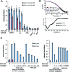
The XRCC1 BRCT1 phosphate-binding pocket binds PAR, in vitro. (A) Binding of His-XRCC1161–406 and His-XRCC1161–406 RK to the indicated mock-ribosylated (-NAD+) or ribosylated (1–50 μM NAD+) proteins was measured as indicated in Figure 1D. Data are the mean (±1 SD) of at least three experiments. (B) Top, thermal stability of recombinant His-XRCC1161–406 and His-XRCC1161–406 RK. 2 μM XRCC1 protein was assayed in the presence of SYPRO Orange and unfolding temperatures determined as described in materials and methods. Data are the mean (±1SD) of four independent measurements. Bottom, circular dichroism of His-XRCC1161–406 and His-XRCC1161–406 RK. Data are the average of 10 sequential scans, with the spectrum from sample buffer alone subtracted. (C) Binding of His-XRCC1161–406 and His-XRCC1161–406 RK to calf thymus histone mock-ribosylated in the absence of NAD+ (‘0’) or ribosylated in the presence of either 0.5 μM NAD+ (left panel) or 1 μM NAD+ (right panel). Where indicated, XRCC1 binding was measured in the presence of 43 nM PARP1 competitor that was first autoribosylated in the presence of 0, 0.2, 0.4, 0.8 or 2 μM NAD+, as indicated. Alternatively, His-XRCC1161–406 and His-XRCC1161–406 RK binding was measured the presence of the indicated concentration of either poly (ADP-ribose) (‘PAR’, left) or mono (ADP-ribose) (‘MAR’, right) competitor. PAR/MAR competitor concentrations are total ADP-ribose units (μM) present as PAR (2–300 subunit lengths) or MAR. Data are the mean (±1SD) of at least three experiments.
Figure 3.
Figure 3.
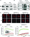
The XRCC1 BRCT1 phosphate-binding pocket mediates PAR-dependent interaction with PARP1 and recruitment at sites of UVA laser induced damage. (A) EGFP-XRCC1 was affinity purified from cell extract from U2OSGFP-XRCC1 cells using GFP-Trap beads. Aliquots of the column input, unbound, last wash, and eluate samples were fractionated by SDS-PAGE and immunoblotted with pS485/pT488 anti-XRCC1 polyclonal antibody or anti-PARP1 antibody. Where indicated (‘+PARPi’), PARP inhibitor (500 nM Ku-58948) was included in the cell lysis buffer and was present in the cell culture medium for 1 h at 37°C prior to lysis. (B) U2OS cells were transiently co-transfected with expression vector encoding either EGFP, EGFP-XRCC1161–406, or EGFP-XRCC1161–406 RK and with expression vector encoding either mCherry-PARP1 or mCherry-PARP1E988K. EGFP-XRCC1 was recovered from whole cell extract and aliquots of column input, unbound, last wash and eluate (bound material) fractionated by SDS-PAGE and immunoblotted with anti-GFP or anti-PARP1 antibody. The position of mCherry-PARP1 and endogenous PARP1 are indicated by black and red arrows, respectively. (C) XRCC1-mutant EM9 cells were transiently transfected with pmRFP-XRCC1, pmRFP-XRCC1RK, pmRFP-XRCC1161–406, or pmRFP-XRCC1161–406 RK and treated with UVA laser. mRFP fluorescence was measured at the indicated times (seco) following microirradiation in the presence or absence of 500nM PARP inhibitor (Ku-58948). Representative images are shown. (D) Left, quantitation of the mRFP-XRCC1 fluorescence proteins at sites of 405 nm UVA laser-induced DNA damage in the above experiments. Inset, pmRFP-XRCC1 and pmRFP-XRCC1RK expression levels in the transfected cells, as measured by immunoblotting with pS485/pT488 anti-XRCC1 polyclonal antibody. Right quantitation of the mRFP-XRCC1161406 fluorescence at sites of 351 nm UVA laser damage. Data is expressed as change in mean fluorescence in ten or more cells per construct ± SEM.
Figure 4.
Figure 4.
The XRCC1 BRCT1 phosphate-binding pocket is important for XRCC1 accumulation at sites of H2O2-induced damage and for colocalization with PCNA. (A) U2OS cells were transfected with pEGFP-XRCC1 or pEGFP-XRCC1RK, mock-treated or treated with 10 mM H2O2 for 10 min, and after 15 min recovery in drug-free medium fixed and analysed by fluorescence microscopy. (B) Left, U2OSGFP-XRCC1 cells were transfected with pCCC-TagRFP to detect endogenous PCNA in the presence or absence of the PARP inhibitor olaparib (100 nM) and analysed as above 24 h later. Right, cells were transfected as above and additionally pulse labelled with EdU (blue) to identify sites of DNA replication. Dotted square denotes the area expanded on the right. (C) U2OS cells were co-transfected with either pEGFP-XRCC1WT or pEGFP-XRCC1RK and pCCC-TagRFP plasmid to detect endogenous PCNA. Representative images are shown.
Figure 5.
Figure 5.
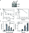
XRCC1-mediated acceleration of SSBR and cell survival requires the XRCC1 BRCT1 phosphate-binding pocket. (A) XRCC1 protein expression in XRCC1-mutant EM9 cells stably transfected with empty expression vector (EM9-V) or expression vector encoding either XRCC1-His (EM9-XH) or XRCC1-HisRK (EM9-XHRK). Cell extracts were fractionated by SDS-PAGE and immunoblotted with anti-XRCC1 Mab (33–2–5) and anti-Actin antibodies. (B) Clonogenic survival of XRCC1-mutant EM9 cells stably transfected with empty expression vector (EM9-V) or expression vector encoding either XRCC1-His (EM9-XH) or XRCC1-HisRK (EM9-XHRK). Cells were treated with the indicated concentrations of H2O2 (left) or MMS (right) for 15 min and then in drug free medium for 10–14 days to allow colony formation. Data are the mean (±SEM) of three independent experiments. Where not visible, error bars are smaller than the symbols. (C) Chromosomal SSBR rates were measured in the above EM9 cell lines in alkaline comet assays following treatment with 150 μM H2O2 for 20 min on ice, followed by recovery in drug-free medium for the indicated time at 37°C, or with the indicated concentration of MMS for 15 min at 37°C to measure the accumulation of SSB intermediates during BER. Data are the mean (±SEM) of three independent experiments.

References

    1. Caldecott K.W. Single-strand break repair and genetic disease. Nat. Rev. Genet. 2008;9:619–631. - PubMed
    1. Caldecott K.W. XRCC1 and DNA strand break repair. DNA Repair (Amst.) 2003;2:955–969. - PubMed
    1. Thompson L.H., West M.G. XRCC1 keeps DNA from getting stranded. Mutat. Res. 2000;459:1–18. - PubMed
    1. Thompson L.H., Brookman K.W., Dillehay L.E., Carrano A.V., Mazrimas J.A., Mooney C.L., Minkler J.L. A CHO-cell strain having hypersensitivity to mutagens, a defect in DNA strand-break repair, and an extraordinary baseline frequency of sister-chromatid exchange. Mutat. Res. 1982;95:427–440. - PubMed
    1. Zdzienicka M.Z., van der Schans G.P., Natarajan A.T., Thompson L.H., Neuteboom I., Simons J.W. A Chinese hamster ovary cell mutant (EM-C11) with sensitivity to simple alkylating agents and a very high level of sister chromatid exchanges. Mutagenesis. 1992;7:265–269. - PubMed

Publication types

Substances