. 2015 Aug 18;43(14):7021-31.
doi: 10.1093/nar/gkv652. Epub 2015 Jun 29.

Human DNA ligase III bridges two DNA ends to promote specific intermolecular DNA end joining

Affiliations

Human DNA ligase III bridges two DNA ends to promote specific intermolecular DNA end joining

Vandna Kukshal et al. Nucleic Acids Res. .

Abstract

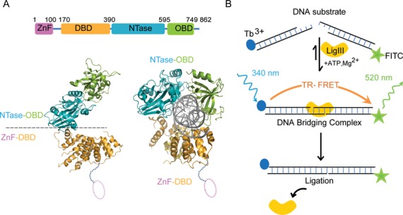
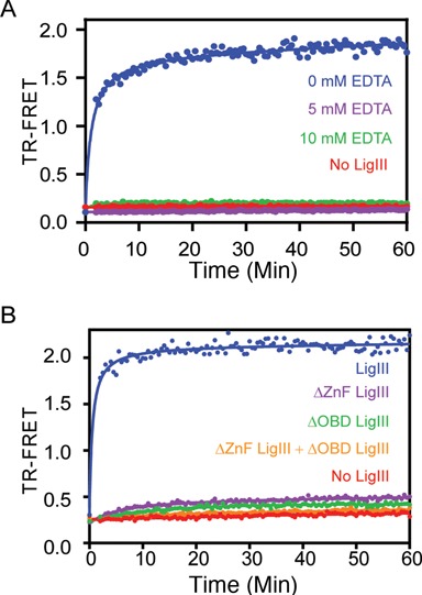
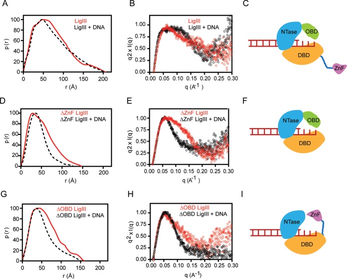
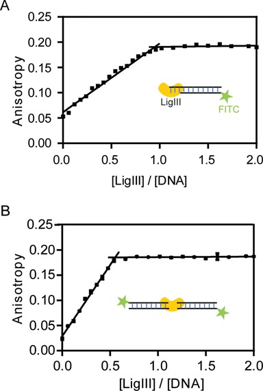
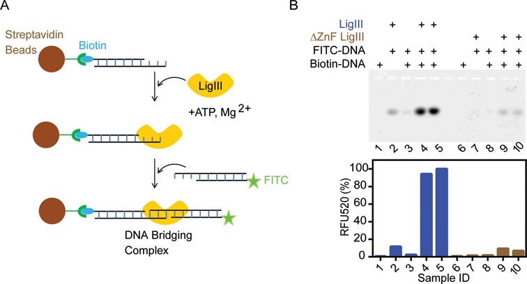
Mammalian DNA ligase III (LigIII) functions in both nuclear and mitochondrial DNA metabolism. In the nucleus, LigIII has functional redundancy with DNA ligase I whereas LigIII is the only mitochondrial DNA ligase and is essential for the survival of cells dependent upon oxidative respiration. The unique LigIII zinc finger (ZnF) domain is not required for catalytic activity but senses DNA strand breaks and stimulates intermolecular ligation of two DNAs by an unknown mechanism. Consistent with this activity, LigIII acts in an alternative pathway of DNA double strand break repair that buttresses canonical non-homologous end joining (NHEJ) and is manifest in NHEJ-defective cancer cells, but how LigIII acts in joining intermolecular DNA ends versus nick ligation is unclear. To investigate how LigIII efficiently joins two DNAs, we developed a real-time, fluorescence-based assay of DNA bridging suitable for high-throughput screening. On a nicked duplex DNA substrate, the results reveal binding competition between the ZnF and the oligonucleotide/oligosaccharide-binding domain, one of three domains constituting the LigIII catalytic core. In contrast, these domains collaborate and are essential for formation of a DNA-bridging intermediate by adenylated LigIII that positions a pair of blunt-ended duplex DNAs for efficient and specific intermolecular ligation.


Figures

Figure 1.
Figure 1.
Tandem DNA binding surfaces of LigIII support DNA bridging. (A) The LigIIIβ protein (LigIII, 862 residues) spans four structural domains, an N-terminal zinc finger (ZnF; residues 1–100), a DNA binding domain (DBD; residues 170–390), a nucleotidyl transferase domain (NTase; residues 391–595) and the OB-fold domain (OBD; residues 596–749) that are required for efficient DNA end joining activity. Two pairs of domains, the ZnF–DBD and the NTase–OBD, comprise independent DNA binding modules (29) that are required in DNA bridging activity (cf. Figure 4B). The crystal structure of ΔZnF LigIII bound to a nicked DNA reveals a compact structure that would preclude the ZnF domain from binding to the nick (PDB 3L2P) (30). (B) We developed a time resolved FRET (TR-FRET) assay to measure intermolecular ligation of two DNAs by LigIII. In this assay, LigIII brings together DNAs labeled with Tb3+ (donor) and FITC (acceptor) to produce a FRET signal. DNA bridging activity and efficient intermolecular ligation of DNAs depend on the presence of the ZnF domain. We present evidence that this DNA bridging complex is a reaction intermediate of the intermolecular ligation pathway.
Figure 2.
Figure 2.
DNA bridging precedes intermolecular DNA ligation. (A) The real time measurement of TR-FRET (520/495 nm) signals the assembly of a DNA bridging complex, in which LigIII juxtaposes two DNAs labeled with donor and acceptor labels. Ligation reactions containing a pair of DNAs with complementary 3-nt overhangs were assembled in the absence (red) or presence (blue) of LigIII and the TR-FRET signal was monitored. The TR-FRET traces shown in Figures 2–5 are the average values of assays performed in triplicate. DNA ligation activity measured under the same reaction conditions (B) is plotted as the percentage of total DNA in the ligation product (green). (B) The time course of DNA ligation under the same reaction conditions as in (A) was monitored by running timed reaction aliquots on a 18% denaturing urea Polyacrylamide gelelectrophoresis (PAGE). DNA ligation activity is significantly slower than the TR-FRET signal, which results from the LigIII dependent juxtaposition of the labeled DNAs. The intermolecular ligation of a blunt-end DNA (Supplementary Figure S5) is markedly slower than ligation of overhanging DNA ends, suggesting that the end structure of the DNAs influences the rate-limiting step of ligation. (C) The same rapid onset of the TR-FRET signal is observed upon addition of LigIII (blue) to a pair of DNAs with 5′-OH groups that cannot be ligated, whereas the TR-FRET value remains at baseline in the absence of LigIII (red). Thus, DNA bridging complex formation is rapid and precedes DNA ligation (A). (D) PAGE analysis of the reaction with non-ligatable DNAs used in (C) shows that no ligated product is formed under conditions that produce the TR-FRET signal corresponding to DNA bridging activity.
Figure 3.
Figure 3.
Bridging requires compatible pairs of DNA ends. Pairs of non-ligatable DNAs with either complementary three nucleotide overhangs (blue) or with blunt ends (green) generated a FRET signal in the presence of LigIII, indicative of DNA bridging activity. In contrast, two mispaired combinations consisting of two DNAs with blunt and overhanging ends (orange, magenta) did not support TR-FRET and resulted in a baseline level of fluorescence.
Figure 4.
Figure 4.
Specificity of DNA bridging by LigIII. (A) The DNA binding activity of LigIII in the DNA bridging assay is effectively competed by an unlabeled DNA, saturable manner, as expected for the formation of a discrete protein–DNA complex. A pair of labeled, non-ligatable DNAs (500 nM of each) was incubated with LigIII (100 nM) in the presence of increasing concentrations of an unlabeled DNA duplex (0–8 μM). See also Supplementary Figure S2. (B) In the absence of added ATP (light blue) the TR-FRET signal amplitude is about 50% of the signal for a reaction containing ATP (100 μM; dark blue). LigIII that was completely deadenylated (orange) did not support DNA bridging and TR-FRET. (C) Addition of ATP to the deadenylated LigIII (shown in B) restored the TR-FRET signal and DNA bridging activity in a concentration-dependent manner. The TR-FRET values 30 min after addition of ATP are shown.
Figure 5.
Figure 5.
DNA bridging requires two independent DNA binding surfaces of LigIII. (A) Addition of EDTA (5 mM) abolishes the DNA bridging activity of LigIII in reaction buffer containing 5 mM MgCl2, indicating that divalent metal(s) are required for DNA binding activity. (B) Deletion mutants of LigIII lacking the ZnF (ΔZnF LigIII) or OBD (ΔOBD LigIII) domains do not form the DNA bridging complex although both mutants can bind to double-stranded DNA (Supplementary Table S1). A mixture of the ΔZnF and ΔOBD LigIII proteins also fails to bridge the labeled DNAs, consistent with a single molecule of LigIII mediating the DNA bridging activity (cf. Figure 7).
Figure 6.
Figure 6.
The ZnF and OBD domains bind to overlapping sites on DNA. Small angle X-ray scattering reveals the dynamic interactions of LigIII and deletion mutants in the 1:1 complex described in Figure 7A. (A) The normalized pair distribution function p(r) is consistent with an elongated conformation of LigIII in the presence or absence of a DNA duplex containing a three nucleotide overhang. (B) A normalized Kratky plot of the scattering data shows evidence of flexible residues in the presence or absence of DNA, which is modeled as a flexible ZnF domain in panel (C). (D) Deletion of the ZnF (ΔZnF LigIII) significantly narrows the p(r) distribution and reveals a large conformational transition upon binding to DNA. (E) The normalized Kratky plot of ΔZnF LigIII in complex with DNA shows a bell shaped Gaussian curve, indicative of a compact structure when bound to DNA. (F) The SAXS data are consistent with a model of ΔZnF LigIII encircling the DNA end as for the crystal structure of ΔZnF LigIII bound to nicked DNA (30). (G) Deletion of the C-terminal OBD domain (ΔOBD LigIII) narrows the pair distribution function p(r) of LigIII in a manner similar to deletion of the ZnF domain. This result suggests that the ZnF can engage DNA when the OBD is deleted, resulting in a compact conformation of ΔOBD LigIII bound to DNA. (H) A normalized Kratky plot of ΔOBD LigIII bound to DNA reveals a Gaussian curve consistent with a compact structure with the ZnF engaging the bound DNA as modeled in panel (I).
Figure 7.
Figure 7.
One molecule of LigIII binds two molecules of DNA in the bridging complex. (A) The DNA binding activity of LigIII was monitored by fluorescence anisotropy of a labeled DNA under conditions of stoichiometric binding. A 5′ FITC labeled duplex (800 nM) containing a three nucleotide overhang that cannot pair with itself was titrated with increasing concentrations of LigIII and the change in fluorescence anisotropy was measured. The DNA concentration in this assay is ∼5-fold higher than the apparent equilibrium binding constant (Kapp ∼ 154 nM) determined with limiting concentrations of DNA and LigIII in excess. The measured binding stoichiometry of 1:1 for the LigIII:DNA complex was additionally confirmed by repeating the titration experiment at a higher concentration of DNA (1600 nM; not shown). (B) A binding stoichiometry of 1:2 (ratio = 0.5) was measured for LigIII in complex with a 5′ FITC labeled, blunt-ended DNA duplex (800 nM) that can form the DNA bridging complex. The same binding stoichiometry was determined in a binding reaction containing a higher concentration of the DNA (1600 nM; not shown).
Figure 8.
Figure 8.
LigIII is stably bound to two DNAs in the bridging complex. (A) Streptavidin magnetic beads coated with a 5′ biotinylated DNA duplex were used to capture LigIII together with a 5′ FITC labeled DNA in a pull-down assay. The FITC labeled DNA on the beads was analyzed by urea PAGE and quantification of fluorescence emission intensity in the gel (see ‘Materials and Methods’ section). (B) Upper panel, denaturing urea PAGE analysis of the FITC labeled DNA recovered from the beads. Lower panel, quantitation of fluorescence intensity in the gel. Samples 4 and 5 are replicates containing LigIII in a bridging complex. Samples 9 and 10 are replicates containing the ΔZnF LigIII mutant protein, which does not form the bridging complex. Figure 8 shows the pull-down of DNAs with complementary 3 nt overhangs in complex with LigIII. Additional controls are shown in the legend above the gel. Supplementary Figure S3 shows the pull-down of a LigIII DNA bridging complex incorporating two blunt-ended DNAs.

References

    1. Tomkinson A.E., Vijayakumar S., Pascal J.M., Ellenberger T. DNA ligases: structure, reaction mechanism, and function. Chem. Rev. 2006;106:687–699. - PubMed
    1. Ellenberger T., Tomkinson A.E. Eukaryotic DNA ligases: structural and functional insights. Annu. Rev. Biochem. 2008;77:313–338. - PMC - PubMed
    1. Lakshmipathy U., Campbell C. Antisense-mediated decrease in DNA ligase III expression results in reduced mitochondrial DNA integrity. Nucleic Acids Res. 2001;29:668–676. - PMC - PubMed
    1. Simsek D., Furda A., Gao Y., Artus J., Brunet E., Hadjantonakis A.-K., Van Houten B., Shuman S., McKinnon P.J., Jasin M. Crucial role for DNA ligase III in mitochondria but not in Xrcc1-dependent repair. Nature. 2011;471:245–248. - PMC - PubMed
    1. Gao Y., Katyal S., Lee Y., Zhao J., Rehg J.E., Russell H.R., McKinnon P.J. DNA ligase III is critical for mtDNA integrity but not Xrcc1-mediated nuclear DNA repair. Nature. 2011;471:240–244. - PMC - PubMed

Publication types