

Delayed presentation of uterine perforation with ovary migration after dilatation and curettage
- PMID: 26131247
- PMCID: PMC4483796
Delayed presentation of uterine perforation with ovary migration after dilatation and curettage
Abstract
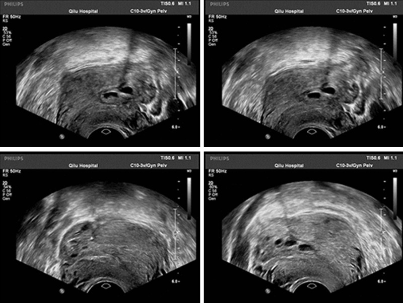
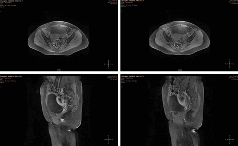
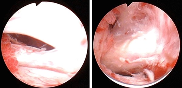
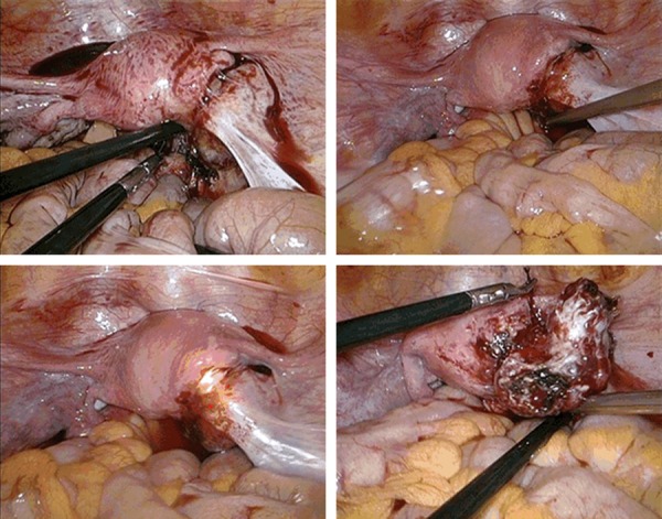
We present a rare but serious uterine perforation. A 31-year-old woman was referred to our department for hyperechogenic mass in uterus on ultrasonography after Dilation and curettage (D&C) for the adherent placenta and retained products of conception. Transvaginal ultrasound examination showed that a mass with several follicles measuring 35×29 mm was seen emanating from the right posterior wall of the uterine cavity, and there was absence of the myometrial tissue. A hysteroscopy and laparoscopy showed a uterine perforation with ovary incarceration. The ovary was rehabilitated, and the uterine perforation site was incised. D&C can not be performed when delayed presentation of uterine perforation with migration of an extrauterine organ is suspected, particularly, some of them are asymptomatic after a difficult intrauterine operation.
Keywords: Uterine perforation; hysteroscopy; laparoscopy; migration; ovary.
Figures
Similar articles
-
Uterine perforation with omentum incarceration after dilatation and evacuation/curettage: magnetic resonance imaging findings.Arch Gynecol Obstet. 2012 Mar;285(3):887-90. doi: 10.1007/s00404-011-2127-z. Epub 2011 Nov 3. Arch Gynecol Obstet. 2012. PMID: 22048784
-
Hysteroscopic Diagnosis of Omentum Incarceration Subsequent to an Iatrogenic Uterine Perforation.J Minim Invasive Gynecol. 2019 Jan;26(1):29-30. doi: 10.1016/j.jmig.2018.02.020. Epub 2018 Mar 7. J Minim Invasive Gynecol. 2019. PMID: 29524723
-
Laparoscopic Management of an Intrauterine Fallopian Tube Incarceration After Curettage for a Non-progressing Pregnancy.J Minim Invasive Gynecol. 2019 Jul-Aug;26(5):805. doi: 10.1016/j.jmig.2018.09.770. Epub 2018 Sep 19. J Minim Invasive Gynecol. 2019. PMID: 30243687
-
Hysteroscopy combined with laparoscopy in the diagnosis and treatment of omentum majus incarceration secondary to uterine perforation: A case report and literature review.J Obstet Gynaecol Res. 2025 Jan;51(1):e16213. doi: 10.1111/jog.16213. J Obstet Gynaecol Res. 2025. PMID: 39814042 Free PMC article. Review.
-
Complete and partial uterine perforation and embedding following insertion of intrauterine devices. II. Diagnostic methods, prevention, and management.Obstet Gynecol Surv. 1981 Aug;36(8):401-17. doi: 10.1097/00006254-198108000-00001. Obstet Gynecol Surv. 1981. PMID: 6455610 Review.
Cited by
-
A Rare Occurrence of Uterine Perforation Following the Dilation and Curettage for Missed Abortion.Cureus. 2024 Sep 24;16(9):e70079. doi: 10.7759/cureus.70079. eCollection 2024 Sep. Cureus. 2024. PMID: 39449922 Free PMC article.
-
Transvaginal strangulated bowel evisceration through uterine perforation due to unsafe abortion: a case report and literature review.BMC Womens Health. 2021 Mar 5;21(1):98. doi: 10.1186/s12905-021-01247-y. BMC Womens Health. 2021. PMID: 33663467 Free PMC article. Review.
-
Uterine Perforation as a Complication of the Intrauterine Procedures Causing Omentum Incarceration: A Review.Diagnostics (Basel). 2023 Jan 16;13(2):331. doi: 10.3390/diagnostics13020331. Diagnostics (Basel). 2023. PMID: 36673141 Free PMC article. Review.
-
A Delayed Case of Uterine Perforation with Omental Adhesions.Gynecol Minim Invasive Ther. 2021 Aug 3;10(3):174-176. doi: 10.4103/GMIT.GMIT_113_19. eCollection 2021 Jul-Sep. Gynecol Minim Invasive Ther. 2021. PMID: 34485064 Free PMC article.
-
Incidental Diagnosis of Uterine Perforation During Lower Segment Caesarean Section: A Rare Obstetric Complication.Cureus. 2025 May 16;17(5):e84223. doi: 10.7759/cureus.84223. eCollection 2025 May. Cureus. 2025. PMID: 40525055 Free PMC article.
References
-
- Coughlin LM, Sparks DA, Chase DM, Smith J. Incarcerated small bowel associated with elective abortion uterine perforation. J Emerg Med. 2013;44:e303–06. - PubMed
-
- Augustin G, Majerović M, Luetić T. Uterine perforation as a complication of surgical abortion causing small bowel obstruction: a review. Arch Gynecol Obstet. 2013;288:311–23. - PubMed
-
- Ntia IO, Ekele BA. Bowel prolapse through perforated uterus following induced abortion. West Afr J Med. 2000;19:209–11. - PubMed
-
- Dignac A, Novellas S, Fournol M, Caramella T, Bafghi A, Chevallier P. Incarceration of the appendix complicating a uterine perforation following surgical abortion: CT aspects. Emerg Radiol. 2008;15:267–69. - PubMed
-
- Ozaki K, Suzuki S. Uterine perforation with omentum incarceration after dilatation and evacuation/curettage. Arch Gynecol Obstet. 2013;287:607–08. - PubMed
Publication types
LinkOut - more resources
Full Text Sources