

Subcapsular Hepatic Hematoma After Endoscopic Retrograde Cholangiopancreatography: A Case Report and Review of Literature
- PMID: 26131812
- PMCID: PMC4504646
- DOI: 10.1097/MD.0000000000001041
Subcapsular Hepatic Hematoma After Endoscopic Retrograde Cholangiopancreatography: A Case Report and Review of Literature
Abstract
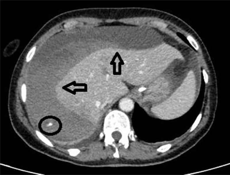
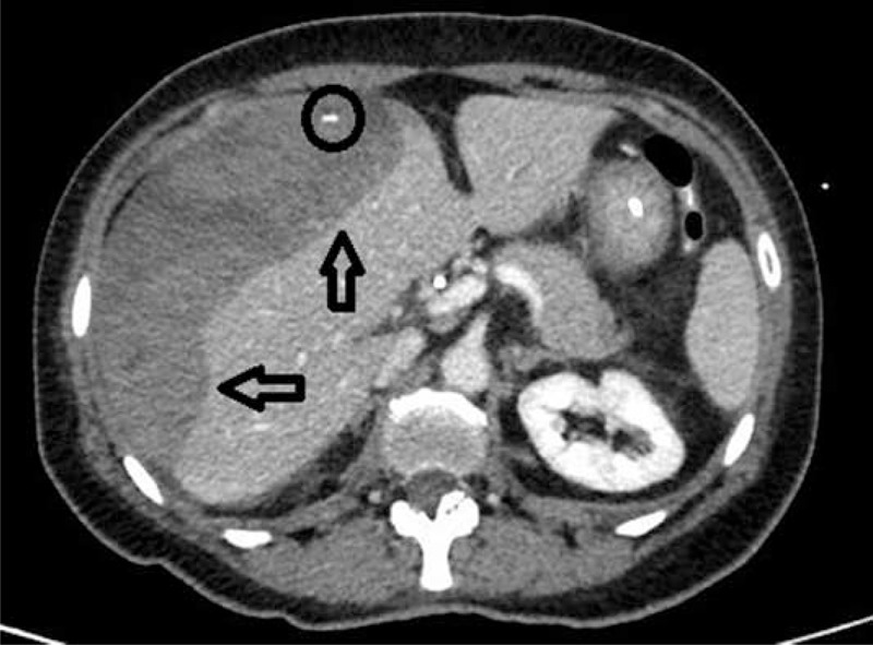
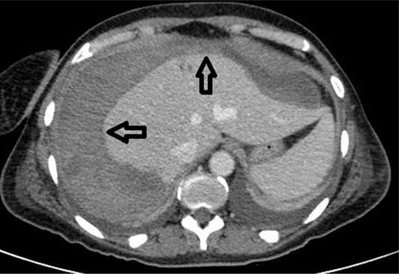
Endoscopic retrograde cholangiopancreatography (ERCP) is one of the most frequently performed procedures for the diagnosis and treatment of biliary-pancreatic diseases. ERCP-related complications total around 2.5% to 8%, with a mortality rate ranging from 0.5% to 1%. An exceptional ERCP complication is subcapsular hepatic hematoma, and few cases are reported worldwide.We present the case of a 52-year-old woman with a history of recurring upper abdominal pain and a clinical and ultrasonographic diagnosis of obstructive jaundice due to common bile duct stones. After 2 difficult endoscopic biliary procedures, common bile duct stones clearance was obtained. Post-ERCP course was symptomatic with upper abdominal pain and anemization with hemodynamic instability.CT scan demonstrated a 15 cm × 11 cm subcapsular hepatic hematoma filled with air and liquid on the surface of the right hepatic lobe. The patient was successfully treated with the embolization of a small branch of right hepatic artery angiographically identified as the cause of bleeding.Subcapsular hepatic hematoma after ERCP is a rare complication that must be taken into account in the differential diagnosis of symptomatic cases after ERCP. Its diagnosis is based on clinical and laboratory data and especially on imaging (ultrasound, CT, or MRI). Treatment is often conservative but, in some cases, embolization or percutaneous drainage or surgery may be necessary.
Conflict of interest statement
The authors have no funding and conflicts of interest to disclose.
Figures
References
-
- Hart R, Classen M. Complications of diagnostic gastrointestinal endoscopy. Endoscopy 1990; 22:229–233. - PubMed
-
- Ortega Deballon P, Fernández Lobato R, García Septern J, et al. Liver hematoma following ERCP. Surg Endosc 2000; 14:767. - PubMed
-
- Horn TL, Peña LR. Subcapsular hepatic hematoma after ERCP: case report and review. Gastrointest Endosc 2004; 59:594–596. - PubMed
-
- Chi KD, Waxman I. Subcapsular hepatic hematoma after Guide Wire Injury during ERCP. Endoscopy 2004; 36:1019–1021. - PubMed
Publication types
MeSH terms
LinkOut - more resources
Full Text Sources
Medical
