

Case Reports
doi: 10.1590/abd1806-4841.20153774.
Epub 2015 Jun 1.
Reflectance confocal microscopy of cutaneous melanoma. Correlation with dermoscopy and histopathology
Affiliations
- PMID: 26131877
- PMCID: PMC4516104
- DOI: 10.1590/abd1806-4841.20153774
Item in Clipboard
Case Reports
Reflectance confocal microscopy of cutaneous melanoma. Correlation with dermoscopy and histopathology
An Bras Dermatol.
2015 May-Jun.
Abstract
In vivo Confocal Microscopy is a method for non-invasive, real-time visualization of microscopic structures and cellular details of the epidermis and dermis, which has a degree of resolution similar to that obtained with histology. We present a case of cutaneous melanoma in which diagnosis was aided by confocal microscopy examination. We also correlate the observed features with the dermoscopic and histopathological findings. Confocal microscopy proved to be an useful adjunct to dermoscopy, playing an important role as a method 'between clinical evaluation and histopathology'.
Conflict of interest statement
Conflict of Interest: None.
Figures
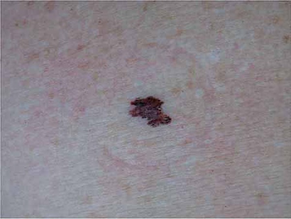
Brownish, hyperchromic, asymmetrical, 6-mm macule with irregular edges and
variable colors in the right deltoid region
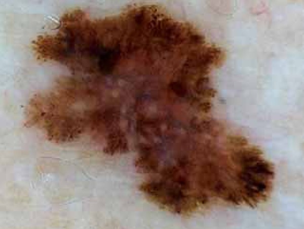
Dermatoscopy of the lesion. Global pattern: asymmetrical structure and varied
colors and structures (multi-components). Specific features: blurs and atypical
pigment network with peripheral projections (pseudopods). Pigmented cells
irregularly distributed in the periphery of the lesion. Blue-gray irregularly
distributed pigmentation
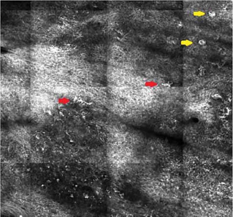
In vivo Confocal Microscopy: Atypical “honeycomb” pattern. Round cells with
bright cytoplasm and dark nucleus (yellow arrow), and dendritic cells (red arrow)
in the epidermis (pagetoid cells)
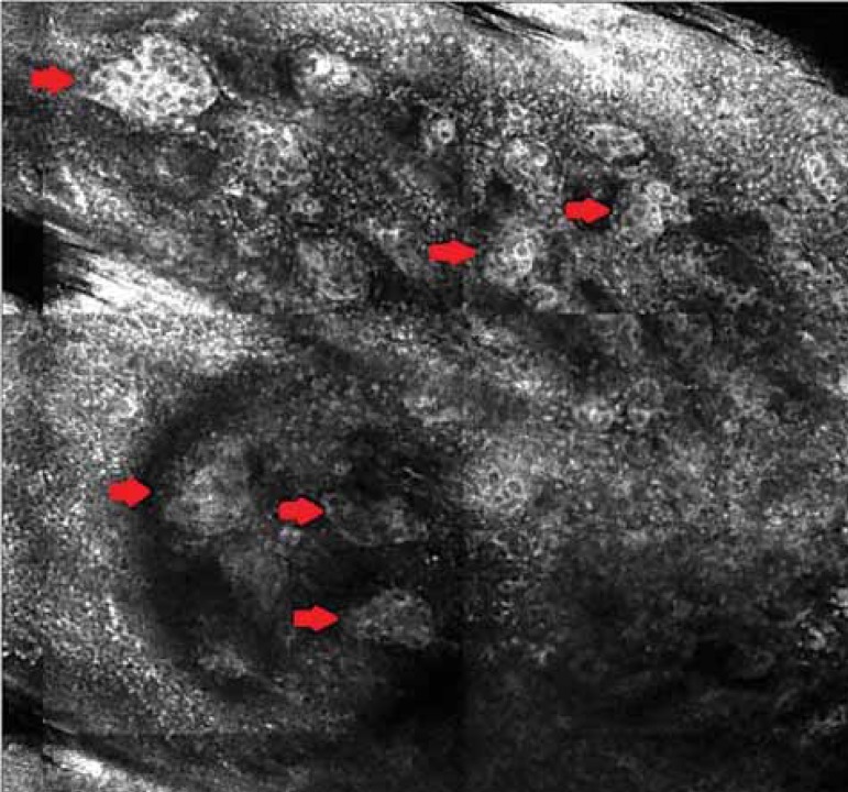
In vivo Confocal Microscopy: Presence of nests with heterogeneous brightness
at the dermo-epidermal junction and papillary dermis (red arrow)
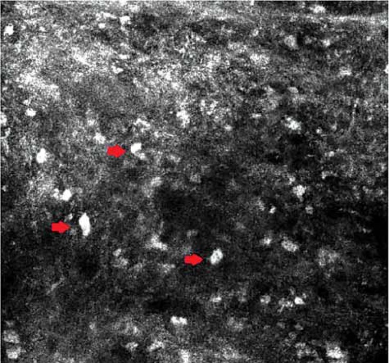
In vivo Confocal Microscopy: Bright cells dispersed in the dermis,
corresponding to melanophages (red arrow)
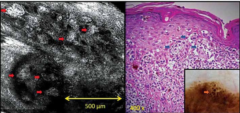
The heterogeneous nests of clear cells seen in ICM (red arrow) are seen in
histopathology as a proliferation of atypical melanocytes arranged in irregular
nests along the DEJ and papillary dermis (blue arrow) In dermoscopy, they are seen
as heterogeneous globules (orange arrow)
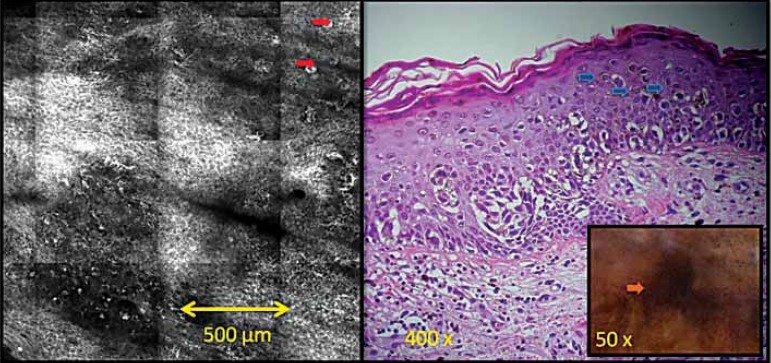
In ICM, round cells with clear cytoplasm and dark nucleus located in the
epidermis (red arrow) correspond to atypical pagetoid melanocytes (blue arrow) in
histopathology. In dermoscopy they may be seen as blurs (orange arrow)
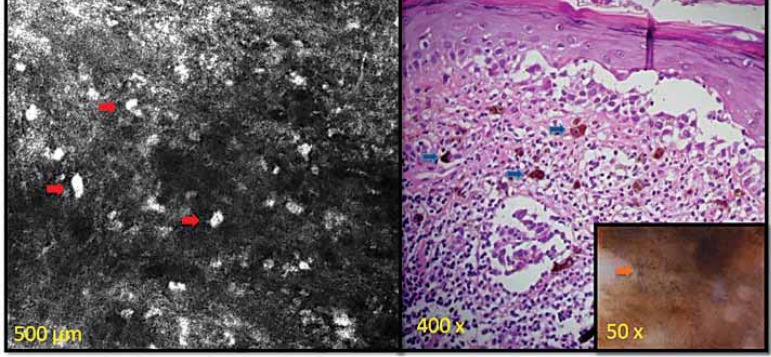
In ICM, the large clear nucleated cells infliltrating the papillary dermis
(red arrow) correspond to the melanophages in the papillary dermis (blue arrow)
seen in histopathology. and to the blue-gray veil seen in dermoscopy (orange
arrow)
Similar articles
-
Role of In Vivo Reflectance Confocal Microscopy in the Analysis of Melanocytic Lesions.Acta Dermatovenerol Croat. 2018 Apr;26(1):64-67. Acta Dermatovenerol Croat. 2018. PMID: 29782304 Review.
-
Morphological classification of melanoma metastasis with reflectance confocal microscopy.J Eur Acad Dermatol Venereol. 2019 Apr;33(4):676-685. doi: 10.1111/jdv.15329. Epub 2018 Dec 4. J Eur Acad Dermatol Venereol. 2019. PMID: 30394598
-
Acral melanoma with network pattern: a dermoscopy-reflectance confocal microscopy and histopathology correlation.Dermatol Surg. 2010 May;36(5):701-3. doi: 10.1111/j.1524-4725.2010.01533.x. Epub 2010 Apr 1. Dermatol Surg. 2010. PMID: 20384739 No abstract available.
-
Remodeling of the dermoepidermal junction in superficial spreading melanoma: insights gained from correlation of dermoscopy, reflectance confocal microscopy, and histopathologic analysis.Arch Dermatol. 2008 Dec;144(12):1644-9. doi: 10.1001/archdermatol.2008.504. Arch Dermatol. 2008. PMID: 19075152 No abstract available.
-
[Reflectance confocal microscopy in the diagnosis of cutaneous melanoma].An Bras Dermatol. 2009 Nov-Dec;84(6):636-42. doi: 10.1590/s0365-05962009000600009. An Bras Dermatol. 2009. PMID: 20191175 Review. Portuguese.
Cited by
-
Use of Advanced Imaging Techniques for the Characterization of Oily Skin.Front Physiol. 2019 Mar 26;10:254. doi: 10.3389/fphys.2019.00254. eCollection 2019. Front Physiol. 2019. PMID: 30971936 Free PMC article.
References
-
- Scope A, Benvenuto-Andrade C, Agero AL, Halpern AC, Gonzalez S, Marghoob AA. Correlation of dermoscopic structures of melanocytic lesions to reflectance confocal microscopy. Arch Dermatol. 2007;143:176–185. - PubMed
-
- Pellacani G, Longo C, Malvehy J, Puig S, Carrera C, Segura S, et al. In vivo confocal microscopic and histopathologic correlations of dermoscopic features in 202 melanocytic lesions. Arch Dermatol. 2008;144:1597–1608. - PubMed
-
- Nobre Moura F, Dalle S, Depaepe L, Durupt F, Balme B, Thomas L. Melanoma: early diagnosis using in vivo reflectance confocal microscopy. Clin Exp Dermatol. 2011;36:209–211. - PubMed
-
- Carrera C, Puig S, Malvehy J. In vivo confocal reflectante microscopy in melanoma. Dermatol Ther. 2012;25:410–422. - PubMed
-
- Rito C, Maceira JP. Reflectance confocal microscopy in the diagnosis of cutaneous melanoma. An Bras Dermatol. 2009;86:636–642. - PubMed
Publication types
MeSH terms
LinkOut - more resources
Full Text Sources
Other Literature Sources
Medical