

An Optimized Fluorescence-Based Bidimensional Immunoproteomic Approach for Accurate Screening of Autoantibodies
- PMID: 26132557
- PMCID: PMC4489013
- DOI: 10.1371/journal.pone.0132142
An Optimized Fluorescence-Based Bidimensional Immunoproteomic Approach for Accurate Screening of Autoantibodies
Abstract
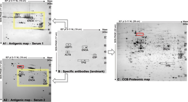
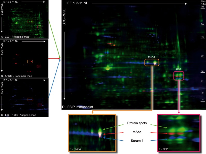
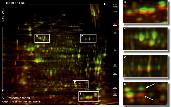
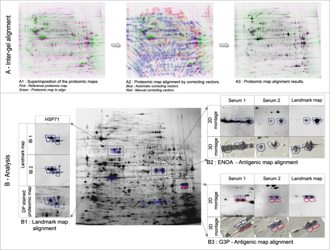
Serological proteome analysis (SERPA) combines classical proteomic technology with effective separation of cellular protein extracts on two-dimensional gel electrophoresis, western blotting, and identification of the antigenic spot of interest by mass spectrometry. A critical point is related to the antigenic target characterization by mass spectrometry, which depends on the accuracy of the matching of antigenic reactivities on the protein spots during the 2D immunoproteomic procedures. The superimposition, based essentially on visual criteria of antigenic and protein spots, remains the major limitation of SERPA. The introduction of fluorescent dyes in proteomic strategies, commonly known as 2D-DIGE (differential in-gel electrophoresis), has boosted the qualitative capabilities of 2D electrophoresis. Based on this 2D-DIGE strategy, we have improved the conventional SERPA by developing a new and entirely fluorescence-based bi-dimensional immunoproteomic (FBIP) analysis, performed with three fluorescent dyes. To optimize the alignment of the different antigenic maps, we introduced a landmark map composed of a combination of specific antibodies. This methodological development allows simultaneous revelation of the antigenic, landmark and proteomic maps on each immunoblot. A computer-assisted process using commercially available software automatically leads to the superimposition of the different maps, ensuring accurate localization of antigenic spots of interest.
Conflict of interest statement
Figures
Similar articles
-
What's the Difference? 2D DIGE Image Analysis by DeCyderTM versus SameSpotsTM.J Mol Microbiol Biotechnol. 2018;28(3):128-136. doi: 10.1159/000494083. Epub 2018 Nov 14. J Mol Microbiol Biotechnol. 2018. PMID: 30428476
-
Comparative Testis Tissue Proteomics Using 2-Dye Versus 3-Dye DIGE Analysis.Methods Mol Biol. 2018;1664:185-202. doi: 10.1007/978-1-4939-7268-5_15. Methods Mol Biol. 2018. PMID: 29019134
-
2-D gel electrophoresis: constructing 2D-gel proteome reference maps.Methods Mol Biol. 2012;815:163-73. doi: 10.1007/978-1-61779-424-7_13. Methods Mol Biol. 2012. PMID: 22130991
-
2D-DIGE and Fluorescence Image Analysis.Methods Mol Biol. 2018;1664:25-39. doi: 10.1007/978-1-4939-7268-5_3. Methods Mol Biol. 2018. PMID: 29019122 Review.
-
Cancer biomarker development and two-dimensional difference gel electrophoresis (2D-DIGE).Biochim Biophys Acta Proteins Proteom. 2019 Jan;1867(1):2-8. doi: 10.1016/j.bbapap.2018.07.002. Epub 2018 Jul 7. Biochim Biophys Acta Proteins Proteom. 2019. PMID: 30392560 Review.
Cited by
-
Serological Proteome Analysis for the Characterization of Secreted Fungal Protein Antigens.Methods Mol Biol. 2021;2260:15-26. doi: 10.1007/978-1-0716-1182-1_2. Methods Mol Biol. 2021. PMID: 33405028
-
Partial Immunoblotting of 2D-Gels: A Novel Method to Identify Post-Translationally Modified Proteins Exemplified for the Myelin Acetylome.Proteomes. 2017 Jan 12;5(1):3. doi: 10.3390/proteomes5010003. Proteomes. 2017. PMID: 28248254 Free PMC article.
-
Immunoproteomics to identify Staphylococcus aureus antigens expressed in bovine milk during mastitis.J Dairy Sci. 2018 Jul;101(7):6296-6309. doi: 10.3168/jds.2017-14040. Epub 2018 May 3. J Dairy Sci. 2018. PMID: 29729920 Free PMC article.
References
-
- Lefranc D, Almeras L, Dubucquoi S, de Seze J, Vermersch P, Prin L. Distortion of the self-reactive IgG antibody repertoire in multiple sclerosis as a new diagnostic tool. J Immunol Baltim Md 1950. 2004;172: 669–678. - PubMed
-
- Almeras L, Lefranc D, Drobecq H, de Seze J, Dubucquoi S, Vermersch P, et al. New antigenic candidates in multiple sclerosis: identification by serological proteome analysis. Proteomics. 2004;4: 2184–2194. - PubMed
Publication types
MeSH terms
Substances
LinkOut - more resources
Full Text Sources
Other Literature Sources
