. 2015 Jul;56(8):4186-97.
doi: 10.1167/iovs.14-15496.

IL-13 Stimulates Proliferation and Expression of Mucin and Immunomodulatory Genes in Cultured Conjunctival Goblet Cells

Affiliations

IL-13 Stimulates Proliferation and Expression of Mucin and Immunomodulatory Genes in Cultured Conjunctival Goblet Cells

Johanna Tukler Henriksson et al. Invest Ophthalmol Vis Sci. 2015 Jul.

Abstract

Purpose: To investigate the effects of IL-13 on goblet cell proliferation, differentiation, and expression of mucin and immunomodulatory genes.

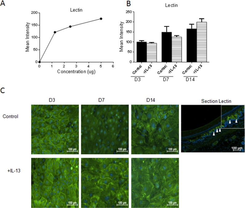
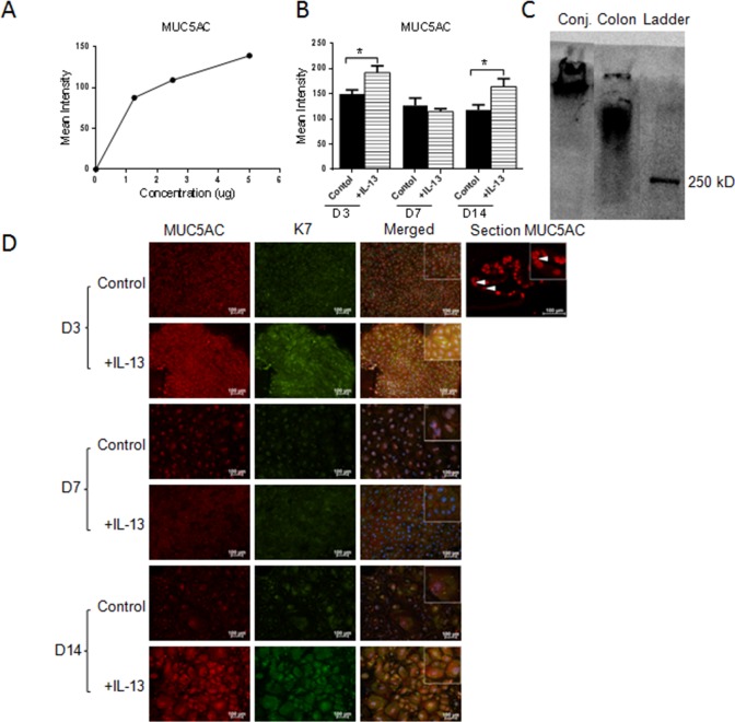
Methods: Explants were excised from the conjunctiva of young C57BL/6 mice. Cultures received 200 μL per week of either Keratinocyte media (KSFM) or KSFM supplemented with 10 ng/mL IL-13 and were incubated for 3 (D3), 7 (D7), or 14 (D14) days. Subsequently, cell proliferation was assessed or cultures were immunostained, collected for dot blot, or for reverse transcription (RT) and quantitative real-time PCR (qPCR) or for RT-PCR gene array.

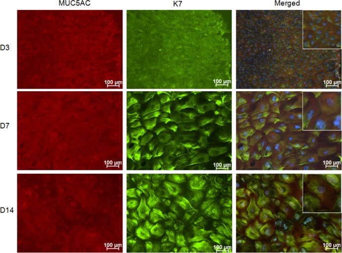
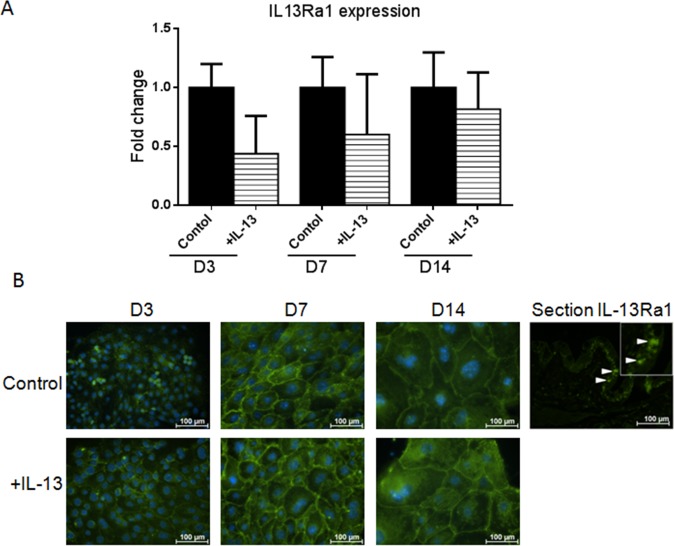
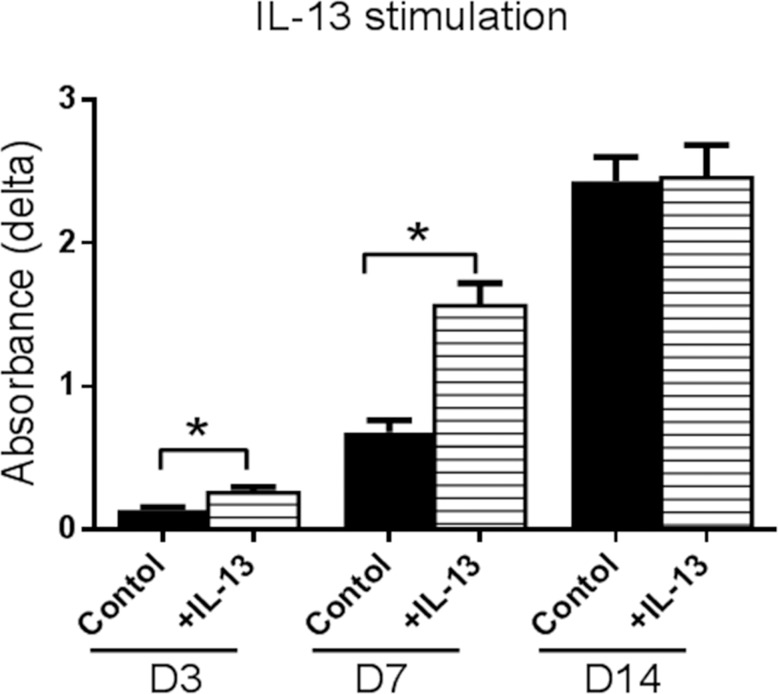
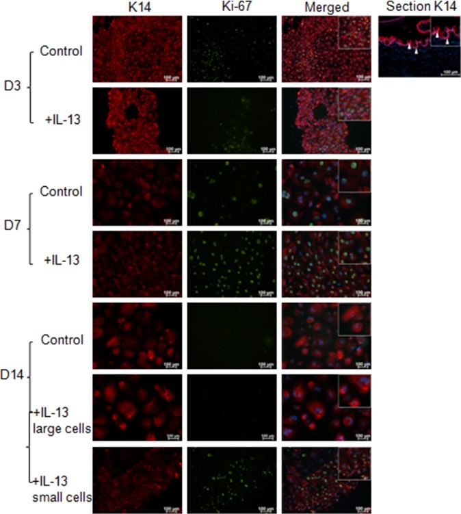
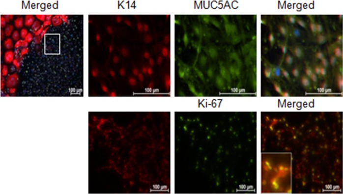
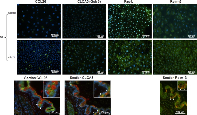
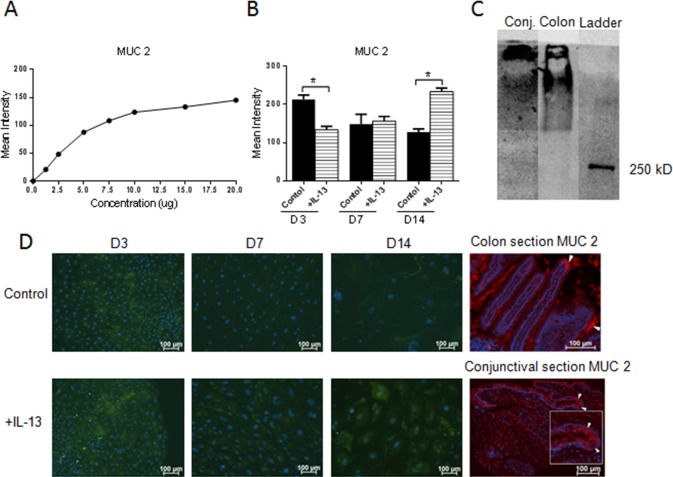
Results: The cultured conjunctival epithelium expressed goblet cell associated keratin 7 and mucins MUC5AC and MUC2 and when stimulated with IL-13 showed increased proliferation at D3 and D7 (P < 0.05) compared with control. MUC5AC expression was increased in the IL-13-treated group at D3 and D14 (P < 0.05). IL-13-treated cultures showed increased chemokine ligand 26 (CCL26), chloride channel calcium activated channel 3 (CLCA3), fas ligand (FasL), and Relm-β at D7. All conjunctival cultures expressed MUC2, and its expression was decreased at D3 (P < 0.05) and increased at D14 (P < 0.05) with IL-13 treatment.

Conclusions: This study demonstrated that conjunctival goblet cells are IL-13 responsive cells that produce factors known to maintain epithelial barrier, stimulate mucin production, and modulate immune response in nonocular mucosa when treated with IL-13. The functional significance of IL-13-stimulated factors remains to be determined.


Figures

Figure 1
Figure 1
MUC5AC and K7 immunostaining in control conjunctival explant cultures. The staining confirms that all cells growing from the mouse conjunctival explants express the goblet cell markers MUC5AC and K7. Hoechst (DNA stain), blue; MUC5AC, red; and K7, green.
Figure 2
Figure 2
Interleukin-13Rα1 expression in primary conjunctival goblet cell cultures. Change in mRNA transcripts over D14 in culture (A). Interleukin-13Rα1 immunostaining in control and IL-13 treated cells (B). Hoechst (DNA stain), blue; IL-13Rα1, green. Arrows, IL-13Ra1 stained goblet cells in conjunctival tissue section.
Figure 3
Figure 3
Effects of IL-13 stimulation on proliferation of cultured conjuntival goblet cells measured by WST assay. Cultures stimulated with IL-13 showed a statistically significant increase in cell number at D3 and D7 compared with control. *P < 0.05.
Figure 4
Figure 4
K14 and Ki-67 immunostaining cultured conjuntival goblet cells. The increased proliferation with IL-13 stimulation at D3 and D7 detected by the WST assay was confirmed at the protein level with K14 (red) and Ki-67 (green) staining. At D14, the majority of the cultured cells were Ki-67 negative; however, in approximately 25% of the cultures a mixture of large, Ki-67 negative and small, Ki-67 positive cells were observed. Arrows, K14 epithelial staining in conjunctival tissue section.
Figure 5
Figure 5
Higher magnification of small proliferating cells noted in the D14 IL-13–treated conjunctival goblet cell cultures (area surrounded by white box in upper left). Staining illustrates that these small cells are K14, MUC5AC, and KI-67 positive. Inset in bottom panel shows higher magnification of K14 and Ki-67 and dual positive cells. Hoechst (DNA stain), blue; K14, red; Ki-67, or MUC5AC, green.
Figure 6
Figure 6
Glycoprotein production in cultured conjunctival goblet cells in response to IL-13 treatment. Wheat germ agglutinin lectin showed a positive correlation between concentration and mean intensity in dot-blot assay (A). No difference was found in WGA staining between any of the groups (B, C). Hoechst (DNA stain), blue; WGA, green. Arrows, Lectin staining in goblet cells in conjunctival tissue section.
Figure 7
Figure 7
MUC5AC production in cultured conjunctival goblet cells in response to IL-13 treatment. MUC5AC showed a positive correlation between concentration and mean intensity in dot blot assay (A). Increased expression of MUC5AC was seen at D3 and D14 in the IL-13 treated samples compared with control (B, D). Immunoreactivity of the MUC5AC antibody in mouse conjunctiva was confirmed by Western blot using colon as a positive control (C). Hoechst (DNA stain), blue; MUC5AC, red; WGA or K7, green. Arrows, MUC5AC staining in goblet cells in conjunctival tissue section. *P < 0.05.
Figure 8
Figure 8
Immunofluorescent staining of goblet cell produced products that showed marked changes with IL-13 stimulation in the PCR array performed on cultured conjunctival goblet cells. Hoechst (DNA stain), blue; CCL26, CLCA3, Fas-L, or Relm-β, green. In conjunctival tissue sections (bottom row): MUC5AC, Relm-β, red; CCL26, CLCA3, and K7, green (arrows indicate goblet cells).
Figure 9
Figure 9
MUC2 glycoprotein production in cultured conjunctival goblet cells in response to IL-13 treatment in dot-blot assay. Positive correlation between concentration and mean intensity of positive colon control (A). In cultured cells, MUC2 expression decreased at D3, showed no difference at D7, and increased at D14 (B). Immunoreactivity of the MUC2 antibody in mouse conjunctiva was confirmed by Western blot using colon as a positive control (C). Immunofluorescent staining of MUC2, colon, and conjunctival sections were used as control (D). Hoechst (DNA stain), blue; MUC2 in culture, green; MUC2 in section, red. *P < 0.05. Arrow(s) in the colon and conjunctival sections indicate positively-stained goblet cells.

References

    1. Watsky MA,, Jablonski MM,, Edelhauser HF. Comparison of conjunctival and corneal surface areas in rabbit and human. Curr Eye Res. 1988; 7: 483–486. - PubMed
    1. Govindarajan B,, Gipson IK. Membrane-tethered mucins have multiple functions on the ocular surface. Exp Eye Res. 2010; 90: 655–663. - PMC - PubMed
    1. Gipson IK. Distribution of mucins at the ocular surface. Exp Eye Res. 2004; 78: 379–388. - PubMed
    1. Zhang X,, Volpe EA,, Gandhi NB,, et al. NK cells promote Th-17 mediated corneal barrier disruption in dry eye. PLoS One. 2012; 7: e36822. - PMC - PubMed
    1. Pflugfelder SC,, Tseng SCG,, Yoshino K,, Monroy D,, Felix C,, Reis BL. Correlation of goblet cell density and mucosal epithelial membrane mucin expression with rose bengal staining in patients with ocular irritation. Ophthalmology. 1997; 104: 223–235. - PubMed

Publication types

MeSH terms