

The role of computed tomography in the diagnosis of acute and chronic pulmonary embolism
- PMID: 26133321
- PMCID: PMC4498425
- DOI: 10.5152/dir.2015.14403
The role of computed tomography in the diagnosis of acute and chronic pulmonary embolism
Abstract
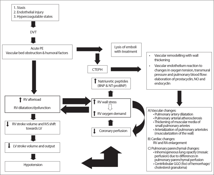
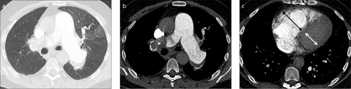
Pulmonary embolism (PE) is a potentially life threatening condition requiring adequate diagnosis and treatment. Computed tomography pulmonary angiography (CTPA) is excellent for including and excluding PE, therefore CT is the first-choice diagnostic imaging technique in patients suspected of having acute PE. Due to its wide availability and low invasiveness, CTPA tends to be overused. Correct implementation of clinical decision rules in diagnostic workup for PE improves adequate use of CT. Also, CT adds prognostic value by evaluating right ventricular (RV) function. CT-assessed RV dysfunction and to lesser extent central emboli location predicts PE-related mortality in normotensive and hypotensive patients, while PE embolic obstruction index has limited prognostic value. Simple RV/left ventricular (LV) diameter ratio measures >1.0 already predict risk for adverse outcome, whereas ratios <1.0 can safely exclude adverse outcome. Consequently, assessing the RV/LV diameter ratio may help identify patients who are potential candidates for treatment at home instead of treatment in the hospital. A minority of patients develop chronic thromboembolic pulmonary hypertension (CTEPH) following acute PE, which is a life-threatening condition that can be diagnosed by CT. In proximal CTEPH, involving the more central pulmonary arteries, thrombectomy usually results in good outcome in terms of both functional status and long-term survival rate. CT is becoming the imaging method of choice for diagnosing CTEPH as it can identify patients who may benefit from thrombectomy. New CT developments such as distensibility measurements and dual-energy or subtraction techniques may further refine diagnosis and prognosis for improved patient care.
Figures
References
-
- Giuntini C, Di Ricco G, Marini C, Melillo E, Palla A. Pulmonary embolism: epidemiology. Chest. 1995;107(Suppl 1):3S–9S. http://dx.doi.org/10.1378/chest.107.1_Supplement.3S. - DOI - PubMed
-
- Stein PD, Woodard PK, Hull RD, et al. Gadolinium-enhanced magnetic resonance angiography for detection of acute pulmonary embolism: an in-depth review. Chest. 2003;124:2324–2328. - PubMed
-
- Wintersperger BJ, Nikolaou K. Basics of cardiac MDCT: techniques and contrast application. Eur Radiol. 2005;15( Suppl 2):B2–B9. http://dx.doi.org/10.1007/s10406-005-0090-0. - DOI - PubMed
-
- Remy-Jardin M, Pistolesi M, Goodman LR, et al. Management of suspected acute pulmonary embolism in the era of CT angiography: a statement from the Fleischner Society. Radiology. 2007;245:315–329. http://dx.doi.org/10.1148/radiol.2452070397. - DOI - PubMed
-
- Carson JL, Kelley MA, Duff A, et al. The clinical course of pulmonary embolism. N Engl J Med. 1992;326:1240–1245. http://dx.doi.org/10.1056/NEJM199205073261902. - DOI - PubMed
Publication types
MeSH terms
LinkOut - more resources
Full Text Sources
Other Literature Sources
Medical
