

Comparison of breast specific gamma imaging and molecular breast tomosynthesis in breast cancer detection: Evaluation in phantoms
- PMID: 26133623
- PMCID: PMC4474957
- DOI: 10.1118/1.4922398
Comparison of breast specific gamma imaging and molecular breast tomosynthesis in breast cancer detection: Evaluation in phantoms
Abstract
Purpose: Breast specific gamma imaging or molecular breast imaging (BSGI) obtains 2D images of (99m)Tc sestamibi distribution in the breast. Molecular breast tomosynthesis (MBT) maps the tracer distribution in 3D by acquiring multiple projections over a limited angular range. Here, the authors compare the performance of the two technologies in terms of spatial resolution, lesion contrast, and contrast-to-noise ratio (CNR) in phantom studies under conditions of clinically relevant sestamibi dose and imaging time.
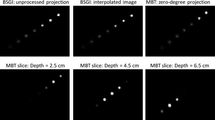
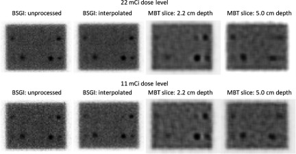
Methods: The systems tested were a Dilon 6800 and a MBT prototype developed at the University of Virginia. Both systems comprise a pixelated sodium iodide scintillator, an array of position sensitive photomultipliers, and a parallel hole collimator. The active areas and energy resolution of the systems are similar. System sensitivity, spatial resolution, lesion contrast, and CNR were measured using a Petri dish, a point source phantom, and a breast phantom containing simulated lesions at two depths, respectively. A single BSGI projection was acquired. Five MBT projections were acquired over ±20°. For both modalities, the total scan count density was comparable to that observed for each in typical 10 min human scans following injection of 22 mCi (814 MBq) of (99m)Tc-sestamibi. To assess the impact of reducing the tracer dose, the pixel counts of projection images were later binomially subsampled by a factor of 2 to give images corresponding to an injected activity of approximately 11 mCi (407 MBq). Both unprocessed (pixelated) BSGI projections and interpolated (smoothed) BSGI images displayed by default on the Dilon 6800 workstation were analyzed. Volumetric images were reconstructed from the MBT projections using a maximum likelihood expectation maximization algorithm and extracted slices were analyzed.
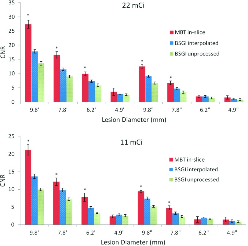
Results: Over a depth range of 1.5-7.5 cm, BSGI spatial resolution was 5.6-11.5 mm in unprocessed projections and 5.7-12.0 mm in interpolated images. Over the same range, the in-slice MBT spatial resolution was 6.7-9.4 mm. Lesion contrast was significantly improved with MBT relative to BSGI for five out of eight lesions imaged at either the 22 mCi or the 11 mCi dose level (p < 0.05). At both dose levels, significant improvements in CNR with MBT were also found for five out of eight lesions (9.8, 7.8, 6.2 mm lesions at water depth of 1.7 cm and 9.8, 7.8 mm lesions at water depth of 4.5 cm, p < 0.05). The 6.2 and 4.9 mm lesions located at 4.5 cm below the water surface were not visible in either modality at either activity level.
Conclusions: Under conditions of equal dose, imaging time and similar detectors, compared to BSGI, MBT provided higher lesion contrast, higher CNR, and spatial resolution that was less depth dependent.
Figures


Similar articles
-
Evaluation of a novel collimator for molecular breast tomosynthesis.Med Phys. 2017 Nov;44(11):5740-5748. doi: 10.1002/mp.12564. Epub 2017 Oct 13. Med Phys. 2017. PMID: 28877351
-
Proof of concept for low-dose molecular breast imaging with a dual-head CZT gamma camera. Part I. Evaluation in phantoms.Med Phys. 2012 Jun;39(6):3466-75. doi: 10.1118/1.4718665. Med Phys. 2012. PMID: 22755726 Free PMC article.
-
Performance characteristics of dedicated molecular breast imaging systems at low doses.Med Phys. 2016 Jun;43(6):3062-3070. doi: 10.1118/1.4950873. Med Phys. 2016. PMID: 27277053
-
Molecular Breast Imaging: A Comprehensive Review.Semin Ultrasound CT MR. 2018 Feb;39(1):60-69. doi: 10.1053/j.sult.2017.10.001. Epub 2017 Oct 20. Semin Ultrasound CT MR. 2018. PMID: 29317040 Review.
-
Breast-specific gamma camera imaging with 99mTc-MIBI has better diagnostic performance than magnetic resonance imaging in breast cancer patients: A meta-analysis.Hell J Nucl Med. 2017 Jan-Apr;20(1):26-35. doi: 10.1967/s002449910503. Epub 2017 Mar 20. Hell J Nucl Med. 2017. PMID: 28315905 Review.
Cited by
-
Mammography with deep learning for breast cancer detection.Front Oncol. 2024 Feb 12;14:1281922. doi: 10.3389/fonc.2024.1281922. eCollection 2024. Front Oncol. 2024. PMID: 38410114 Free PMC article. Review.
-
Molecular Breast Imaging using Synthetic Projections from High-Purity Germanium Detectors: A Simulation Study.IEEE Trans Radiat Plasma Med Sci. 2017 Sep;1(5):405-415. doi: 10.1109/TRPMS.2017.2725310. Epub 2017 Jul 11. IEEE Trans Radiat Plasma Med Sci. 2017. PMID: 28989990 Free PMC article.
-
Dedicated Breast Gamma Camera Imaging and Breast PET: Current Status and Future Directions.PET Clin. 2018 Jul;13(3):363-381. doi: 10.1016/j.cpet.2018.02.008. PET Clin. 2018. PMID: 30100076 Free PMC article. Review.
-
Design and characterization of a low profile NaI(Tl) gamma camera for dedicated molecular breast tomosynthesis.Proc SPIE Int Soc Opt Eng. 2016 Aug 28;9969:99690O. doi: 10.1117/12.2246561. Epub 2016 Oct 3. Proc SPIE Int Soc Opt Eng. 2016. PMID: 28835730 Free PMC article.
-
Current State of Breast Cancer Diagnosis, Treatment, and Theranostics.Pharmaceutics. 2021 May 14;13(5):723. doi: 10.3390/pharmaceutics13050723. Pharmaceutics. 2021. PMID: 34069059 Free PMC article. Review.
References
-
- Rosenberg R. D. et al., “Effects of age, breast density, ethnicity, and estrogen replacement therapy on screening mammographic sensitivity and cancer stage at diagnosis: Review of 183,134 screening mammograms in Albuquerque, New Mexico,” Radiology 209, 511–518 (1998).10.1148/radiology.209.2.9807581 - DOI - PubMed
Publication types
MeSH terms
Substances
Grants and funding
LinkOut - more resources
Full Text Sources
Other Literature Sources
Medical
