. 2015 Sep;138(Pt 9):2493-504.
doi: 10.1093/brain/awv185. Epub 2015 Jun 30.

Mutations in GMPPB cause congenital myasthenic syndrome and bridge myasthenic disorders with dystroglycanopathies

Affiliations

Mutations in GMPPB cause congenital myasthenic syndrome and bridge myasthenic disorders with dystroglycanopathies

Katsiaryna Belaya et al. Brain. 2015 Sep.

Abstract

Congenital myasthenic syndromes are inherited disorders that arise from impaired signal transmission at the neuromuscular junction. Mutations in at least 20 genes are known to lead to the onset of these conditions. Four of these, ALG2, ALG14, DPAGT1 and GFPT1, are involved in glycosylation. Here we identify a fifth glycosylation gene, GMPPB, where mutations cause congenital myasthenic syndrome. First, we identified recessive mutations in seven cases from five kinships defined as congenital myasthenic syndrome using decrement of compound muscle action potentials on repetitive nerve stimulation on electromyography. The mutations were present through the length of the GMPPB, and segregation, in silico analysis, exon trapping, cell transfection followed by western blots and immunostaining were used to determine pathogenicity. GMPPB congenital myasthenic syndrome cases show clinical features characteristic of congenital myasthenic syndrome subtypes that are due to defective glycosylation, with variable weakness of proximal limb muscle groups while facial and eye muscles are largely spared. However, patients with GMPPB congenital myasthenic syndrome had more prominent myopathic features that were detectable on muscle biopsies, electromyography, muscle magnetic resonance imaging, and through elevated serum creatine kinase levels. Mutations in GMPPB have recently been reported to lead to the onset of muscular dystrophy dystroglycanopathy. Analysis of four additional GMPPB-associated muscular dystrophy dystroglycanopathy cases by electromyography found that a defective neuromuscular junction component is not always present. Thus, we find mutations in GMPPB can lead to a wide spectrum of clinical features where deficit in neuromuscular transmission is the major component in a subset of cases. Clinical recognition of GMPPB-associated congenital myasthenic syndrome may be complicated by the presence of myopathic features, but correct diagnosis is important because affected individuals can respond to appropriate treatments.

Keywords: GMPPB; congenital myasthenic syndrome; dystroglycan; glycosylation; neurotransmission defect.


Figures

None
Congenital myasthenic syndromes are associated with impairments in neuromuscular transmission. Belaya et al. show that mutations of the glycosylation pathway enzyme GMPPB, which has previously been implicated in muscular dystrophy dystroglycanopathy, also cause a congenital myasthenic syndrome. This differential diagnosis is important to ensure that affected individuals receive appropriate medication.
Figure 1
Figure 1
Simplified scheme of N-linked and O-linked glycosylation. The scheme shows five glycosylation genes associated with CMS.
Figure 2
Figure 2
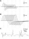
Neurophysiological examination of Case 3 using repetitive nerve stimulation and concentric needle EMG. Repetitive nerve stimulation studies performed on right anconeus muscle showed 50% amplitude decrement (A) in compound muscle action potentials while there was no change in compound muscle action potential amplitude with repetitive stimulation of right abductor digiti minimi muscle (B). Concentric needle EMG examination of the right biceps muscle at low force of contraction showed low amplitude, polyphasic motor unit action potentials.
Figure 3
Figure 3
Co-segregation of CMS phenotype with GMPPB mutations in one family (Cases 4–6). Pedigree symbols are shaded according to the presence of clinical CMS symptoms.
Figure 4
Figure 4
Mutation c.130-3C>G disrupts wild-type splicing pattern of GMPPB. (A) Schematic structure of GMPPB gene, the pET01 exon trap vector, and the exon trap vector with inserted GMPPB exons 2–4. (B) Gel electrophoresis of amplicons generated using vector-specific primers. The wild-type (WT) construct generated one transcript, whereas c.130-3C>G mutant construct generates two shorter transcripts. (C) Sequencing data and schematic diagrams showing aberrant splicing from the mutant construct. The nucleotide sequence around each splice site is shown.
Figure 5
Figure 5
GMPPB domain structure and conservation. GMPPB is a 360-aa polypeptide and consists of nine exons (shown with blocks on the scheme). It has two predicted PFAM domains: nucleotidyl transferase domain and bacterial transferase hexapeptide domain (shown with yellow blocks). The scheme shows CMS-associated mutations (above GMPPB scheme), and mutations associated with muscular dystrophy (underneath GMPPB scheme). Mutations described in this paper are shown in black, whereas mutations published previously are shown in grey for comparison. Protein alignment was performed in ClustalW2.
Figure 6
Figure 6
Effect of different variants on GMPPB expression and localization. (A) GMPPB constructs were transfected into HEK293 cells, protein lysates were prepared 48 h after transfection and analysed by western blot using anti-GMPPB antibody. (B) GMPPB constructs were transfected into C2C12 cells. Permeabilized cells were stained with anti-GMPPB antibody. Scale bar = 20 µm.
Figure 7
Figure 7
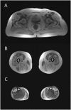
Muscle MRI from Case 2. The muscle MRI study (T1-weighted sequences) showed consistent abnormalities in gluteal (A), thigh (B) and calf (C) muscles, with confluent areas of increased signal or end-stage appearance but relative sparing of certain muscles, especially at the distal level, in keeping with the clinical and electrophysiological findings.

References

    1. Adzhubei IA, Schmidt S, Peshkin L, Ramensky VE, Gerasimova A, Bork P, et al. A method and server for predicting damaging missense mutations. Nat Methods 2010; 7: 248–9. - PMC - PubMed
    1. Belaya K, Finlayson S, Slater CR, Cossins J, Liu WW, Maxwell S, et al. Mutations in DPAGT1 cause a limb-girdle congenital myasthenic syndrome with tubular aggregates. Am J Hum Genet 2012; 91: 193–201. - PMC - PubMed
    1. Carss KJ, Stevens E, Foley AR, Cirak S, Riemersma M, Torelli S, et al. Mutations in GDP-mannose pyrophosphorylase B cause congenital and limb-girdle muscular dystrophies associated with hypoglycosylation of alpha-dystroglycan. Am J Hum Genet 2013; 93: 29–41. - PMC - PubMed
    1. Chaouch A, Müller J, Lochmüller H. A retrospective clinical study of the treatment of slow-channel congenital myasthenic syndrome. J Neurol 2012; 259: 474. - PubMed
    1. Compton A, Albrecht DE, Seto JT, Cooper ST, Ilkovski B, Jones KJ, et al. Mutations in contactin-1, a neural adhesion and neuromuscular junction protein, cause a familial form of lethal congenital myopathy. Am J Hum Genet 2008; 83: 714–24. - PMC - PubMed

Publication types

MeSH terms

Substances