. 2015 Aug;114(2):1146-57.
doi: 10.1152/jn.00355.2015. Epub 2015 Jul 1.

pigk Mutation underlies macho behavior and affects Rohon-Beard cell excitability

Affiliations

pigk Mutation underlies macho behavior and affects Rohon-Beard cell excitability

V Carmean et al. J Neurophysiol. 2015 Aug.

Abstract

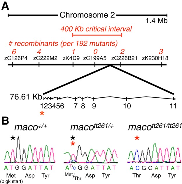
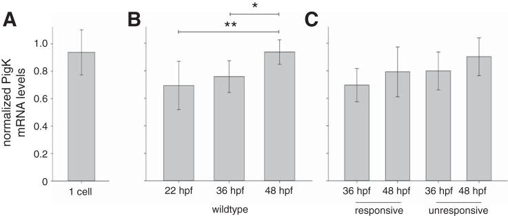
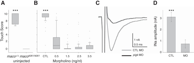
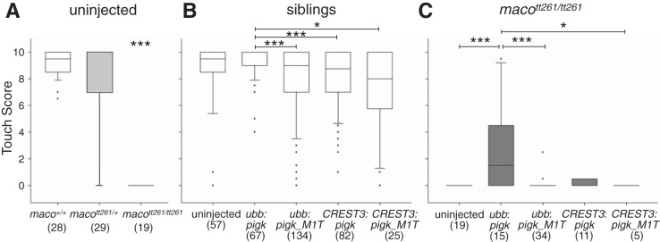
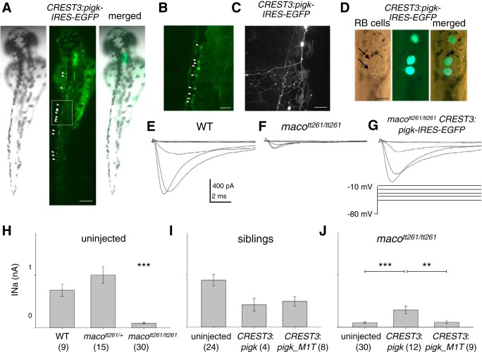
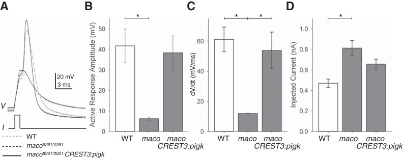
The study of touch-evoked behavior allows investigation of both the cells and circuits that generate a response to tactile stimulation. We investigate a touch-insensitive zebrafish mutant, macho (maco), previously shown to have reduced sodium current amplitude and lack of action potential firing in sensory neurons. In the genomes of mutant but not wild-type embryos, we identify a mutation in the pigk gene. The encoded protein, PigK, functions in attachment of glycophosphatidylinositol anchors to precursor proteins. In wild-type embryos, pigk mRNA is present at times when mutant embryos display behavioral phenotypes. Consistent with the predicted loss of function induced by the mutation, knock-down of PigK phenocopies maco touch insensitivity and leads to reduced sodium current (INa) amplitudes in sensory neurons. We further test whether the genetic defect in pigk underlies the maco phenotype by overexpressing wild-type pigk in mutant embryos. We find that ubiquitous expression of wild-type pigk rescues the touch response in maco mutants. In addition, for maco mutants, expression of wild-type pigk restricted to sensory neurons rescues sodium current amplitudes and action potential firing in sensory neurons. However, expression of wild-type pigk limited to sensory cells of mutant embryos does not allow rescue of the behavioral touch response. Our results demonstrate an essential role for pigk in generation of the touch response beyond that required for maintenance of proper INa density and action potential firing in sensory neurons.

Keywords: Nav; Pigk; Rohon-Beard cells; glycophosphatidylinositol-anchored protein; touch response.


Figures

Fig. 1.
Fig. 1.
macott261/tt261 embryos have a mutation in the pigk start codon. A: bulked segregant analysis was first used to map the macott261 mutation to a region of Chromosome 2. Subsequent fine mapping with Simple Sequence Length Polymorphism (SSLP) markers identified a region with no recombinants. This area was sequenced, revealing the macott261 locus in the pigk start codon. Asterisks designate the locus of the maco mutation. B: sequence chromatograms revealed a thymidine to cytosine substitution in the pigk start codon. Asterisks denote the residue of interest (black asterisk, wild-type sequence; red asterisk, mutant sequence).
Fig. 2.
Fig. 2.
pigk mRNA is maternally expressed and detected during embryonic stages of development. To test for the presence of pigk mRNA during embryonic development, we performed qPCR using RNA extracted from wild-type (WT) embryos, touch-unresponsive maco embryos, and their responsive siblings. Data are presented as means ± SD, with pigk mRNA levels normalized to those of actb2 mRNA (see materials and methods). A: qPCR detected pigk mRNA at the one-cell stage, indicating that the transcripts are maternally provided. B: in WT embryos, pigk expression levels increased between 22 and 48 h postfertilization (hpf). *P < 0.05, **P < 0.01. C: both responsive (maco+/+ and macott261/+, pooled) and unresponsive (macott261/tt261) sibling embryos expressed pigk mRNA at 36 and 48 hpf. At both time points, unresponsive and responsive siblings had similar pigk expression levels.
Fig. 3.
Fig. 3.
PigK knockdown phenocopies maco behavior. A: maco+/+ embryos had significantly higher touch response scores than did macott261/tt261 embryos. (n: 10, maco+/+; 12, macott261/tt261). B: PigK morpholino (MO) led to a dose-dependent decrease in touch scores. In contrast, injection of CTL MO had no effect on touch sensitivity. (n = 73, 3.5 ng/nl CTL MO; 76, 0.5 ng/nl PigK MO; 61, 1.5 ng/nl PigK MO; 71, 2.5 ng/nl PigK MO; 72, 3.5 ng/nl PigK MO). For the graphs of A and B, solid horizontal lines designate the median, boxes indicate the 25th–75th percentiles, and whiskers show 90th percentile. C: injection of PigK MO also led to reduction of RB INa amplitudes. D: INa amplitudes were decreased significantly in embryos injected with PigK MO. Data are presented as means ± SE. ***P < 0.001 (n = 3, 3.5 ng/nl MO CTL; 5, 3.5 ng/nl PigK MO).
Fig. 4.
Fig. 4.
Ubiquitous but not RB-restricted expression of WT pigk rescues touch responsiveness in macott261/tt261 embryos. One-cell-stage embryos were either not injected or injected with the indicated transgenic constructs. Touch responsiveness was assayed at 48 hpf, and genotyping was performed after behavioral testing. In the graphs, the solid horizontal lines designate the median score, boxes indicate the 25th–75th percentile range, and whiskers show the 90th percentile. Sample size (n) is indicated within the figure. The data presented in A for uninjected embryos are reshown in B and C as the uninjected conditions. A: uninjected WT (maco+/+; column 1) and heterozygote (macott261/+; column 2) embryos have significantly higher touch scores than do sibling macott261/tt261 mutants (column 3). ***P < 0.001. B: data were pooled for maco+/+ and macott261/+ embryos injected with the same transgene. Regardless of transgene injection, the different sibling groups all had touch scores that were closer to 10 than 0. However, sibling embryos injected with ubb:pigk-IRES-EGFP (column 2) had slightly higher median scores than those injected with ubb:pigk_M1T-IRES-eGFP (column 3) or either of the CREST3 driven transgenes (columns 4 and 5). *P < 0.05, ***P < 0.001. C: data for embryos that were genotypically homozygous for the maco mutation (macott261/tt261). macott261/tt261 embryos injected with ubb:pigk-IRES-EGFP (column 2) had significantly greater touch scores than did uninjected macott261/tt261 embyros (column 1) or those injected with ubb:pigk_M1T-IRES-EGFP (column 3), CREST3:pigk-IRES-EGFP (column 4), or CREST3:pigk_M1T-IRES-EGFP (column 5). *P < 0.05, ***P < 0.001.
Fig. 5.
Fig. 5.
Overexpression of WT pigk in RB cells partially rescues RB INa. A: in a single embryo injected with CREST3:pigk-IRES-EGFP, several GFP+ RB cells are present (left, bright field; center, fluorescent image; right, merge). Arrowheads point to GFP+ RB cells. Scale Bar, 250 μm. B: the GFP+ RB cells in the box in the middle panel of A are shown at higher magnification. This region corresponds to that assayed for touch. Scale bar, 50 μm. C: a GFP+ RB cell in an embryo injected with CREST3:pigk-IRES-EGFP has a relatively normal extensive peripheral arbor. Scale Bar, 50 μm. D: the spinal cord of a CREST3:pigk-IRES-EGFP injected embryo is mounted and minimally dissected in preparation for electrophysiology. The left panel is a bright field image, with arrows indicating RB cells that are identified on the basis of position and size of soma (Ribera and Nüsslein-Volhard 1998). The middle panel shows the GFP fluorescence of several RB cells. The right panel is a merged image of the brightfield and fluorescent images. Scale bar, 20 μm. E–G: INa was recorded from RB cells in embryos that were either uninjected WT (E), uninjected macott261/tt261 (F), or macott261/tt261 injected (G) with the CREST3:pigk-IRES-EGFP transgene. For injected embryos, currents were recorded from GFP+ RB cells. H–J: the graphs of H–J present the absolute peak INa amplitude values as means ± SE. Samples size (n) information is provided within the figure. H: RB cells of uninjected maco+/+ (WT) and macott261/+ sibling embryos had INa amplitudes that were significantly larger than those recorded from RB cells in macott261/tt261 uninjected embryos. ***P < 0.001. I: for the comparisons of this graph, data obtained from RBs in maco+/+ (WT) and macott261/+ siblings were pooled. Peak INa amplitudes recorded from RBs in uninjected siblings (column 1) or those injected with either the CREST3:pigk-IRES-EGFP (column 2) or CREST3:pigk_M1T-IRES-EGFP (column 3) transgenes were compared. Transgene injection did not have a significant effect on peak INa amplitude. J: peak INa amplitudes recorded from RBs in uninjected macott261/tt261 embryos (column 1) or those in macott261/tt261 embryos injected with either the CREST3:pigk-IRES-EGFP (column 2) or the CREST3:pigk_M1T-IRESeGFP (column 3) transgenes are shown. Injection of the CREST3:pigk-IRES-EGFP, but not CREST3:pigk_M1T-IRESeGFP, transgene led to a significant increase in RB peak INa amplitude. **P < 0.01, ***P < 0.001.
Fig. 6.
Fig. 6.
Overexpression of WT pigk in macott261/tt261 RB cells restores the ability to fire an action potential. A: action potentials were elicited by current injection (I), and the membrane potential responses (V) of RB cells in 48 hpf embryos were recorded. Exemplar responses are shown for RBs from embryos that were either uninjected WT (dashed gray line), or macott261/tt261 (dashed black line) or macott261/tt261 injected with the CREST3:WTPigk-IRES-EGFP transgene (solid black line). B: the amplitudes of the active membrane responses recorded from RBs in WT (column 1) were significantly larger than those recorded from RBs in uninjected macott261/tt261 embryos (column 2). *P < 0.05. C: the rates of rise (dV/dt) of the active membrane responses recorded from RBs in either WT (column 1) or macott261/tt261 injected with CREST3:pigk-IRES-EGFP (column 3) embryos were significantly faster than those of RBs in uninjected macott261/tt261 embryos (column 2). *P < 0.05. D: to elicit an active response, significantly more current was required for RBs of macott261/tt261 (column 2) vs. than that required for RBs in WT embryos (column 1). In addition, as shown in B and C and Table 1, the active membrane responses of RB cells in maco mutants did not meet the criteria for being considered an action potential (see materials and methods). *P < 0.05.

Similar articles

Cited by

References

    1. Almeida AM, Murakami Y, Layton DM, Hillmen P, Sellick GS, Maeda Y, Richards S, Patterson S, Kotsianidis I, Mollica L, Crawford DH, Baker A, Ferguson M, Roberts I, Houlston R, Kinoshita T, Karadimitris A. Hypomorphic promoter mutation in PIGM causes inherited glycosylphosphatidylinositol deficiency. Nat Med 12: 846–851, 2006. - PubMed
    1. Amores A, Force A, Yan YL, Joly L, Amemiya C, Fritz A, Ho RK, Langeland J, Prince V, Wang YL, Westerfield M, Ekker M, Postlethwait JH. Zebrafish hox clusters and vertebrate genome evolution. Science 282: 1711–1714, 1998. - PubMed
    1. Bernhardt RR, Chitnis AB, Lindamer L, Kuwada JY. Identification of spinal neurons in the embryonic and larval zebrafish. J Comp Neurol 302: 603–616, 1990. - PubMed
    1. Bosch DG, Boonstra FN, Kinoshita T, Jhangiani S, de Ligt J, Cremers FP, Lupski JR, Murakami Y, de Vries BB. Cerebral visual impairment and intellectual disability caused by PGAP1 variants. Eur J Hum Genet 2015. March 25. doi:10.1038/ejhg.2015.42 [Epub ahead of print]. - DOI - PMC - PubMed
    1. Casadei R, Pelleri MC, Vitale L, Facchin F, Lenzi L, Canaider S, Strippoli P, Frabetti F. Identification of housekeeping genes suitable for gene expression analysis in the zebrafish. Gene Express Patterns 11: 271–276, 2011. - PubMed

Publication types

MeSH terms

LinkOut - more resources