Review
. 2015 Jul;95(3):1025-109.
doi: 10.1152/physrev.00028.2014.

The Sick and the Weak: Neuropathies/Myopathies in the Critically Ill

Affiliations
Review

The Sick and the Weak: Neuropathies/Myopathies in the Critically Ill

O Friedrich et al. Physiol Rev. 2015 Jul.

Abstract

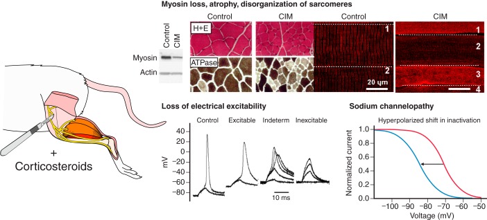
Critical illness polyneuropathies (CIP) and myopathies (CIM) are common complications of critical illness. Several weakness syndromes are summarized under the term intensive care unit-acquired weakness (ICUAW). We propose a classification of different ICUAW forms (CIM, CIP, sepsis-induced, steroid-denervation myopathy) and pathophysiological mechanisms from clinical and animal model data. Triggers include sepsis, mechanical ventilation, muscle unloading, steroid treatment, or denervation. Some ICUAW forms require stringent diagnostic features; CIM is marked by membrane hypoexcitability, severe atrophy, preferential myosin loss, ultrastructural alterations, and inadequate autophagy activation while myopathies in pure sepsis do not reproduce marked myosin loss. Reduced membrane excitability results from depolarization and ion channel dysfunction. Mitochondrial dysfunction contributes to energy-dependent processes. Ubiquitin proteasome and calpain activation trigger muscle proteolysis and atrophy while protein synthesis is impaired. Myosin loss is more pronounced than actin loss in CIM. Protein quality control is altered by inadequate autophagy. Ca(2+) dysregulation is present through altered Ca(2+) homeostasis. We highlight clinical hallmarks, trigger factors, and potential mechanisms from human studies and animal models that allow separation of risk factors that may trigger distinct mechanisms contributing to weakness. During critical illness, altered inflammatory (cytokines) and metabolic pathways deteriorate muscle function. ICUAW prevention/treatment is limited, e.g., tight glycemic control, delaying nutrition, and early mobilization. Future challenges include identification of primary/secondary events during the time course of critical illness, the interplay between membrane excitability, bioenergetic failure and differential proteolysis, and finding new therapeutic targets by help of tailored animal models.


Figures

FIGURE 1.
FIGURE 1.
Risk factors contributing to the development of peripheral nervous system dysfunction seen as muscle weakness in ICU patients and potential differences in disease entities. A: in intensive care unit (ICU) patients, several conditions and risk factors have been associated with the development of muscle weakness pointing to a failure in the peripheral nervous system. Clinically, in the past, the presence of “weakness” has pragmatically been lumped together in the term ICU-acquired weakness (ICUAW), although this term neither discriminates between primary neuropathy or primary myopathy, nor does it account for potentially different mechanisms leading to neuropathy/myopathy. Apart from steroids, denervation, and sepsis, mechanical ventilation and immobilization itself is a prominent risk factor that contributes to the development of ICU-related myopathies. Critical illness myopathy (CIM) seen in critically ill patients is almost exclusively associated with altered muscle excitability, severe atrophy, and a preferential myosin loss, usually seen in critically ill patients who are exposed to several trigger factors at once. ICUAW, on the other hand, is a clinically pragmatic term that can be applied in the absence of any further special electrophysiology or biopsy testing. B: the separation of the distinct disease entities summarized under the term ICUAW as well as unraveling the specific differences of the underlying pathophysiological mechanisms defining those separate disease entities is still a matter of active research, and results from animal models suggest that some of those trigger factors on their own may be able to produce CIM while others may not (e.g., pure sepsis without steroid/denervation and/or mechanical ventilation/immobilization). In ICU patients suffering ICUAW, trigger factors usually combine, and septic ICU patients that are ususally subjected to mechanical ventilation and muscle unloading will present with CIM rather than SIM.
FIGURE 2.
FIGURE 2.
Specific pathophysiological mechanisms in ICUAW may either evolve in time independently or trigger subsequent pathways to present with phases of myopathy during critical illness. Early phases may begin as early as from 24 h of ICU treatment and are characterized by a dysfunction of membrane excitability (hypoexcitability), followed by subcellular putative alterations in Ca2+ homeostasis, bioenergetics, and motor protein function, whereas later phases after several days to 1 wk are marked by hyperproteolysis of myofibrillar proteins (atrophy) and/or preferential myosin loss. Severe loss of sarcomeric proteins may also be responsible for the structural fixation of the disease seen as altered cytoarchitecture and loss of contractile filaments that are only slowly regenerated over months. Note that each mechanism is not limited to a particular phase, and mechanisms may coexist at late stages (i.e., membrane hypoexcitability, myosin loss, impaired autophagy, etc.).
FIGURE 3.
FIGURE 3.
Pro- and anti-inflammatory cytokines and their role in sepsis/critical illness and the systemic inflammation-muscle axis. A: time course of pro- and anti-inflammatory cytokines in the systemic circulation in critical illness as best studied in the scenario of sepsis. During sepsis, pro-inflammatory IL-1 and TNF-α rise early, followed by an increase in IL-1, which is then followed by a rise in anti-inflammatory mediators to initiate the compensatory anti-inflammatory response syndrome (CARS). IL-6 both exerts pro- and anti-inflammatory effects. For example, exercise lacks the pro-inflammatory response and is purely characterized by anti-inflammatory response triggered by almost sole IL-6 release from exercising muscle (“myokine”; note: IL-6 amplitude in both sepsis and exercise scaled to maximum; absolute IL-6 amplitudes are much larger in exercise). Individual time courses may vary depending on the model (e.g., CLP shown in the right panel) and outcome. [Right panel according to data from Chensue et al. (119). Left panel modified from Petersen and Pedersen (545).] B: systemic inflammation-muscle axis during sepsis and critical illness. Activated macrophages in inflamed tissue as well as monocytes and neutrophils in the blood secrete large amounts of pro-inflammatory IL-1 and TNF-α that drive the pro-inflammatory systemic response. These mediators also stimulate myokine production in skeletal muscle, of which IL-6 is the most predominant to initiate the systemic CARS by inhibiting further IL-1/TNF-α release and to stimulate production of IL-10 and IL-1RA, for example.
FIGURE 4.
FIGURE 4.
Mechanosignaling in skeletal muscle. Mechanosignaling represents an emerging and dynamic field in biomedical science, but the complexity of the different pathways involved, and how they are interrelated, is not fully resolved. Different pathways involved in mechanosensing and tensegrity in skeletal muscle are briefly summarized, and those reported to be altered in CIM are highlighted in red. However, mechanosignaling has only recently been forwarded as a factor triggering CIM, and the mechanosensing pathways are most probably far more complex than those indicated in red. Multiple signaling pathways influence protein synthesis and degradation in the muscle fiber spanning from the muscle membrane and extracellular matrix to the M-band in the center of the sarcomere. Insulin-like growth factor I (IGF-I) has been suggested to play an important role for the muscle hypertrophy induced by mechanical overload, but a maintained hypertrophy response has been reported in transgenic mice with an IGF-I receptor lacking the ability to bind IGF-I, suggesting multiple other mechanosensitive pathways (651). Calcium- and sodium-permeable stretch-activated channels (SAC) respond to mechanical stimuli and various intracellular signaling cascades. In addition, membrane invaginations, i.e., caveolae, respond to cell stress and stretch-induced signaling, and many different proteins involved in cell signaling bind to caveolins, such as neural nitric oxide synthase (nNOS), G protein subunits, tyrosine kinases, small GTPases, and growth receptors (240, 641). The cytokine leukemia inhibitory factor (LIF) plays an important role in muscle hypertrophy in response to mechanical loading (651). Integrins spanning from the extacellular matrix to the interior of the muscle cell, linked to cytoskeletal actin, directly connect to the nuclei and mitochondria, thus allowing a “hard-wired” and rapid signal propagation to nuclear and mitochondrial DNA (754). The subsarcolemmal dystrophin sarcoglycan complex (DSG) is involved in mechanosensing, and a deficiency in this complex is a feature of muscular dystrophies with defects in the signaling related to mechanical load, resulting in muscle degeneration (793). A number of sarcomeric proteins are involved in mechanosensing, and there is emerging evidence of a very dynamic exchange of multiple sarcomeric proteins to the cytoplasmic pool affecting muscle gene expression in response to mechanical load from the Z-line to the center of the sarcomere in the M-band (76, 234, 390). Multiple major signaling cascades are downstream effectors of mechanosensing, such as PI-3K, MAPKs, calmodulin, calcineurin, glycogen synthase kinase, AMP activated kinase, and AKT/mTOR (86).
FIGURE 5.
FIGURE 5.
Alterations at the level of neuromuscular transmission contributing to ICU-acquired weakness. ICUAW is usually a mixed syndrome of either neuropathy- or myopathy-induced weakness during critical illness either due to sepsis (or even sepsis-unrelated conditions, e.g., burns), immobilization, and eventual denervation contributing to alterations to neuromuscular function. Typical effects on either presynaptic (neuropathy) or postsynaptic (myopathy) functions are shown and, where available from literature, dissected into the single confounding conditions where pure immobilization or denervation studies were performed. See text for details. For pure denervation and immobilization, data were also extracted from References 375, 668, and 782.
FIGURE 6.
FIGURE 6.
Alterations of ion channels in models of ICU-related myopathies. Graphical summary of documented mechanisms contributing to altered membrane excitability through ion channel dysfunction in ICU-related myopathies. Most research performed points towards reduction in ion current densities, e.g., for Na+, Ca2+, and chloride conductances. For voltage-gated Na+ channels, a hyperpolarizing shift of inactivation curves is induced in chronic sepsis and steroid-denervation models of critical illness, thus reducing availability of Na+ channels for action potential generation at resting potentials, which is even worsened by additional membrane depolarizations. Results shown are taken from References , , , and . [Top middle and right panels from Rich and Pinter (580). Copyright John Wiley and Sons. Middle right panel from Friedrich et al. (218). Copyright Springer Science + Business Media.]
FIGURE 7.
FIGURE 7.
Key findings involved in the reconstruction of the cellular pathology related to Ca2+ homeostasis, EC coupling, and motor protein function in sepsis-related myopathies. A: macrophages (CD68-positive) within muscle tissue in a biopsy from a CIPNM patient. [From De Letter et al. (150). Copyright 2000 Elsevier.] B: Ca2+ flux into soleus muscles from septic (fecal pellet implantation) and sham-operated animals either pretreated or not with diltiazem. [From Bhattacharyya et al. (57).] C: resting fura-2 Ca2+ levels in whole EDL muscles from sham or CLP-septic rats (202). D: depolarization potency of plasma from a sheep in endotoxin shock on rat diaphragm in vitro, compared with direct E. coli LPS depolarizing effect on rat diaphragm (90). E: membrane integrity is compromised differentially in different muscles and sepsis models. Myofiber membrane damage allows passage of extracellular cations and small molecules into the myoplasm (427). F: force recordings in a single skinned fiber bathed in a low Mg2+ (10 μM) environment under control conditions (no IL-1) and in the presence of 25 ng/l IL-1 (α isoform). As long as IL-1 is present, no force is produced. Washing out IL-1 while still in low Mg2+ environment then restores a force transient indicative of IL-1 directly stabilizing the inhibitory Mg2+ site on RyR1. [From Friedrich et al. (220). Reprinted with permission of the American Thoracic Society. Copyright 2015 American Thoracic Society.]
FIGURE 8.
FIGURE 8.
Cellular mechanisms that contribute to muscle dysfunction in the phase determined by altered Ca2+ homeostasis, EC coupling, and motor protein function during critical illness. For details, see section IX. CLP, cecal ligation and puncture; LPS, lipopolysaccharides; DHPR, dihydropyridine receptor; SR, sarcoplasmic reticulum; RyR1, ryanodine receptor 1; SERCA, sarco/endoplasmic reticulum Ca2+-ATPase; l-NMMA: NG-monomethyl-l-arginine; iNOS, inducible nitric oxide synthase.
FIGURE 9.
FIGURE 9.
Mechanisms of compromised energy production in muscle during critical illness. A: normal mitochondrial respiratory chain function. Complexes I–V are shown. NAD+ and NADH, nicotinamide dinucleotide, oxidized and reduced form, respectively; FAD and FADH2, flavine adenine dinucleotide, oxidized and reduced form, respectively; ATP, adenosine triphosphate; ADP, adenosine diphosphate; Pi, inorganic phosphate. B: disturbed mitochondrial function in critical illness. A vicious cycle of increased free radical generation resulting from enhanced nitric oxide synthase and from increased escape of electrons from the respiratory chain with formation of superoxide anion radicals reversibly or irreversibly inhibits several mitochondrial respiratory chain enzyme complexes. Consequently, the supply of energy-rich phosphates is compromised.
FIGURE 10.
FIGURE 10.
Alterations in the mitochondrial repair mechanisms in muscle during critical illness. Mitochondrial repair mechanisms include three major processes: mitochondrial fusion and fission, removal of damaged mitochondria by autophagy (mitophagy), and generation of new mitochondria via mitochondrial biogenesis. Mitochondrial fusion and fission are important in determining the overall shape of these organelles, have the potential to dilute damage over different organelles, and are also involved in mitochondrial biogenesis and autophagy. Mitofusins and optic atrophy-1 (OPA1) are important in mitochondrial fusion, whereas dynamin-related protein-1 (Drp1) and Fission-1 (Fis1) are required for mitochondrial fission. Asymmetric fission can generate uneven daughter organelles and, thus, can segregate the daughter unit that contains most of the defects. Due to low membrane potential ΔΨm and low OPA1 content, this daughter organelle will have an impaired fusion capacity, hence predisposing the organelle to removal by autophagy. Autophagy is a multiphase process by which portions of cytoplasm and/or organelles are degraded, and this can be either selective or nonselective. In the initiation phase, a phagophore or isolation membrane is formed, which elongates in the elongation phase to surround the material that is to be degraded. This phase ends with the formation of a double-membrane structure, the autophagosome, which in the maturation phase fuses with a lysosome to form an autolysosome. This structure contains all the enzymes that are needed to degrade the sequestered content. Microtubule-associated protein-1 light chain-3 (LC3) plays a central role in the autophagy process. LC3 needs to be converted from its cytosolic precursor form LC3-I to its active mature form LC3-II by lipidation. LC3-II is translocated to the growing autophagosomal membrane where it probably functions as a scaffold protein that supports membrane expansion and is used as an autophagosomal marker. The LC3-II/LC3-I ratio is often used as a marker for autophagosome formation. Mitochondrial biogenesis is controlled by both nuclear and mitochondrial gene expression. Receptor inhibitory protein-140 (RIP-140) is an inhibitor of this process. In contrast, peroxisome proliferator-activated receptor-gamma coactivator-1α (PGC-1α) stimulates mitochondrial biogenesis via regulation of the nuclear respiratory factors NRF-1 and NRF-2. The NRFs control mitochondrial transcription factor-A (TFAM), mitochondrial DNA polymerase-γ (Pol-γ), and single-strand binding protein (SSB), which in turn stimulate mtDNA replication and transcription of several mtDNA-encoded respiratory chain complex subunits. The NRFs also regulate other proteins, including several subunits of the respiratory chain complexes that are encoded by the nuclear DNA. The colored signs indicate the reported impact of critical illness on these pathways in different types of skeletal muscle during critical illness (increases indicated by the arrows, equal signs indicate no change).
FIGURE 11.
FIGURE 11.
Critical illness and skeletal muscle proteolysis. A: primary data from critically ill patients document decreased nitrogen balance and an increased rate of muscle protein breakdown in individuals with sepsis; the rate of muscle protein synthesis was not different between septic and nonseptic individuals. [From Klaude et al. (366).] B: Western blot analyses show higher levels of E3 proteins (MuRF1, MAFbx) and 20S proteasome subunits in muscle biopsies from critically ill patients. [From Constantin et al. (130). Copyright John Wiley and Sons.] C: immunocytochemical staining illustrates greater ubiquitin density in small vs. large muscle fibers of ICU patients, consistent with fiber atrophy via the ubiquitin-proteasome pathway. [From Helliwell et al. (298). Copyright John Wiley and Sons.] D: enzymatic assay data demonstrate greater 20S proteasome activity in muscle of septic individuals. [From Klaude et al. (366).] E: proteasome inhibition lessens degradation of muscle protein in the rat CLP model of sepsis. [From Hobler et al. (315).] F: calpain activity and the rate of protein degradation are elevated in limb muscles of septic rats; proteolysis is partially inhibited by pharmacologic calpain blockade. [From Fareed et al. (191).]
FIGURE 12.
FIGURE 12.
Muscle protein homeostasis in critical illness. Diagram depicts changes to protein metabolism caused by pathophysiological processes in critical illness including sepsis, inflammation, and prolonged exposure to pro-inflammatory cytokines. Upper light grey boxes denote altered regulation of major steps in protein synthesis (left) and degradation (right). Blue boxes identify the downstream effects of altered pathway regulation. PI3K, phosphatidylinositol-4,5-bisphosphate 3-kinase; AKT, also known as protein kinase B; GSK, glycogen synthase kinase; mTOR, mechanistic target of rapamycin; p70S6K, p70S6 kinase; S6, ribosomal protein S6; elF-4E, eukaryotic translation initiation factor-4E; 4E-BP1, elF-4E binding protein-1; MAPK, mitogen-activated protein kinase; NF-κB, nuclear factor-κB; FOXO, forkhead box-O; HDAC, histone deacetylase; DNA, deoxyribonucleic acid; RNA, ribonucleic acid; mRNA, messenger RNA; tRNA, transfer RNA; Ca2+, calcium ions; E2, ubiquitin-conjugating enzyme; E3, ubiquitin ligase; U, ubiquitin; ATP, adenosine triphosphate; ADP, adenosine diphosphate.
FIGURE 13.
FIGURE 13.
Rat steroid-denervation model of CIM and phenotypic characterization. The steroid-denervation (SD) model involves a combined surgical denervation (partial sciatic nerve removal) and a pharmacological steroid treatment (e.g., dexamethasone 5 mg/kg ip daily). This model produces reduction/loss of electrical excitability in limb muscle and Na+ channelopathy within a few days. In addition, severe atrophy (as seen in reduced fiber cross-sectional areas), preferential myosin loss (as detected in gel electrophoresis), and sarcomere disorganization (as visualized by confocal microscopy) provide most of the phenotypic changes to model CIM as seen in critically ill patients. [Top right panel from Kraner et al. (377). Left panel from Rich et al. (582). Copyright John Wiley and Sons. Bottom middle panel from Rich and Pinter (580). Copyright John Wiley and Sons.]
FIGURE 14.
FIGURE 14.
Rat and porcine ICU animal models of CIM. A: settings of the rodent (top) and porcine (bottom) experimental ICU models. B: single muscle (EDL, soleus) cross-sectional area (CSA) and specific force progressively declined in mechanically ventilated rats with duration of treatment and fell to ∼50% after 2 wk (518). The data show prominent muscle weakness beyond pure atrophy. C: electrical hypoexcitability of porcine tibialis anterior muscle seen as marked drop in CMAP amplitudes 5 days following mechanical ventilation (MV) alone and in combination with NMBAs, corticosteroids (CS), sepsis, or all triggering factors (517). D: preferential myosin loss during mechanical ventilation, documented in Coomassie-stained 12% SDS-PAGE stained gels from rat soleus muscle in a control animal (1) and in a rat mechanically ventilated and immobilized for 10 days (2). The myosin-to-actin ratio was 2.1 in the control and 0.5 after 10 days of complete immobilization. The myosin-to-actin ratio in the diaphragm (3) from a rat mechanically ventilated and immobilized for 10 days was 2.0, i.e., similar to control values in both limb and respiratory muscles. E: electron micrographs from soleus and diaphragm muscles from control rats and rats mechanically ventilated for 10 days. In the soleus muscle, 10 days immobilization and mechanical ventilation (MV + IM) resulted in a disorganization of the A-band in the sarcomere, while an intact A-band is observed in the diaphragm after 10 days of mechanical ventilation, in accordance with the maintained stoichiometric relationship between myosin and actin. In spite of a maintained myosin-to-actin ratio in the diaphragm, a slight general myofibrillar protein loss was observed in response to 10 days of mechanical ventilation. In the diaphragm, mitochondria appeared swollen with disorganized cristae after 10 days of mechanical ventilation (mitochondria are indicated by the arrows). Horizontal bars denote 1 μm.
FIGURE 15.
FIGURE 15.
Rodent animal models of sepsis. Cecum ligation and puncture (CLP) (A) and lipopolysaccharide challenge (LPS) (B) are the most commonly used animal models to mimic various degrees of sepsis from mild to moderate sublethal sepsis and septic shock, depending on details of the techniques. In CLP, the prominent rodent cecum is exposed after median laparatomy, ligated with suture, and punctured with a needle. After gently squeezing the cecum to allow feces to penetrate, the cecum is relocated back into the abdomen and the incision closed. [Photographs from Rittirsch et al. (588). Reprinted by permission from Macmillan Publishers Ltd.] CLP aims to model human peritonitis sepsis. In muscle, myofibrillar protein loss (actin and myosin) is detected from ∼4 h post-CLP. [Data from Williams et al. (776), with permission from The FASEB Journal (www.fasebj.org).] In LPS challenge, lipopolysaccharides from various bacterial sources (mostly E. coli) are injected as single doses either intravenously, intraperitoneally, or subcutaneously. Depending on the doses, the degree of sepsis can be roughly controlled. LPS-induced sepsis results in marked protein loss in skeletal muscle as early as 2 h postinjection. [Modified according to data from Chai et al. (113).]
FIGURE 16.
FIGURE 16.
Flow chart of proposed mechanisms and their interactions contributing to the development of CIP and CIM. [Extended from Hermans et al. (302).] Summary of pathophysiological mechanisms in ICU-related weakness affecting the nerve/muscle function, demonstrating the various pathways affected and mechanistic explanations for the development of CIP and CIM.

References

    1. Aare S, Ochala J, Norman HS, Radell P, Eriksson LI, Goransson H, Chen YW, Hoffman EP, Larsson L. Mechanisms underlying the sparing of masticatory versus limb muscle function in an experimental critical illness model. Physiol Genomics 43: 1334–1350, 2011. - PubMed
    1. Aare S, Radell P, Eriksson LI, Chen YW, Hoffman EP, Larsson L. The role of sepsis in the development of limb muscle weakness in a porcine intensive care unit model. Physiol Genomics 44: 865–877, 2012. - PubMed
    1. Abdellaoui A, Prefaut C, Gouzi F, Couillard A, Coisy-Quivy M, Hugon G, Molinari N, Lafontaine T, Jonquet O, Laoudj-Chenivesse D, Hayot M. Skeletal muscle effects of electrostimulation after COPD exacerbation: a pilot study. Eur Respir J 38: 781–788, 2011. - PubMed
    1. Adams V, Nehrhoff B, Späte U, Linke A, Schulze PC, Baur A, Gielen S, Hambrecht R, Chuler G. Induction of iNOS expression in skeletal muscle by IL-1beta and NFkappaB activation. An in vitro and in vivo study. Cardiovasc Res 54: 95–104, 2002. - PubMed
    1. Adhihetty PJ, O'Leary MFN, Chabi B, Wicks KL, Hood DA. Effect of denervation on mitochondrially mediated apoptosis in skeletal muscle. J Appl Physiol 102: 1143–1151, 2007. - PubMed

Publication types

MeSH terms