

Review
doi: 10.1101/cshperspect.a015784.
Emergent Properties of the Metaphase Spindle
Affiliations
- PMID: 26134313
- PMCID: PMC4484974
- DOI: 10.1101/cshperspect.a015784
Item in Clipboard
Review
Emergent Properties of the Metaphase Spindle
Cold Spring Harb Perspect Biol.
.
Abstract
A metaphase spindle is a complex structure consisting of microtubules and a myriad of different proteins that modulate microtubule dynamics together with chromatin and kinetochores. A decade ago, a full description of spindle formation and function seemed a lofty goal. Here, we describe how work in the last 10 years combining cataloging of spindle components, the characterization of their biochemical activities using single-molecule techniques, and theory have advanced our knowledge. Taken together, these advances suggest that a full understanding of spindle assembly and function may soon be possible.
Copyright © 2015 Cold Spring Harbor Laboratory Press; all rights reserved.
Figures
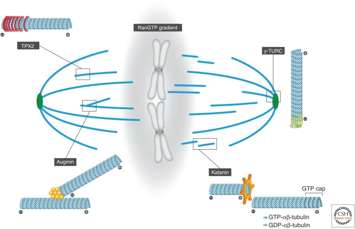
Microtubule nucleation, stabilization, and amplification. The metaphase spindle is a complex structure consisting of microtubules (blue) that nucleate from centrosomes (green) and chromatin (gray). A central centrosomal component is the γ-tubulin ring complex (γ-TuRC), which templates the nucleation of microtubules. The spatial cue necessary to nucleate microtubules around chromatin is mediated by a diffusion-limited RanGTP gradient, the first identified direct effector of which is TPX2. The eight-subunit complex augmin nucleates microtubules parallel to existing microtubules, while katanin severs and disassembles microtubules. GDP, Guanosine diphosphate; GTP, guanosine triphosphate.
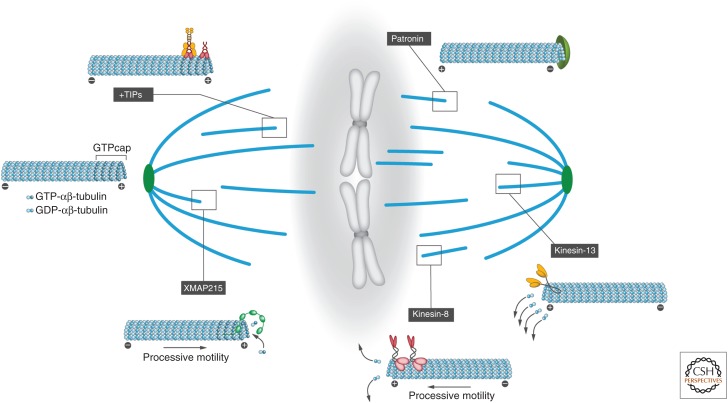
Microtubule dynamics. The intrinsic dynamic instability of microtubules is generated by guanosine triphosphate (GTP) hydrolysis at the nucleotide exchangeable site in β-tubulin. In addition, various proteins regulate the dynamic behavior of microtubules. Although microtubule − ends are specifically stabilized, for example, by patronin, the + ends switch stochastically between growing (regulated by polymerases such as XMAP215) and shrinking phases (regulated by depolymerases such as kinesin-8 and -13). Growing microtubule + ends are further regulated by so-called microtubule plus-end tracking proteins (+TIPs). GDP, Guanosine diphosphate.
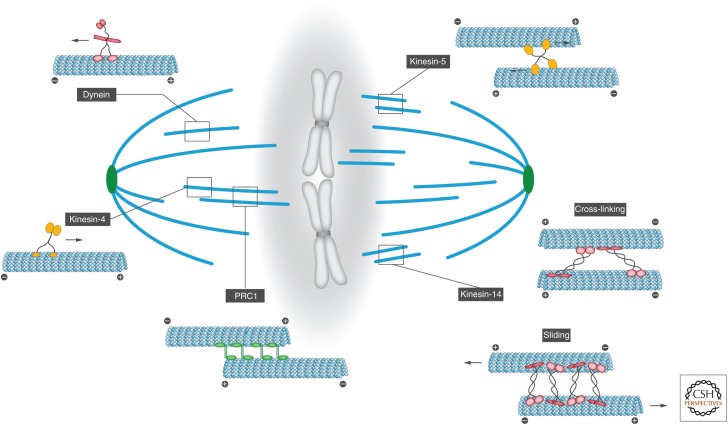
Spindle forces. During assembly and function, the spindle passes through several steady states, each relying on a distinct balance of complementary and antagonistic forces. The homotetramer kinesin-5 is a highly conserved plus-end-directed motor optimized to cross-link and slide antiparallel microtubules, thereby producing outward forces that drive centrosome separation during spindle assembly. Kinesin-4 is a dimeric plus-end-directed motor. Together with PRC1, it forms antiparallel microtubule overlaps with precisely defined lengths; while PRC1 marks the microtubule overlap region and recruits kinesin-4, the motor protein walks processively to microtubule ends in the overlap region, where its accumulation leads to the inhibition of microtubule growth. In contrast to the plus-end-directed motility of other kinesin proteins, kinesin-14 is a minus-end-directed motor that can either slide antiparallel microtubules or cross-link parallel microtubules (adapted from Fink et al. 2009). Cytoplasmic dynein is the major motor responsible for microtubule minus-end-directed movements.
References
-
- Akhmanova A, Hoogenraad CC, Drabek K, Stepanova T, Dortland B, Verkerk T, Vermeulen W, Burgering BM, De Zeeuw CI, Grosveld F, et al. 2001. Clasps are CLIP-115 and -170 associating proteins involved in the regional regulation of microtubule dynamics in motile fibroblasts. Cell 104: 923–935. - PubMed
-
- Al-Bassam J, Larsen NA, Hyman AA, Harrison SC. 2007. Crystal structure of a TOG domain: Conserved features of XMAP215/Dis1-family TOG domains and implications for tubulin binding. Structure 15: 355–362. - PubMed
-
- Ananthanarayanan V, Schattat M, Vogel SK, Krull A, Pavin N, Tolić-Nørrelykke IM. 2013. Dynein motion switches from diffusive to directed upon cortical anchoring. Cell 153: 1526–1536. - PubMed
-
- Asenjo AB, Chatterjee C, Tan D, DePaoli V, Rice WJ, Diaz-Avalos R, Silvestry M, Sosa H. 2013. Structural model for tubulin recognition and deformation by kinesin-13 microtubule depolymerases. Cell Rep 3: 759–768. - PubMed
Publication types
MeSH terms
Substances
LinkOut - more resources
Full Text Sources
Other Literature Sources