Review
. 2015 Jul 1;7(7):a021758.
doi: 10.1101/cshperspect.a021758.

Structural Components of Synaptic Plasticity and Memory Consolidation

Affiliations
Review

Structural Components of Synaptic Plasticity and Memory Consolidation

Craig H Bailey et al. Cold Spring Harb Perspect Biol. .

Abstract

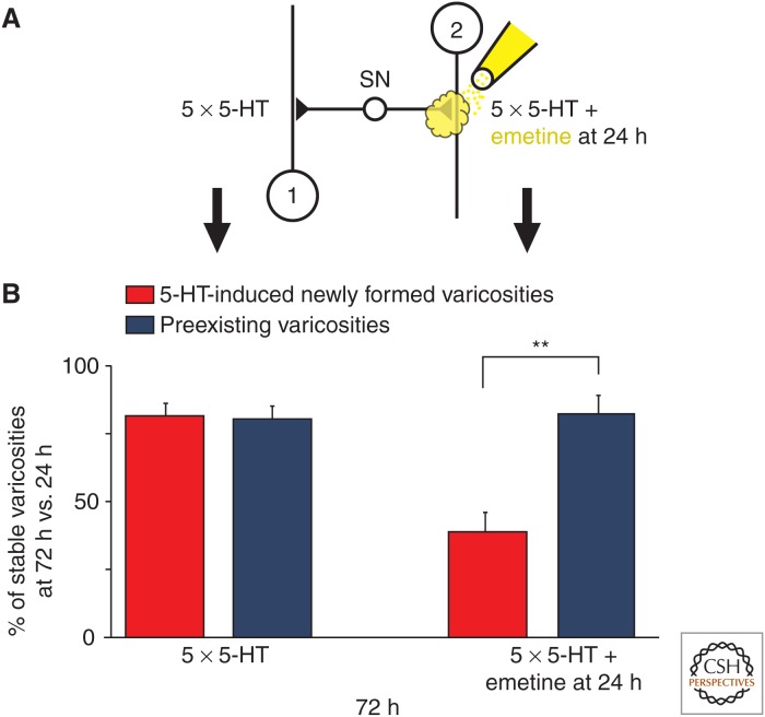
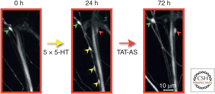
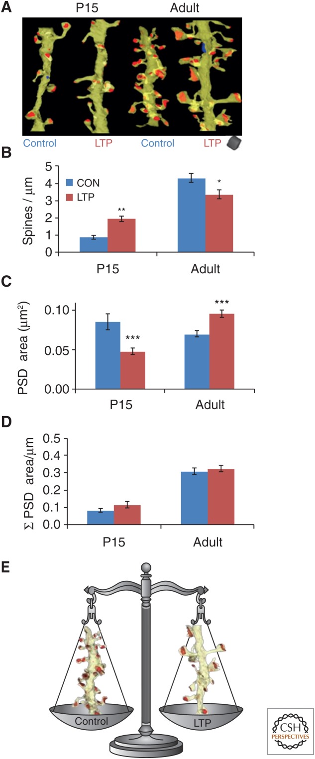
Consolidation of implicit memory in the invertebrate Aplysia and explicit memory in the mammalian hippocampus are associated with remodeling and growth of preexisting synapses and the formation of new synapses. Here, we compare and contrast structural components of the synaptic plasticity that underlies these two distinct forms of memory. In both cases, the structural changes involve time-dependent processes. Thus, some modifications are transient and may contribute to early formative stages of long-term memory, whereas others are more stable, longer lasting, and likely to confer persistence to memory storage. In addition, we explore the possibility that trans-synaptic signaling mechanisms governing de novo synapse formation during development can be reused in the adult for the purposes of structural synaptic plasticity and memory storage. Finally, we discuss how these mechanisms set in motion structural rearrangements that prepare a synapse to strengthen the same memory and, perhaps, to allow it to take part in other memories as a basis for understanding how their anatomical representation results in the enhanced expression and storage of memories in the brain.


Figures

Figure 1.
Figure 1.
Fine structure of an identified sensory neuron presynaptic varicosity. A thin section containing a sensory neuron varicosity (SNV) labeled with horseradish peroxidase (HRP) (Bailey et al. 1979) is shown. The density of the HRP reaction product allows one to clearly distinguish the labeled sensory neuron profile from unlabeled profiles while still being able to visualize the intracellular contents of the identified varicosity. This portion of the identified sensory neuron presynaptic compartment contains three dense core vesicles and a population of electron-lucent vesicles, some of which cluster at the electron-dense specializations that define the active zone (between arrow heads). In this thin section, the labeled sensory neuron presynaptic varicosity forms a synaptic contact with an unlabeled postsynaptic dendritic spine (Sp) of a follower neuron. By combining this selective intracellular-labeling technique with the analysis of serial thin sections and transmission EM, complete 3D reconstructions of active zone morphology (number, size, and vesicle complement) in unequivocally identified sensory neuron synapses were quantitatively analyzed from both control and behaviorally modified animals. (Unpublished electron micrograph courtesy of Mary Chen and Craig Bailey.)
Figure 2.
Figure 2.
Learning-related growth of the sensory neuron synaptic arbor induced by long-term sensitization in Aplysia. Serial 3D reconstructions of identified sensory neurons labeled with horseradish peroxidase (HRP) from long-term sensitized and control animals are shown. Total extent of the synaptic neuropil arbors of sensory neurons from one control (untrained) and two long-term sensitized animals are shown. In each case, the rostral (row 3) to caudal (row 1) extent of the arbor is divided roughly into thirds. Each panel was produced by the superimposition of camera lucida tracings of all HRP-labeled processes present in 17 consecutive slab-thick Epon sections and represents a linear segment through the ganglion of roughly 340 µm. For each composite, ventral is up, dorsal is down, lateral is to the left, and medial is to the right. By examining images across each row (rows 1, 2, and 3), the viewer is comparing similar regions of each sensory neuron. In all cases, the synaptic arbor of long-term sensitized cells is markedly expanded compared with cells from control (untrained) animals, and parallels the concomitant twofold increase in the total number of sensory neuron presynaptic varicosities. (From Bailey and Chen 1988a; modified, with permission.)
Figure 3.
Figure 3.
Time course and functional contribution of two distinct presynaptic structural changes associated with intermediate-term facilitation and long-term facilitation (LTF) in Aplysia. Repeated pulses of 5-HT in sensory to motor neuron cocultures trigger two distinct classes of learning-related presynaptic structural changes: (1) the rapid filling of synaptic vesicles and active zone material to preexisting silent sensory neuron varicosities (3–6 h), and (2) the slower generation of new sensory neuron synaptic varicosities (12–18 h). The resultant newly filled and newly formed varicosities are functionally competent (capable of evoked transmitter release) and contribute to the synaptic enhancement that underlies LTF measured at 24 h. The rapid filling and activation of silent presynaptic terminals at 3 h suggests that, in addition to its role in LTF, this remodeling of preexisting varicosities may also contribute to the intermediate phase of synaptic plasticity. Red triangles represent transmitter-release sites (active zones). (From Kim et al. 2003; modified, with permission.)
Figure 4.
Figure 4.
Differential down-regulation of the glycosyl-phosphoinositol (GPI)-linked versus transmembrane isoforms of cell-adhesion molecule in Aplysia (apCAM): the role in learning-related synaptic growth. Some of the first evidence for a role of cell-adhesion molecules (CAMs) during learning and memory came from studies of an immunoglobulin-related CAM in Aplysia, designated apCAM, which is homologous to neural cell–adhesion molecules (NCAMs) in vertebrates and Fasciclin II in Drosophila. To determine the fate of the two isoforms of apCAM in learning-related synaptic growth, gold-conjugated epitope-tagged constructs of either the transmembrane (TM) or GPI-linked isoforms were overexpressed in Aplysia sensory neurons. (A) Neurite of a sensory neuron expressing the GPI-linked isoform of apCAM following a 1-h exposure to 5-HT. Note, virtually all of the gold complexes (black dots) remain on the surface membrane with none inside despite a robust 5-HT-induced activation of the endosomal pathway leading to significant accumulations of internal membranous profiles. (B) Neurite of a sensory neuron expressing the transmembrane isoform of apCAM following a 1-h exposure to 5-HT. In contrast to the lack of down-regulation of the GPI-linked isoform, 5-HT has a dramatic effect on the transmembrane isoform of apCAM, removing most of it from the surface membrane, resulting in heavy accumulations of gold complexes within presumptive endocytic compartments. This 5-HT-induced, clathrin-mediated selective internalization of the transmembrane isoform of apCAM in the presynaptic sensory neuron leads to: (1) defasciculation, a process that destabilizes adhesive contacts normally inhibiting synaptic growth, (2) endocytic activation that results in a redistribution of membrane components to sites in which new synapses form, and, finally, (3) the normal expression of long-term facilitation (LTF) and synaptic growth. These findings also suggest that previously established connections might remain intact following exposure to 5-HT because they would be held in place by the adhesive, homophilic interactions of the GPI-linked isoforms, and the process of outgrowth from sensory neuron axons would be initiated by down-regulation of the transmembrane form at extrasynaptic sites of membrane apposition. (From Bailey et al. 1997; modified, with permission.)
Figure 5.
Figure 5.
Local perfusion of emetine at 24 h leads to a selective retraction of 5-HT-induced newly formed sensory neuron varicosities. (A) Diagram of a single bifurcated sensory neuron (SN) in contact with two spatially separated L7 gill-motor neurons (1 and 2) and experimental protocol. (B) To assess the dynamic properties of the 5-HT-induced newly formed varicosities, their stability was compared under two different experimental conditions: 5-HT (left) and 5-HT + emetine (right). Culture dishes containing the bifurcated sensory neuron–motor neuron preparation were treated with five pulses of 5-HT at time 0 and 24 h later, one of the two branches was perfused locally with emetine. Each individual fluorescently labeled 5-HT-induced newly formed and preexisting varicosity was imaged at 24 h and then the exact target field was reimaged to determine the presence or absence of the same individual varicosities at 72 h. The number of 5-HT-induced newly formed and preexisting varicosities that were present at 72 h were compared with the number of varicosities in the same respective class observed at 24 h. At the branch that only received 5-HT, 81.3% of the 5-HT-induced newly formed varicosities (red, left) and 80.3% of the preexisting varicosities (blue, left) were maintained at 72 h when compared with 24 h. In contrast, at the branch that received emetine 24 h after 5-HT treatment, only 38.1% of the 5-HT-induced newly formed varicosities (red, right) were maintained at 72 h versus 81.63% of the preexisting varicosities (blue, right). In both cases, the 5-HT-induced new varicosities represent varicosities that formed between 0 and 24 h and remained stable at 72 h. Each histogram illustrates the mean percentage (± SEM) of identified varicosities maintained at 72 h compared with 24 h. The selective retraction of 5-HT-induced newly formed varicosities induced by local application of emetine shows that during the stabilization phase this population of learning-related varicosities is significantly more labile and sensitive to disruption than the population of preexisting sensory neuron varicosities. (From Miniaci et al. 2008; modified, with permission.)
Figure 6.
Figure 6.
A late phase of sustained cytoplasmic polyadenylation element-binding (CPEB) protein-dependent local protein synthesis is required to stabilize learning-related synaptic growth. A specific Aplysia CPEB (ApCPEB) antisense oligonucleotide covalently coupled to an 11-amino-acid peptide derived from the HIV-TAT protein (TAT-antisense [TAT-AS]) was locally perfused to one branch of the bifurcated sensorimotor neuron culture preparation for 30 min at 24 h after 5-HT treatment. This antisense oligo has previously been shown to lead to the depletion of ApCPEB messenger RNA (mRNA) and to a selective decrease in the level of CPEB protein (Si et al. 2003). Local perfusion of the TAT-AS selectively reduced the number of 5-HT-induced newly formed varicosities maintained at 72 h compared with preexisting varicosities, similar to what was observed with the local perfusion of emetine (see Fig. 5). This figure contains confocal images of green fluorescent protein (GFP)-labeled sensory neuron presynaptic varicosities in contact with the postsynaptic motor neuron L7 (not labeled), and illustrates the results of three imaging sessions of a representative example of the entire sensory neuron–motor neuron synaptic field. Before the application of 5-HT, a single preexisting sensory neuron varicosity is present (green arrowhead) in this field of view. After repeated applications of 5-HT for 24 h, four newly formed sensory neuron varicosities (one red and three yellow arrowheads) are present along with the single preexisting varicosity seen at time 0. The local perfusion of TAT-AS at 24 h to this synaptic area induces the selective pruning of three newly formed varicosities (yellow arrowheads) without affecting the preexisting varicosity (green arrowhead). The red arrowhead represents the only 5-HT-induced newly formed varicosity in this field that is maintained at 72 h. (From Miniaci et al. 2008; modified, with permission.)
Figure 7.
Figure 7.
Small and large dendritic spines and associated structures in the mature rat hippocampus. These spines are from the middle of stratum radiatum of area CA1 of a perfusion-fixed preparation. (Top) Electron micrograph (EM) illustrating a small (S) and large (L) dendritic spine, the postsynaptic density (PSD, red) of the large spine, presynaptic axon (green) and vesicles it contains, as well as the perisynaptic astroglial processes (light blue). The presynaptic axon of the small spine also contains a small dense-core vesicle, which is usually associated with transport packets involved in delivering presynaptic active zone proteins to growing synapses. (Bottom) These two spines (yellow) are illustrated in 3D reconstructions with their associated PSDs at the same scale as in the top EMs. DCV, dense-core vesicle; SA, sample area.
Figure 8.
Figure 8.
Age differences in the structural correlates of long-term potentiation (LTP) in acute rat hippocampal slices. (A) 3D electron microscopy (EM) of representative dendrites that received control (CON) stimulation versus induction of LTP by θ-burst stimulation (TBS). (B) Opposite effects of TBS on spine density and (C) synaptic surface area (postsynaptic density [PSD]) at P15 versus adult (P60–70) dendrites. (D) Yet, the summed surface area of the synapses per micron length of dendrite was unchanged by LTP at either age. These graphs also illustrate that neither spine density nor summed synapse area has reached adult levels by P15. (E) Thus, as illustrated for adult dendrites, either a dendritic segment supports more, smaller, and presumably less-effective synapses or more, larger, and presumably more-effective synapses.
Figure 9.
Figure 9.
Plasticity of synaptic nascent zones at the edges of synapses from the mature rat hippocampus. (A–D) Electron micrographs (EMs) and 3DEM through representative sections of a synapse to distinguish active zones (AZ, red) from nascent zones (NZ, aqua). Synaptic vesicles are colorized to distinguish docked vesicles (dark blue) from vesicles in a pool within 94 nm of the presynaptic membrane (light purple) from the reserve pool (green). NZs had no presynaptic vesicles located within 94 nm perpendicular to them. (E) Model of the sequence of morphological changes associated with different times following the induction of long-term potentiation (LTP) by theta-burst stimulation (TBS), which could participate in the preparation of synapses for subsequent augmentation of LTP. DCV, dense-core vesicle; syns, synapses; ssv, small synaptic vesicle; STP, short-term potentiation.

References

    1. Abraham WC, Bear MF. 1996. Metaplasticity: The plasticity of synaptic plasticity. Trends Neurosci 19: 126–130. - PubMed
    1. Abraham WC, Huggett A. 1997. Induction and reversal of long-term potentiation by repeated high-frequency stimulation in rat hippocampal slices. Hippocampus 7: 137–145. - PubMed
    1. Abraham WC, Tate WP. 1997. Metaplasticity: A new vista across the field of synaptic plasticity. Prog Neurobiol 52: 303–323. - PubMed
    1. Abraham WC, Mason-Parker SE, Bear MF, Webb S, Tate WP. 2001. Heterosynaptic metaplasticity in the hippocampus in vivo: A BCM-like modifiable threshold for LTP. Proc Natl Acad Sci 98: 10924–10929. - PMC - PubMed
    1. Abraham WC, Logan B, Greenwood JM, Dragunow M. 2002. Induction and experience-dependent consolidation of stable long-term potentiation lasting months in the hippocampus. J Neurosci 22: 9626–9634. - PMC - PubMed

Publication types

LinkOut - more resources