. 2015 Aug 14;290(33):20360-73.
doi: 10.1074/jbc.M115.648907. Epub 2015 Jul 1.

Structure and Function of the Hypertension Variant A486V of G Protein-coupled Receptor Kinase 4

Affiliations

Structure and Function of the Hypertension Variant A486V of G Protein-coupled Receptor Kinase 4

Samantha J Allen et al. J Biol Chem. .

Abstract

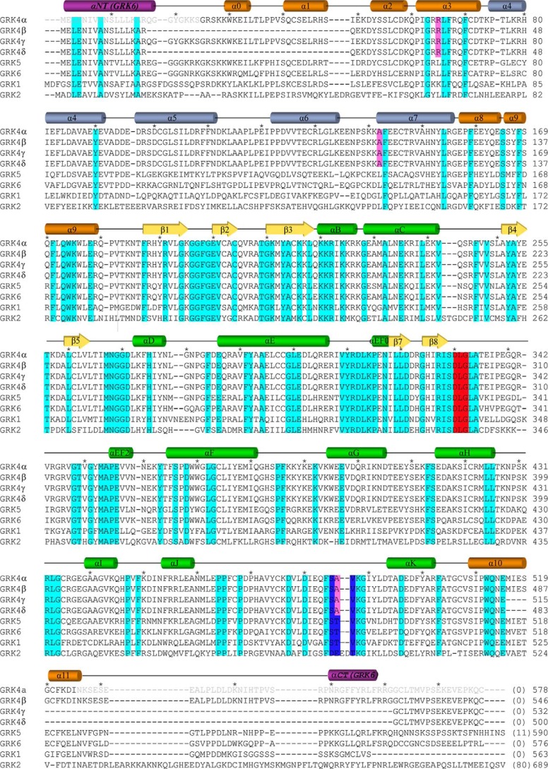
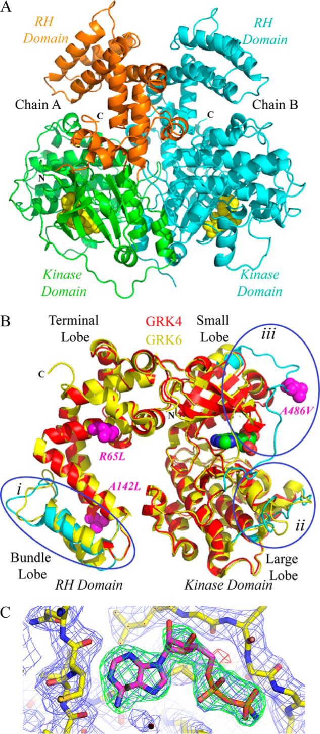
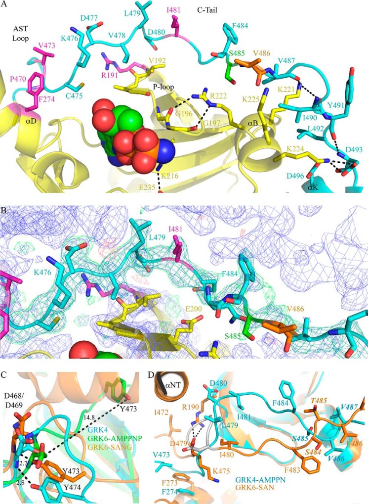
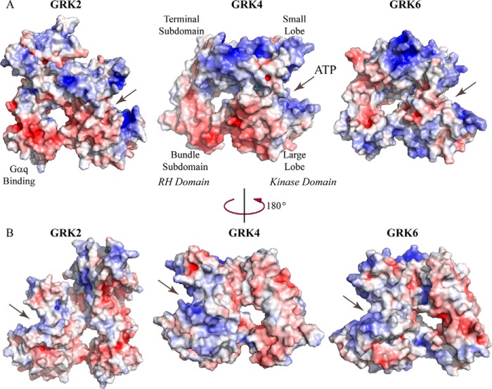
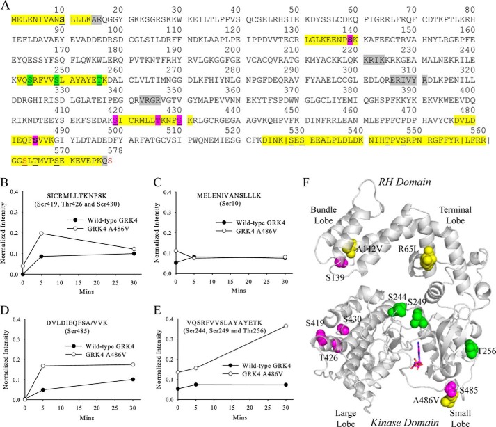
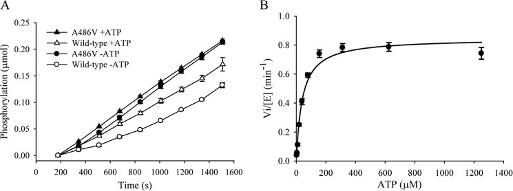
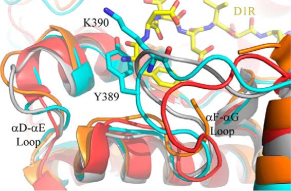
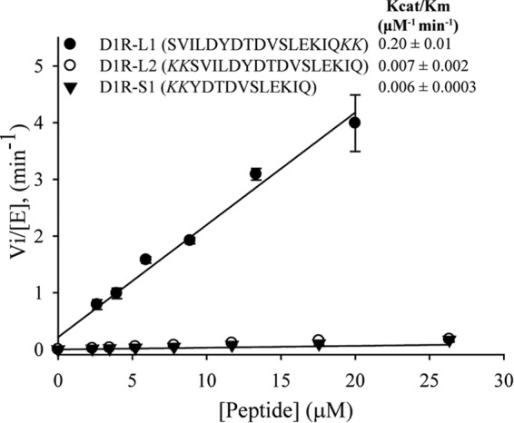
G-protein-coupled receptor (GPCR) kinases (GRKs) bind to and phosphorylate GPCRs, initiating the process of GPCR desensitization and internalization. GRK4 is implicated in the regulation of blood pressure, and three GRK4 polymorphisms (R65L, A142V, and A486V) are associated with hypertension. Here, we describe the 2.6 Å structure of human GRK4α A486V crystallized in the presence of 5'-adenylyl β,γ-imidodiphosphate. The structure of GRK4α is similar to other GRKs, although slight differences exist within the RGS homology (RH) bundle subdomain, substrate-binding site, and kinase C-tail. The RH bundle subdomain and kinase C-terminal lobe form a strikingly acidic surface, whereas the kinase N-terminal lobe and RH terminal subdomain surfaces are much more basic. In this respect, GRK4α is more similar to GRK2 than GRK6. A fully ordered kinase C-tail reveals interactions linking the C-tail with important determinants of kinase activity, including the αB helix, αD helix, and the P-loop. Autophosphorylation of wild-type GRK4α is required for full kinase activity, as indicated by a lag in phosphorylation of a peptide from the dopamine D1 receptor without ATP preincubation. In contrast, this lag is not observed in GRK4α A486V. Phosphopeptide mapping by mass spectrometry indicates an increased rate of autophosphorylation of a number of residues in GRK4α A486V relative to wild-type GRK4α, including Ser-485 in the kinase C-tail.

Keywords: G-protein-coupled receptor (GPCR); GRK4; dopamine D1 receptor; hypertension; kinetics; phosphorylation; serine/threonine protein kinase; x-ray crystallography.


Figures

FIGURE 1.
FIGURE 1.
Sequence alignment of GRK4 splice variants α, β, γ, and δ with representative members of the GRK family. All sequences are human. Every 10th residue in GRK4α is denoted by a black asterisk above the alignment. The four GRK4 splice variants result from alternative splicing of exons 2 and 15 in the RH domain, with GRK4β missing exon 2 (residues 18–49), GRK4γ missing exon 15 (residues 516–561), and GRK4δ missing exons 2 and 15 (residues 18–49 and 516–561) relative to full-length GRK4 (GRK4α). The sequence of the kinase domain is identical for all four GRK4 splice variants. Conserved residues are shown in cyan; the DFG motif at the start of the activation loop (DLG in the GRK family) is shown in red, and GRK4 hypertension-related mutations are in magenta. The SA/VV motif (residues 485–487 in human GRK4α) is in blue. This motif is STV in human GRK5, human GRK6, human GRK1, dog GRK4, and bovine GRK4; SVI in rabbit GRK4; and SVV in rat, chimp, and mouse GRK4. The secondary structural elements of GRK4α are shown above the alignment, with cylinders representing α-helices, arrows representing β-strands, and lines representing unstructured residues. Secondary structural elements are colored as follows: RH terminal subdomain in orange; RH bundle subdomain in blue; kinase domain β-strands in yellow; and kinase domain α-helices in green. Disordered residues at the N and C termini in the structure of GRK4α are in gray. Additional helices at the N and C termini of GRK6 bound to sangivamycin (αNT and αCT) (41) are shown in purple.
FIGURE 2.
FIGURE 2.
Crystal structure of human GRK4α A486V. A, crystal structure contains two molecules in the asymmetric unit. The N terminus (N) and C terminus (C) of chain A are labeled, and the C terminus is labeled for chain B. The N terminus of chain B is on the back of the structure as shown and is not visible. The RH domain of chain A is shown in orange, and the kinase domain of chain A is in green. Chain B is shown in cyan. AMPPN is in space-fill. B, GRK4α chain A (shown in red) overlaid with chain A of GRK6 bound to AMPPNP (26) (shown in yellow). Regions of the proteins that vary between the two structures are highlighted on the GRK4α structure in cyan. These are as follows: (i) the bundle subdomain of the RH domain; (ii) the substrate-binding channel of the kinase domain large lobe (e.g. αD–αE and αF-αG loops), and (iii) the region around the C-tail of the kinase domain. Hypertension-related mutations of GRK4 (R65L, A142L, and A486V) are shown in magenta. C, simulated annealing omit maps for AMPPN. 2m|Fo| − D|Fc|map (blue mesh) contoured at 1σ. The m|Fo| − D|Fc| maps are contoured at ±3σ and shown as green (positive density) and red (negative density) meshes.
FIGURE 3.
FIGURE 3.
Key interactions in the GRK4α kinase domain. A, C-tail of GRK4α is shown in cyan, and the rest of the kinase domain is in yellow. Key residues and secondary structure elements in the C-tail and small lobe are labeled, and notable salt bridges and hydrogen bonds are shown. Conserved residues associated with GPCR and peptide phosphorylation in GRK1, GRK2, or GRK6 are highlighted in magenta. Ser-485 (autophosphorylation site) is in green, and the A486V hypertension-related polymorphism is shown in orange. AMPPN is shown in space-fill. B, simulated annealing omit map for the C-tail of GRK4α. The 2m|Fo| − D|Fc| map (blue mesh) contoured at 1σ. The m|Fo| − D|Fc| maps are contoured at ±3σ and shown as green (positive density) and red (negative density) meshes. Residues are colored as in A. C, structural overlay of the AST loop of GRK4α with two previous structures of GRK6. GRK4α is shown in cyan; GRK6 bound to AMPPNP (26) is in green, and GRK6 bound to sangivamycin (41) is in orange. Distances (Å) between specific atoms are shown. A hydrogen bond between the backbone amide of Asp-468 and the side chain hydroxyl of Tyr-473 previously observed in the structure of GRK6-sangivamycin (41) is conserved in GRK4α (corresponding residues for GRK4α are Asp-469 and Tyr-474). This is in contrast to GRK6-AMPPNP, where the distance between these atoms is over 14 Å. D, C-tail of GRK4α A486V (shown in cyan) overlaid with GRK6-sangivamycin (41) (shown in orange). The hydrogen bond between Lys-475 and Arg-190 in GRK6-sangivamycin is shown with black dashed lines and the Asp-476–Asp-479 turn in GRK6-sangivamycin is shown in gray. The SA/VV motif in GRK4α (STV in GRK6) is highlighted in italics.
FIGURE 4.
FIGURE 4.
Electrostatic surface of GRK4α compared with GRK2 and GRK6. The electrostatic surface is contoured to ±6 kT, with negative surface charge shown in red and positive surface charge in blue. The ATP binding pocket is highlighted by arrows. A, GRK2 (28) and GRK4α and GRK6 (26) are oriented as in Fig. 2B, with the RH domain on the left and kinase domain on the right. B, rotation by 180° around the vertical relative to A.
FIGURE 5.
FIGURE 5.
Comparison of GRK4α and GRK6. A, chains A of GRK4α (kinase domain shown in yellow and RH domain in red) and GRK6 (26) (shown in green) are overlaid. The corresponding B chains are shown in light blue for GRK4α and magenta for GRK6. The ligand is shown as space-fill. B, activation loop of one monomer (highlighted in blue and the rest of kinase domain in yellow) interacts with residues in the RH domain, kinase N-lobe, and kinase C-lobe of the other monomer (shown in cyan). C, Arg-237 of each αC helix monomer stacked on top of each other. D, interactions between residues in the α7 helix of the RH domain in monomers A (cyan) and B (red). E, alignment of residues in the RH domain (α0-α1 helix, α9 helix, and C-terminal end of α11 helix) that form a hydrophobic domain-swap interface in GRK6 and GRK1 crystal structures (26, 27, 41) or a C-terminal/RH domain packing interface within monomeric GRK5 structures (30, 31). Conserved residues in the domain-swap interface of GRK6-AMPPNP (26) are highlighted in yellow, and those also interacting in the GRK5 monomer structures (30, 31) are in green. The LXXDL motif (residues 534–538 of human GRK5) is conserved in GRK4α as described previously (30) and is underlined.
FIGURE 6.
FIGURE 6.
Phosphopeptide mapping of wild-type GRK4α and GRK4α A486V with LC-MS/MS. A, phosphorylated peptides are highlighted in yellow, and phosphorylated residues in the basal state are underlined. Residues that show an increase in phosphorylation after incubation with ATP in GRK4α A486V (but not wild-type GRK4) are highlighted in green, and those that increase in both GRK4α variants are in magenta. Non-native serine residues C563S and C578S are shown in red. Residues that can be unambiguously assigned as phosphorylation sites are shown in boldface. For peptides where unambiguous assignment is not possible, all potential phosphorylation sites are highlighted. Vertical lines indicate instances where multiple versions of the same peptide are observed due to incomplete cleavage. Residues that were not observed in zero time point in-gel trypsin digest samples are highlighted in gray. B–E, effects of ATP incubation upon intensities of four representative phosphopeptides. Phosphopeptide intensities were normalized using the sum of the three most intense nonphosphorylated peptides. F, phosphorylation sites mapped on the crystal structure of GRK4α. Phosphorylated residues are shown as space-fill and colored as in A. Hypertension-related mutations are shown in yellow.
FIGURE 7.
FIGURE 7.
Kinetic studies of wild-type GRK4α and GRK4α A486V upon incubation with ATP. A, rate of D1R peptide phosphorylation is affected by preincubation with ATP for wild-type GRK4α (open circles versus open triangles) but not for GRK4α A486V (black circles versus black triangles). B, Km for ATP is 40 μm.
FIGURE 8.
FIGURE 8.
Overlay of the αD–αE and αF–αG loops of GRK kinase large lobes. GRK4α A486V is shown in cyan; GRK6-AMPPMP (26) is in gray; GRK6-sangivamycin (41) is in orange, and GRK2 (28) is in red. The D1R peptide (shown in yellow) was docked into GRK4α by structural alignment to the GSK3β peptide-bound structure of PKB (37). Docking based on this structure results in multiple clashes at the N-terminal side of the peptide with Tyr-389 and Lys-390 in the αF–αG loop.
FIGURE 9.
FIGURE 9.
Initial rates of D1R peptide phosphorylation by GRK4α. kcat/Km values for D1R-L1, D1R-L2, and D1R-S1 are displayed on the figure. The data are derived from three independent experiments and the means ± S.D. are shown. Initial rate of D1R-S2 (YDTDVSLEKIQ) was very similar to D1R-S1 (kcat/Km of D1R-S2 = 0.0076 ± 0.0013 μm−1 min−1).

References

    1. Ribas C., Penela P., Murga C., Salcedo A., García-Hoz C., Jurado-Pueyo M., Aymerich I., Mayor F., Jr. (2007) The G protein-coupled receptor kinase (GRK) interactome: role of GRKs in GPCR regulation and signaling. Biochim. Biophys. Acta 1768, 913–922 - PubMed
    1. Gurevich E. V., Tesmer J. J., Mushegian A., Gurevich V. V. (2012) G protein-coupled receptor kinases: more than just kinases and not only for GPCRs. Pharmacol. Ther. 133, 40–69 - PMC - PubMed
    1. Homan K. T., Glukhova A., Tesmer J. J. (2013) Regulation of G protein-coupled receptor kinases by phospholipids. Curr. Med. Chem. 20, 39–46 - PubMed
    1. Pitcher J. A., Fredericks Z. L., Stone W. C., Premont R. T., Stoffel R. H., Koch W. J., Lefkowitz R. J. (1996) Phosphatidylinositol 4,5-bisphosphate (PIP2)-enhanced G protein-coupled receptor kinase (GRK) activity. Location, structure, and regulation of the PIP2 binding site distinguishes the GRK subfamilies. J. Biol. Chem. 271, 24907–24913 - PubMed
    1. Pronin A. N., Satpaev D. K., Slepak V. Z., Benovic J. L. (1997) Regulation of G protein-coupled receptor kinases by calmodulin and localization of the calmodulin binding domain. J. Biol. Chem. 272, 18273–18280 - PubMed

MeSH terms

Substances