. 2015 Jul 1;35(26):9622-31.
doi: 10.1523/JNEUROSCI.3973-14.2015.

Differential Effect of Neuropeptides on Excitatory Synaptic Transmission in Human Epileptic Hippocampus

Affiliations

Differential Effect of Neuropeptides on Excitatory Synaptic Transmission in Human Epileptic Hippocampus

Marco Ledri et al. J Neurosci. .

Abstract

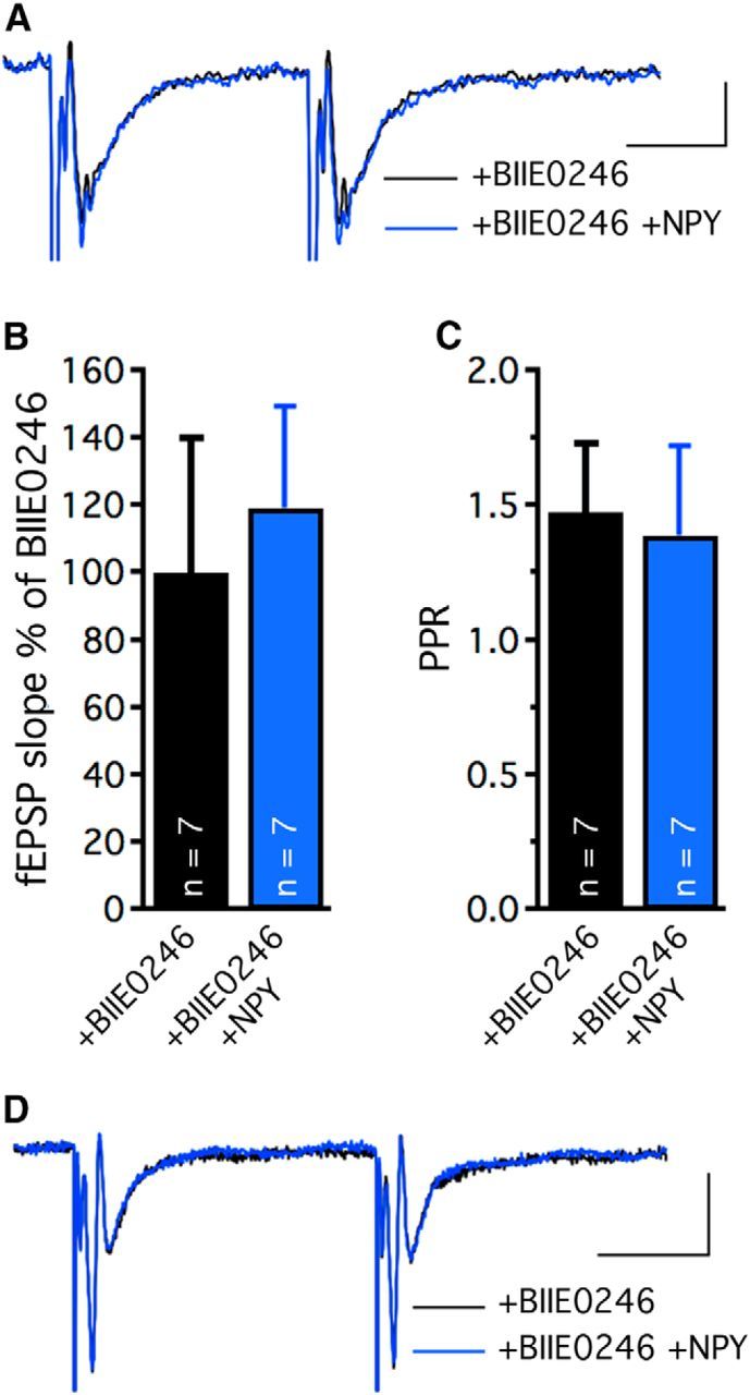
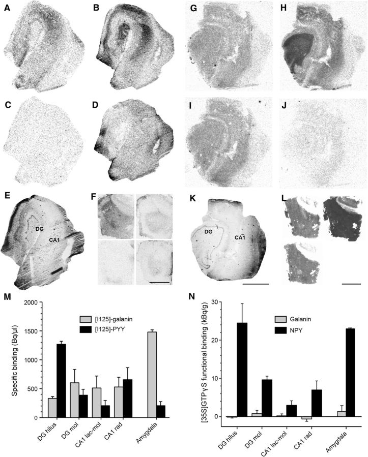
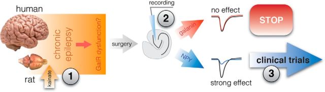
Development of novel disease-modifying treatment strategies for neurological disorders, which at present have no cure, represents a major challenge for today's neurology. Translation of findings from animal models to humans represents an unresolved gap in most of the preclinical studies. Gene therapy is an evolving innovative approach that may prove useful for clinical applications. In animal models of temporal lobe epilepsy (TLE), gene therapy treatments based on viral vectors encoding NPY or galanin have been shown to effectively suppress seizures. However, how this translates to human TLE remains unknown. A unique possibility to validate these animal studies is provided by a surgical therapeutic approach, whereby resected epileptic tissue from temporal lobes of pharmacoresistant patients are available for neurophysiological studies in vitro. To test whether NPY and galanin have antiepileptic actions in human epileptic tissue as well, we applied these neuropeptides directly to human hippocampal slices in vitro. NPY strongly decreased stimulation-induced EPSPs in dentate gyrus and CA1 (up to 30 and 55%, respectively) via Y2 receptors, while galanin had no significant effect. Receptor autoradiographic binding revealed the presence of both NPY and galanin receptors, while functional receptor binding was only detected for NPY, suggesting that galanin receptor signaling may be impaired. These results underline the importance of validating findings from animal studies in human brain tissue, and advocate for NPY as a more appropriate candidate than galanin for future gene therapy trials in pharmacoresistant TLE patients.

Keywords: NPY; galanin; gene therapy; hippocampus; temporal lobe epilepsy.


Figures

Figure 1.
Figure 1.
Sprouting and severe degeneration of neurons in hippocampal tissue derived from patients with pharmacoresistant TLE. Example of hematoxylin (A–C) and MAP2 (D–F) staining seen in hippocampal tissue resected from an epileptic patient with severe sclerosis. While the overall architecture of the hippocampus is preserved, the pyramidal cell layer in CA1 is almost completely degenerated with scattered cells seen in surrounding layers (A, C, D, F). The dentate granule cell layer appears intact (A, B), but is accompanied by sprouting (D, E). Boxed areas are magnified. gcl, granule cell layer; ml, molecular layer; so, stratum oriens; pcl, principal cell layer; sr, stratum radiatum; sl-m, stratum lacunosum moleculare. Scale bars: A, D, 1 mm; B, C, E, F, 200 μm.
Figure 2.
Figure 2.
Excitatory neurotransmission in dentate granule cells synapses in sclerotic human hippocampal tissue is attenuated by NPY, but not galanin. A, Whole-cell patch-clamp recording of a dentate granule cell in hippocampal slice preparation derived from a TLE patient shows fast repetitive action potentials upon a 300 pA current ramp depolarization. Calibration: 20 mV and 200 ms. B, Spontaneous postsynaptic currents recorded in a dentate granule cell held at −70 mV. Boxed area is magnified on the right. Calibration: 10 pA, 200 and 20 ms, respectively. C, Post hoc visualization of recorded dentate granule cells revealed by immunohistochemistry. Alexa 488-conjugated streptavidin labels intracellular biocytin and shows apical dendrites extending into the molecular layers. Scale bar, 50 μm. D, Galanin application does not affect the amplitude of evoked EPSCs. Insert, Representative traces of evoked EPSCs during aCSF (black trace) and aCSF + galanin application (red trace). Calibration: 50 pA and 20 ms. E, PPR of EPSCs remains unaltered following galanin application. F, NPY application attenuates evoked EPSC amplitudes. Insert, Representative traces of evoked EPSCs during aCSF (black trace) and aCSF + NPY (blue trace) application. Calibration: 50 pA and 20 ms. G, NPY application increases the PPR, suggesting decreased release probability of glutamate. H, I, During 40 Hz stimulation, only NPY suppresses consecutive evoked EPSCs. EPSC amplitudes are normalized to baseline values for each condition. J, Examples of EPSC traces evoked by 40 Hz stimulation shown in aCSF (black), aCSF + galanin (red), and aCSF + NPY (blue), respectively. Calibration: 25 ms, 100 pA; *p < 0.05, **p < 0.01.
Figure 3.
Figure 3.
Excitatory synaptic transmission onto human dentate granule cells is strongly inhibited by NPY, but not by galanin. Examples of fEPSPs evoked by high-frequency stimulation before and after galanin (A) and NPY (E) application. Merged traces are shown for aCSF (A, black trace) and aCSF + galanin (A, red trace) and aCSF (E, black trace) and aCSF + NPY (E, blue trace) application period, respectively. Calibration: 25 ms, 0.4 mV. Galanin only moderately suppresses the first evoked fEPSP (B), while NPY strongly attenuates the same response (F). fEPSP slopes are normalized to baseline values and compared against the galanin (B) and NPY (F) application period, respectively. PPR of fEPSPs is unaltered by galanin (C), but increased following NPY application (G). During high-frequency stimulation, galanin induces minor effects (significant attenuation at sixth evoked EPSP, D), while NPY prominently suppresses the first two evoked EPSPs (H). Slopes of consecutive fEPSPs are normalized to the first evoked fEPSP during aCSF application. *p < 0.05, **p < 0.01, ***p < 0.001.
Figure 4.
Figure 4.
Excitatory synaptic transmission in Schaffer collateral-CA1 synapses in human epileptic hippocampus is strongly inhibited by NPY, but not by galanin. Representative traces of high-frequency stimulation-induced fEPSPs in CA1 stratum radiatum during aCSF and following galanin (A) and NPY (F) application. Blocking AMPA/kainate receptors with NBQX almost completely inhibits the postsynaptic potential while the presynaptic fiber volley is intact following galanin (B) and NPY (G) application. Calibrations: A, F, 25 ms, 0.5 mV; B, G, 2 ms, 0.5 mV. C–E, fEPSPs evoked by Schaffer collateral stimulation in CA1 area (C), PPR (D), and consecutive evoked fEPSPs (E) during high-frequency stimulation are unaffected by galanin. During NPY application (H, I), a profound inhibitory effect is observed on the first evoked fEPSP (H, J) with a concomitant increase in PPR (I), indicating suppression of glutamate release. *p < 0.05, **p < 0.01.
Figure 5.
Figure 5.
NPY inhibits excitatory transmission in human epileptic hippocampus via Y2 receptors. Representative traces of paired-pulse stimulation-induced fEPSPs in CA1 stratum radiatum (A) or MPP-dentate granule cell synapses (D) before (black) and after (blue) NPY application in presence of the Y2 receptor antagonist BIIE0246. Normalized slopes (B) and PPR (C) of fEPSPs evoked by Schaffer collateral stimulation in CA1 area, showing absence of NPY effect in presence of BIIE0246. Calibrations: A, 20 ms and 0.1 mV; D, 20 ms and 0.4 mV.
Figure 6.
Figure 6.
Galanin and NPY receptor binding and functional binding in the human epileptic hippocampus and amygdala. A, Galanin and (B) NPY receptor binding in the human epileptic hippocampus. C, D, Nonspecific binding corresponding to A and B, respectively. E, Hematoxylin staining of an adjacent section showing the gross morphology of the layers analyzed. F, [125I]-galanin binding (top left), corresponding nonspecific binding (bottom left), [125I]-PYY binding (top right), and corresponding nonspecific binding (bottom right) in sections from the human amygdala. G, Galanin and NPY(H) receptor functional binding. I, Basal and nonspecific (J) binding corresponding to G and H, respectively. K, Hematoxylin staining of an adjacent section showing the gross morphology of the layers analyzed. L, Galanin functional binding (top left), NPY functional binding (top right), and corresponding basal binding (bottom left) in sections from the human amygdala. M, Quantification of specific [125I]-galanin and [125I]-PYY receptor binding measured in hippocampal regions (n = 5) and amygdala (n = 2). N, Quantification of galanin and NPY receptor functional binding (i.e., peptide-stimulated binding minus basal binding) measured in hippocampal regions (n = 5) and amygdala (n = 2). Note almost complete absence of galanin receptor functional binding signal (N), despite specific [125I]-galanin binding found in M. Mol, stratum moleculare; rad, stratum radiatum; lac-mol, stratum lacunosum moleculare. Scale bars: A–E, G–K, 3 mm; F, L, 4 mm.
Figure 7.
Figure 7.
Translational road map for clinical trials with gene therapy in epilepsy. Schematic drawing illustrating the importance of validating results from animal models in human epileptic tissue. Three major points need to be addressed when considering novel therapeutic targets against pharmacoresistant epilepsy. Putative antiepileptic agents need to be tested in epileptic animals with recurrent seizures where their action may differ from that in naive animals (1). The effectiveness of antiepileptic agents needs to be validated in human epileptic tissue, since it may be different from rodent epileptic tissue (2). The decision to continue toward clinical trials needs to be based on results from both animal and human tissue studies to minimize the risk of failure in human trials (3).

Similar articles

Cited by

References

    1. Agasse F, Xapelli S, Coronas V, Christiansen SH, Rosa AI, Sardá-Arroyo L, Santos T, Ferreira R, Schitine C, Harnois T, Bourmeyster N, Bragança J, Bernardino L, Malva JO, Woldbye DP. Galanin promotes neuronal differentiation in murine subventricular zone cell cultures. Stem Cells Dev. 2013;22:1693–1708. doi: 10.1089/scd.2012.0161. - DOI - PubMed
    1. Baraban SC, Hollopeter G, Erickson JC, Schwartzkroin PA, Palmiter RD. Knock-out mice reveal a critical antiepileptic role for neuropeptide Y. J Neurosci. 1997;17:8927–8936. - PMC - PubMed
    1. Christensen DZ, Olesen MV, Kristiansen H, Mikkelsen JD, Woldbye DP. Unaltered neuropeptide Y (NPY)-stimulated [35S]GTPgammaS binding suggests a net increase in NPY signalling after repeated electroconvulsive seizures in mice. J Neurosci Res. 2006;84:1282–1291. doi: 10.1002/jnr.21028. - DOI - PubMed
    1. Christiansen SH, Woldbye DP. Regulation of the galanin system by repeated electroconvulsive seizures in mice. J Neurosci Res. 2010;88:3635–3643. doi: 10.1002/jnr.22517. - DOI - PubMed
    1. Colmers WF, Lukowiak K, Pittman QJ. Neuropeptide Y reduces orthodromically evoked population spike in rat hippocampal CA1 by a possibly presynaptic mechanism. Brain Res. 1985;346:404–408. doi: 10.1016/0006-8993(85)90880-7. - DOI - PubMed

Publication types

MeSH terms