Review
. 2015 Jul 1;5(7):a021386.
doi: 10.1101/cshperspect.a021386.

Hepadnavirus Genome Replication and Persistence

Affiliations
Review

Hepadnavirus Genome Replication and Persistence

Jianming Hu et al. Cold Spring Harb Perspect Med. .

Abstract

Hallmarks of the hepadnavirus replication cycle are the formation of covalently closed circular DNA (cccDNA) and the reverse transcription of a pregenomic RNA (pgRNA) in core particles leading to synthesis of the relaxed circular DNA (rcDNA) genome. cccDNA, the template for viral RNA transcription, is the basis for the persistence of these viruses in infected hepatocytes. In this review, we summarize the current state of knowledge on the mechanisms of hepadnavirus reverse transcription and the biochemical and structural properties of the viral reverse transcriptase (RT). We highlight important gaps in knowledge regarding cccDNA biosynthesis and stability. In addition, we discuss the impact of current antiviral therapies on viral persistence, particularly on cccDNA.


Figures

Figure 1.
Figure 1.
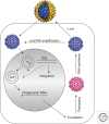
Hepadnavirus life cycle. The figure shows a model for the life cycle of hepadnaviruses RNA- and DNA-containing capsids are shown in red and blue, respectively. For simplicity, only pregenomic RNA (pgRNA) is shown. ccc, covalently closed circular; dsl, double-stranded linear; env, envelope; rc, relaxed circular. (From Seeger et al. 2013; adapted, with permission, from the authors.)
Figure 2.
Figure 2.
Hepatitis B virus (HBV) genome structure. The relaxed circular DNA (rcDNA) genome of HBV with a complete minus strand (black) and incomplete plus strand (cyan) is shown, together with pregenomic RNA (pgRNA) (red) and the core and pol genes required for DNA replication. Reverse transcriptase (RT) and a capped RNA oligomer at the 5′ ends of minus- and plus-strand DNA, respectively, are indicated. The positions of the start sites for the translation of the precore, presurface 1, -2, surface, and X proteins are marked by arrows (PC, PS1, PS2, S, and X, respectively). R, large terminal redundancy on pgRNA; DR, direct repeat; TP, terminal protein of RT.
Figure 3.
Figure 3.
DNA replication cycle. The figure shows a model for the formation of relaxed circular DNA (rcDNA) and double-stranded linear (dsl) from pregenomic RNA (pgRNA). (A) Transfer of the DNA primer from ɛ to DR1 near the 3′ end of pgRNA, (B) synthesis of minus-strand DNA and digestion of pgRNA by RNase H, (C) transfer of the capped RNA primer from DR1 to DR2, (D) synthesis of plus-strand DNA to the 5′ end of minus-strand DNA, (E) template switch of the nascent plus strand with the help of the small terminal redundancies, 5′r and 3′r, resulting in circularization of the genome, (F) completion of plus-strand DNA synthesis, (G) in situ priming of plus-strand DNA, and (H) formation of dslDNA.
Figure 4.
Figure 4.
Ribonucleoprotein complex for the protein-priming reaction. The figure shows a model for the first steps of pregenomic RNA (pgRNA) packaging and protein priming of reverse transcription. Binding of ɛRNA to the terminal protein (TP) and polymerase domains of the reverse transcriptase (RT) is facilitated by chaperones (blue circle). Φ depicts the RNA segment that is believed to base pair with ɛ and is required for circularization of pgRNA. Additional protein factors believed to play a role in RNA packaging by binding to the cap structure are indicated (gray circle). dNTP, deoxyribonucleotide triphosphate; Hsp90, heat shock protein 90.
Figure 5.
Figure 5.
Physical and functional map of the hepatitis B virus (HBV) reverse transcriptase (RT). The figure shows the four domains of the RT (terminal protein, spacer, polymerase, and RNase H) described in the text. The predicted structural subdomains of the polymerase domain (fingers, palm, and thumb) are based on comparison with the crystal structure of human immunodeficiency virus (HIV) RT (Das et al. 2001). The location of mutations that confer drug resistance to nucleotide analogs currently used in antiviral therapy is indicated (drug resistance, DR). The regions of the RT required for RNA packaging, protein priming, and binding to ɛRNA are shown above the figure (see text for details).

References

    1. Abraham TM, Loeb DD. 2006. Base pairing between the 5′ half of ɛ and a cis-acting sequence, Φ, makes a contribution to the synthesis of minus-strand DNA for human hepatitis B virus. J Virol 80: 4380–4387. - PMC - PubMed
    1. Abraham TM, Loeb DD. 2007. The topology of hepatitis B virus pregenomic RNA promotes its replication. J Virol 81: 11577–11584. - PMC - PubMed
    1. Badtke MP, Khan I, Cao F, Hu J, Tavis JE. 2009. An interdomain RNA binding site on the hepadnaviral polymerase that is essential for reverse transcription. Virology 390: 130–138. - PMC - PubMed
    1. Bartenschlager R, Schaller H. 1992. Hepadnaviral assembly is initiated by polymerase binding to the encapsidation signal in the viral RNA genome. EMBO J 11: 3413–3420. - PMC - PubMed
    1. Bartenschlager R, Junker-Niepmann M, Schaller H. 1990. The P gene product of hepatitis B virus is required as a structural component for genomic RNA encapsidation. J Virol 64: 5324–5332. - PMC - PubMed

Publication types

MeSH terms