. 2015 Nov;135(11):2714-2722.
doi: 10.1038/jid.2015.254. Epub 2015 Jul 2.

IsaB Inhibits Autophagic Flux to Promote Host Transmission of Methicillin-Resistant Staphylococcus aureus

Affiliations

IsaB Inhibits Autophagic Flux to Promote Host Transmission of Methicillin-Resistant Staphylococcus aureus

Pei-Feng Liu et al. J Invest Dermatol. 2015 Nov.

Abstract

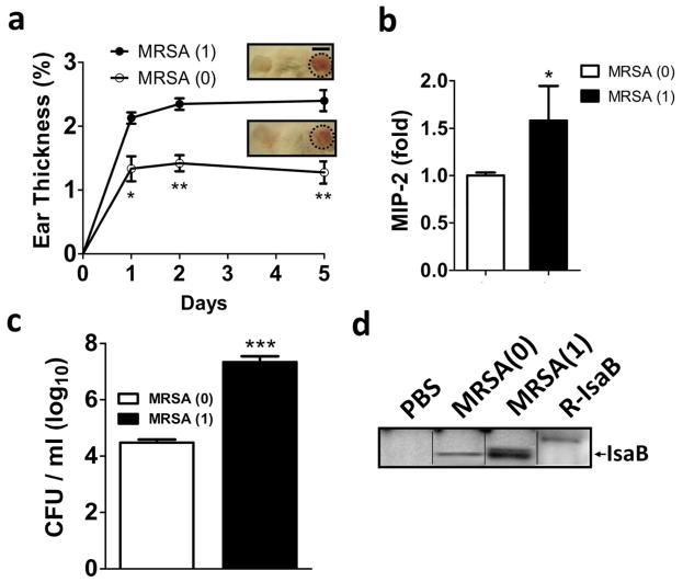
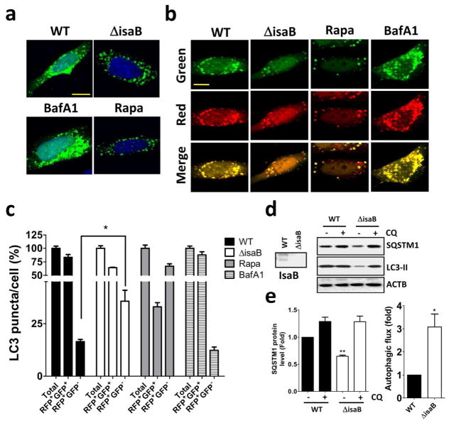
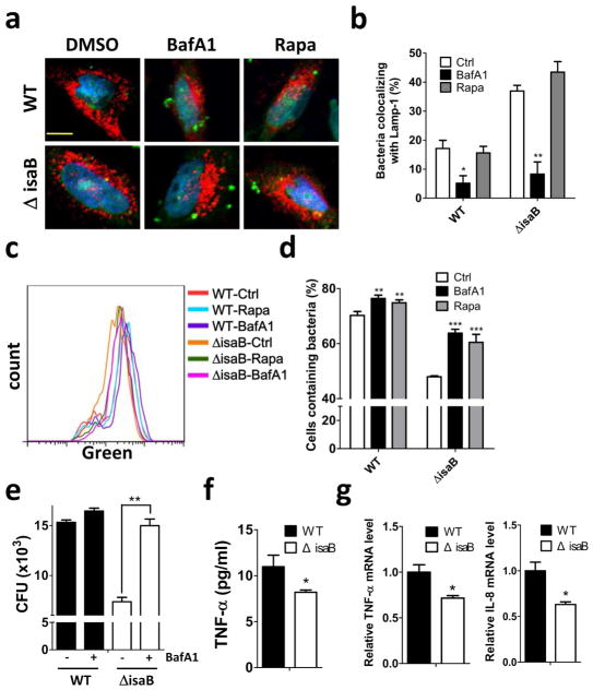
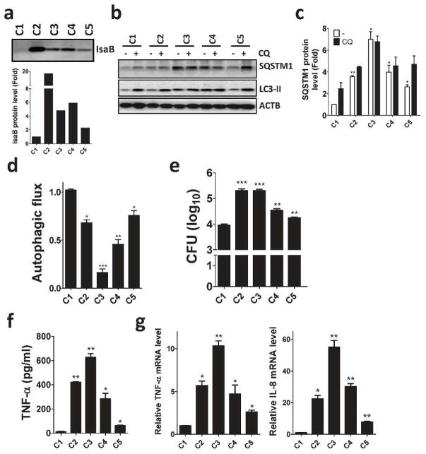
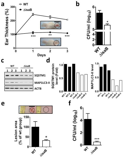
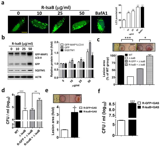
Methicillin-resistant Staphylococcus aureus (MRSA) has emerged as a major nosocomial pathogen that is widespread in both health-care facilities and in the community at large, as a result of direct host-to-host transmission. Several virulence factors are associated with pathogen transmission to naive hosts. Immunodominant surface antigen B (IsaB) is a virulence factor that helps Staphylococcus aureus to evade the host defense system. However, the mechanism of IsaB on host transmissibility remains unclear. We found that IsaB expression was elevated in transmissible MRSA. Wild-type isaB strains inhibited autophagic flux to promote bacterial survival and elicit inflammation in THP-1 cells and mouse skin. MRSA isolates with increased IsaB expression showed decreased autophagic flux, and the MRSA isolate with the lowest IsaB expression showed increased autophagic flux. In addition, recombinant IsaB rescued the virulence of the isaB deletion strain and increased the group A streptococcus (GAS) virulence in vivo. Together, these results reveal that IsaB diminishes autophagic flux, thereby allowing MRSA to evade host degradation. These findings suggest that IsaB is a suitable target for preventing or treating MRSA infection.


Conflict of interest statement

CONFLICT OF INTEREST

The authors state no conflict of interest.

Figures

Figure 1
Figure 1. Transmissible MRSA252 induces IsaB expression and inflammation in vivo
(a) The right ears of mice were injected intradermally with MRSA(0) or MRSA(1) (1 × 10⁷ CFU). Ear thickness was measured and calculated as a percentage, compared to the PBS-injected control, on days 1, 2, and 5 after MRSA injection. Representative images of skin inflammation are shown. Bar = 1 cm. (b) MIP-2 production in mice infected with MRSA(1) was measured by ELISA and normalized to that in mice infected with MRSA(0). (c) The infected ears on day 5 were homogenized for counting MRSA colonies. Data are expressed as the mean ± SEM (n=8). (d) The samples in (c) were subjected to immunoblotting. An arrow indicates IsaB protein, which has a molecular weight of 20 kDa.
Figure 2
Figure 2. IsaB is involved in autophagic flux inhibition in cells
(a) HeLa cells harboring GFP-MAP1LC3 or (b) RFP-GFP- MAP1LC3 were infected with wild-type (WT) S. aureus or an isogenic deletion mutant (ΔisaB) (MOI of 200) for 6 h to observe GFP-MAP1LC3 puncta and autophagic flux using confocal microscopy. Rapamycin (0.5 μM) or BafA1 (100 nM) are used as a control for autophagy induction and inhibition, respectively. Bar = 10 μm. (c) The percentage of autophagosomes (RFP+GFP+) and autolysosomes (RFP+GFP) in each cell as (b) were quantitated from a pool of at least 10 images. (d) Human macrophage-like THP-1 cells were infected with wild-type (WT) S. aureus or an isogenic deletion mutant (ΔisaB) (MOI of 200) for 6 hrs in the presence or absence of 20 μM CQ. The accumulation of SQSTM1 and MAP1LC3-II in infected THP-1 cells was examined by immunoblotting. (e) The quantification of SQSTM1 protein levels and autophagic flux is shown. The results are expressed as the mean±SEM from 3 independent experiments.
Figure 3
Figure 3. Autophagic flux inhibition increases the viability of S. aureus in host cells
HeLa cells harboring Lamp1-RFP (red) were infected with SYTO (green)-labeled wild-type (WT) S. aureus or an isogenic deletion mutant (ΔisaB) (MOI of 200) for 5 hrs in the absence or presence of BafA1 (100 nM) or rapamycin (0.5 μM). The cells were then fixed and observed under confocal microscopy. Bar = 10 μm. (b) Colocalization of bacteria and Lamp1 in cells as (a) was quantitated. (c) Human macrophage-like THP-1 cells were infected with SYTO (green)-labeled bacteria (MOI of 200) for 6 hrs with or without BafA1 (100 nM) or Rapamycin (0.5 μM). The infected cells were harvested to determine the amount of bacteria in the host cells using flow cytometer. (d) The percentage of bacteria in cells was quantitated by FlowJo software and analyzed by Prism 5.0. (e) Colony formation by the cells infected with bacteria (MOI of 200) for 6 hrs in the presence or absence of BafA1 was quantified. (f) The secretion of the inflammatory cytokine TNF-α and (g) the mRNA levels of Tnf-α and Il-8 in infected THP-1 cells were determined by ELISA and real-time PCR, respectively. The results are expressed as the mean ± SEM from 3 independent experiments.
Figure 4
Figure 4. IsaB expression in clinical MRSA isolates is associated with autophagy and inflammation in THP-1 cells
(a) Clinical MRSA isolates were lysed to examine IsaB protein levels by immunoblotting. (b) Human macrophage-like THP-1 cells were infected with clinical MRSA isolates (MOI of 10) for 6 h in the presence (+) or absence (-) of 20 μM CQ for 2 h prior to harvesting. Cells were then lysed to examine SQSTM1, MAP1LC3-II protein levels by immunoblotting. Because the clinical MRSA isolate C1 shows the lowest IsaB expression of the 5 tested clinical MRSA isolates, the protein levels of (c) SQSTM1 and (d) MAP1LC3-II in the infected cells were quantified using the C1-infected cells as a control. (e) Colony formation, (f) secretion of TNF-α and (g) the mRNA levels of Tnf-α and Il-8 in infected THP-1 cells were determined by ELISA and real-time PCR, respectively. The results are expressed as the mean ± SEM from 3 independent experiments.
Figure 5
Figure 5. IsaB inhibits autophagic flux and triggers inflammation in vivo
(a) The isaB wild-type (WT) or the isaB deletion (ΔisaB) S. aureus strain (1 × 10⁷ CFU) was subcutaneously injected into the ears of mice (3 mice for each group). The ear thickness of each infected mouse was measured every day for 3 days. Representative images of ear skin inflammation are shown. Bar = 1 cm. (b) Infected right ears were homogenized on day 3 after injection to determine the level of bacterial colonization. (c) The homogenized ears were analyzed for SQSTM1 and MAP1LC3-II protein levels. (d) Quantified SQSTM1 and MAP1LC3-II protein levels in infected ears are shown. (e) The dorsal skin of mice was injected intradermally with wild type (WT) or isaB-deletion mutant (ΔisaB) strain (4 mice for each group). Representative images of dorsal skin lesions after 3 days of infection are shown and were quantified with ImageJ software. Bar = 0.5 cm. (f) Colonization by wild-type (WT) or isaB-deletion (ΔisaB) strain in the skin was counted on day 3 after injection. The results are expressed as the mean ± SEM.
Figure 6
Figure 6. Recombinant IsaB diminishes autophagic flux and reconstitutes the virulence of the isaB-deficient strain and GAS
(a) HeLa cells harboring GFP-MAP1LC3 were treated with recombinant IsaB for 6 h and fixed to determine the effect of IsaB on GFP-MAP1LC3-II puncta, using florescence microscopy. BafA1 (100 nM) is used as a control for autophagic flux inhibition. Bar = 10 μm. (b) MEF cells stably harboring GFP-MAP1LC3 were treated with recombinant IsaB for 6 h. Protein levels of GFP-MAP1LC3-II, GFP, and SQSTM1 in treated cells were determined by immunoblotting. Protein levels were normalized with ACTB and are shown as quantified in the right panel. (c) Mice were injected with the isaB deletion strain (1 × 10⁷ CFU) and 50 μg recombinant MRSA IsaB (R-IsaB ± ΔisaB) or GFP (R-GFP ±V isaB). Lesion sizes were quantified on day 3 after injection using ImageJ software (n = 4). Bar = 0.5 cm. (d) Bacterial survival on day 3 after injection was quantified in agar plates. (e) Live GAS NZ131 (1 × 10⁷ CFU) mixed with 50 μg recombinant IsaB (R-IsaB+GAS) or GFP (R-GFP+GAS) was subcutaneously injected into the dorsal skin of mice. Lesion sizes and (f) bacterial survival on day 3 after injection were quantified using ImageJ software. Representative images of dorsal skin lesion are shown. Bar = 0.5 cm. The results are expressed as the mean ± SEM.

References

    1. Amano A, Nakagawa I, Yoshimori T. Autophagy in innate immunity against intracellular bacteria. Journal of biochemistry. 2006;140:161–6. - PubMed
    1. Chatterjee SS, Otto M. Improved understanding of factors driving methicillin-resistant Staphylococcus aureus epidemic waves. Clin Epidemiol. 2013;5:205–17. - PMC - PubMed
    1. Dupont N, Jiang S, Pilli M, et al. Autophagy-based unconventional secretory pathway for extracellular delivery of IL-1beta. The EMBO journal. 2011;30:4701–11. - PMC - PubMed
    1. Fuchs S, Pane-Farre J, Kohler C, et al. Anaerobic gene expression in Staphylococcus aureus. J Bacteriol. 2007;189:4275–89. - PMC - PubMed
    1. Garzoni C, Francois P, Huyghe A, et al. A global view of Staphylococcus aureus whole genome expression upon internalization in human epithelial cells. BMC Genomics. 2007;8:171. - PMC - PubMed

Publication types

MeSH terms