. 2015 Nov;135(11):2611-2622.
doi: 10.1038/jid.2015.248. Epub 2015 Jul 2.

Photoactivation of ROS Production In Situ Transiently Activates Cell Proliferation in Mouse Skin and in the Hair Follicle Stem Cell Niche Promoting Hair Growth and Wound Healing

Affiliations

Photoactivation of ROS Production In Situ Transiently Activates Cell Proliferation in Mouse Skin and in the Hair Follicle Stem Cell Niche Promoting Hair Growth and Wound Healing

Elisa Carrasco et al. J Invest Dermatol. 2015 Nov.

Abstract

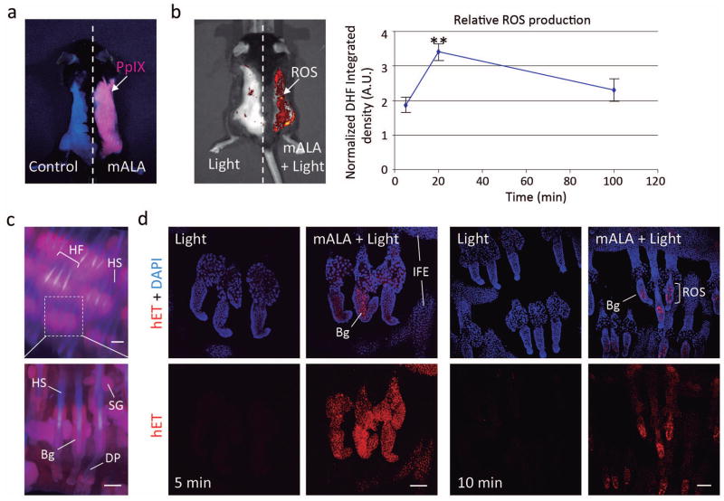
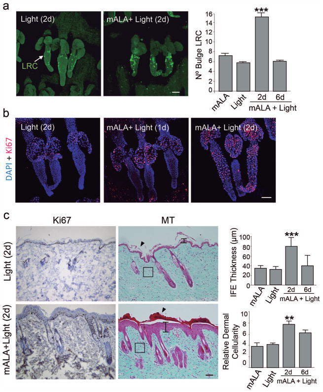
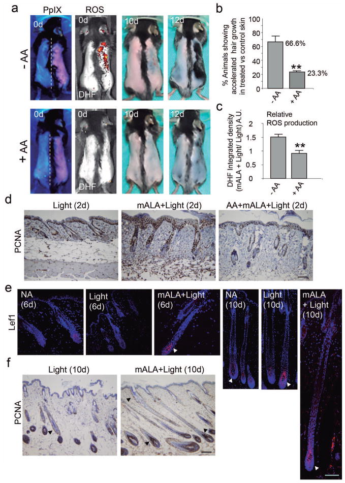
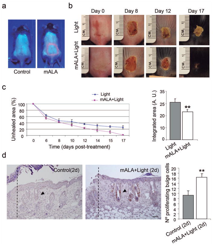
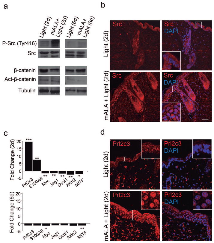
The role of reactive oxygen species (ROS) in the regulation of hair follicle (HF) cycle and skin homeostasis is poorly characterized. ROS have been traditionally linked to human disease and aging, but recent findings suggest that they can also have beneficial physiological functions in vivo in mammals. To test this hypothesis, we transiently switched on in situ ROS production in mouse skin. This process activated cell proliferation in the tissue and, interestingly, in the bulge region of the HF, a major reservoir of epidermal stem cells, promoting hair growth, as well as stimulating tissue repair after severe burn injury. We further show that these effects were associated with a transient Src kinase phosphorylation at Tyr416 and with a strong transcriptional activation of the prolactin family 2 subfamily c of growth factors. Our results point to potentially relevant modes of skin homeostasis regulation and demonstrate that a local and transient ROS production can regulate stem cell and tissue function in the whole organism.


Conflict of interest statement

CONFLICT OF INTEREST

The authors declare no competing financial interest. Clinical and commercial applications of the experimental procedures described in this work have been registered by a CSIC-UAM patent.

Figures

Figure 1
Figure 1. Photodynamic treatment with mALA and red light induces a transient production of ROS in the skin
a) Accumulation of endogenous PpIX after mALA topic treatment in back skin. The left side in the same animal was used as control. b) Left panel: PpIX-dependent ROS (mALA+Light) production monitored by DHF-DA. Right panel: time-course analysis of relative ROS production in back skin; the relative integrated density of DHF-DA fluorescent emission of mALA+Light versus Light regions in each animal was quantified at different times after irradiation and normalized as described in methodology. The mean +/− SE was represented (n=4 for each time point). c) Localization of PpIX in tail skin (fluorescence microscopy images). d) ROS production in tail skin after mALA+Light as revealed by hET showing an increased and sustained accumulation in the bulge region of the hair follicle. Representative confocal microscopy images (maximum projections) are shown. Bars: 100 μm.
Figure 2
Figure 2. Switching on in situ ROS production in the skin activates stem cell proliferation in the hair follicle niche and promotes a transient proliferation of epidermal and dermal cells
a) BrdU label retaining cells (LRC) quantification in the hair follicle bulge region. The mean + SE (n=4) is represented. b) Immunological detection of the Ki67 proliferation marker in the hair follicle bulge region. c) Dorsal skin histological sections stained for inmunohistochemical detection of Ki67 (left panels) or with Masson′s trichrome (middle panels) showing transient effects in the skin induced by mALA+Light treatments, including cell proliferation, hyperplasia in the epidermis (vertical bars), a significant increase in dermal cellularity (squares) and strong cornification (arrowheads). Squares indicate equivalent areas in which cell numbers were quantified. Right panels show the quantification of interfollicular epidermis (IFE) thickness and dermal cellularity in 10 histological fields. The mean + SD (n=3) is represented. In a) and b) representative confocal microscopy images (maximum projections) of tail skin whole-mounts are shown. Bars: 100 μm.
Figure 3
Figure 3. Switching on in situ ROS production in the skin stimulates hair growth
a) Top row: Induction of hair growth during the refractory telogen phase by mALA+Light (right side of dorsal skin) as compared to Light control region (left side). Bottom row: Both ROS production in the skin and the acceleration of hair growth induced by mALA+Light were inhibited by AA anti-oxidant treatment. b) Quantification of the % of animals showing accelerated hair growth in mALA-PT as compared to control region in the absence or presence of the anti-oxidant AA (n=4 in 3 independent experiments). c) Quantification of the ROS production inhibition in dorsal skin induced by AA during mALA-PT (n=4, DHF-DA was applied 20 min after irradiation). d) Inmunohistochemical detection of the PCNA proliferation marker in dorsal skin histological sections 2 days after mALA-PT showing inhibition of cell proliferation by the anti-oxidant AA. e) Inmunolocalization of Lef1 (arrowheads) in equivalent hair follicle whole-length reconstructions generated from confocal microscopy images (maximum projections) 6 and 10 days after treatments in mALA+Light and control Light treated skin and in non-treated skin (normal anagen, NA). f) Dorsal skin histological sections stained for inmunohistochemical detection of the PCNA proliferation marker 10 days after mALA-PT showing extensive cell proliferation in different regions of growing anagen hair follicles (arrowheads). Bars: 100 μm.
Figure 4
Figure 4. Switching on in situ ROS production in the skin accelerates burn healing
a) PpIX production induced by mALA in burn injured regions in treated animals as compared to control samples. b) Burn healing evolution in mALA+Light treated and control animals. c) Time-course quantification of burned areas (left panels) showing accelerated burn healing in mALA+Light treated animals; the mean +/− SE (n=4) of unhealed area is represented. Area-under-the-curve analysis (right panels) demonstrating statistical differences between both time-course curves (p ≤ 0.06). d) Histological images showing BrdU staining of proliferating cells in the bulge region (arrowheads) of hair follicles located in the adjacent area of the burn boundary (dotted lines) in mALA+Light as compared to control samples. Right panels: quantification of the number of proliferating cells in the 3 hair follicles closest to the burn boundary. The mean + SE (n=3) is represented. Bar: 50 μm.
Figure 5
Figure 5. Molecular signatures associated with a transient switching on of ROS production in the skin
a) Immunoblot analysis showing a transient phosphorylation of Src kinase in mALA+Light as compared to Light control samples in back skin. b) Transient Src kinase accumulation in the epidermis of mALA+Light samples. c) Quantitative analysis of mRNA expression levels of the indicated genes in back skin. Fold change represents relative expression in mALA+Light with respect to control samples (*** p ≤ 0.001, ** p ≤ 0.05, *p ≤ 0.1). d) Increased expression and nuclear translocation of Prl2c3 2 days after mALA+Light treatments. Inserts are detailed views of the nuclear distribution of Prl2c3 in epidermal keratinocytes. b) and d) correspond to representative confocal microscopy images (maximum projections). Bars: 50 μm.

References

    1. Baker RE, Murray PJ. Understanding hair follicle cycling: a systems approach. Curr Opin Genet Dev. 2012;22:607–12. - PubMed
    1. Bartosz G. Reactive oxygen species: destroyers or messengers? Biochem Pharmacol. 2009;77:1303–15. - PubMed
    1. Blanpain C, Fuchs E. Epidermal homeostasis: a balancing act of stem cells in the skin. Nat Rev Mol Cell Biol. 2009;10:207–17. - PMC - PubMed
    1. Blazquez-Castro A, Carrasco E, Calvo MI, et al. Protoporphyrin IX-dependent photodynamic production of endogenous ROS stimulates cell proliferation. Eur J Cell Biol. 2012;91:216–23. - PubMed
    1. Braun KM, Niemann C, Jensen UB, et al. Manipulation of stem cell proliferation and lineage commitment: visualisation of label-retaining cells in wholemounts of mouse epidermis. Development. 2003;130:5241–55. - PubMed

Publication types

MeSH terms

Substances