. 2015 Jul 2;10(7):e0131605.
doi: 10.1371/journal.pone.0131605. eCollection 2015.

Co-Expression of Ezrin-CLIC5-Podocalyxin Is Associated with Migration and Invasiveness in Hepatocellular Carcinoma

Affiliations

Co-Expression of Ezrin-CLIC5-Podocalyxin Is Associated with Migration and Invasiveness in Hepatocellular Carcinoma

Teresita N J Flores-Téllez et al. PLoS One. .

Abstract

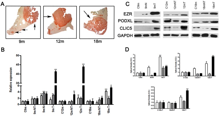
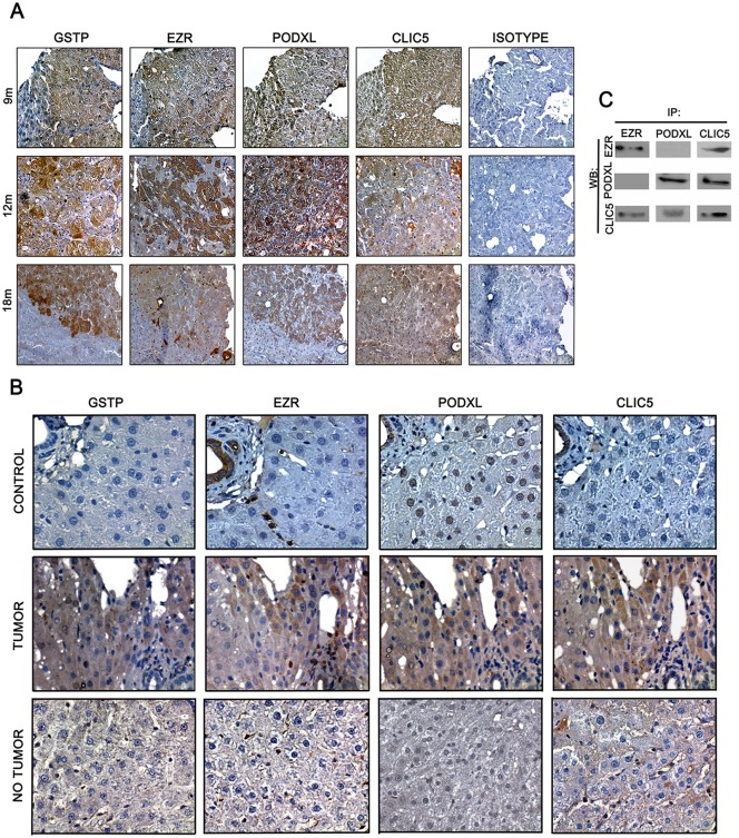
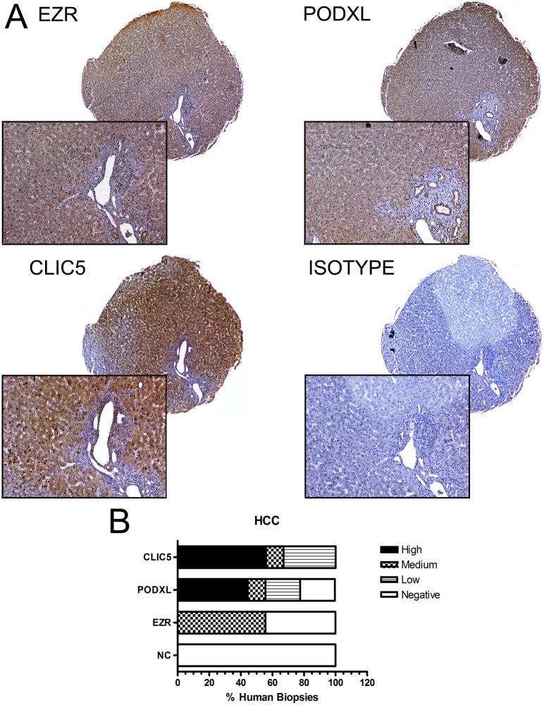
Background and aim: Prognostic markers are important for predicting the progression and staging of hepatocellular carcinoma (HCC). Ezrin (EZR) and Podocalyxin (PODXL) are proteins associated with invasion, migration and poor prognosis in various types of cancer. Recently, it has been observed that chloride intracellular channel 5 (CLIC5) forms a complex with EZR and PODXL and that it is required for podocyte structure and function. In this study, we evaluated the overexpression of EZR, PODXL and CLIC5 in HCC.

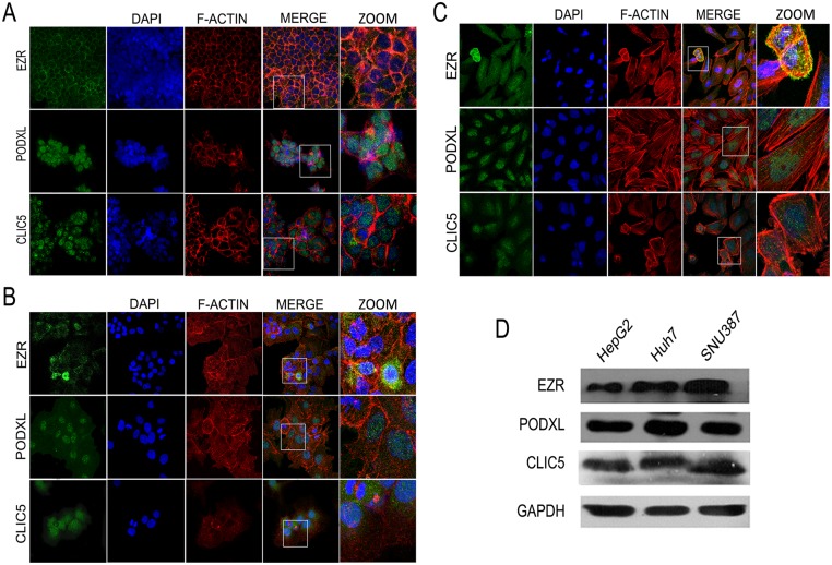
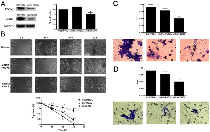
Methods: The modified resistant hepatocyte model (MRHR), human biopsies and HCC cell lines (HepG2, Huh7 and SNU387) were used in this study. Gene and protein expression levels were evaluated in the MRHR by qRT-PCR, Western blot and immunohistochemistry analyses, and protein expression in the human biopsies was evaluated by immunohistochemistry. Protein expression in the HCC cell lines was evaluated by immunofluorescence and Western blot, also the migration and invasive abilities of Huh7 cells were evaluated using shRNA-mediated inhibition.

Results: Our results indicated that these genes and proteins were overexpressed in HCC. Moreover, when the expression of CLIC5 and PODXL was inhibited in Huh7 cells, we observed decreased migration and invasion.

Conclusion: This study suggested that EZR, CLIC5 and PODXL could be biological markers to predict the prognosis of HCC and that these proteins participate in migration and invasion processes.


Conflict of interest statement

Competing Interests: The authors have declared that no competing interests exist.

Figures

Fig 1
Fig 1. Expression of EZR, PODXL and CLIC5 in rat livers after carcinogenic treatment.
Male rats used in the MRHM were sacrificed at 9 (9 m), 12 (12 m) and 18 (18 m) months. A) Tumor and preneoplastic lesions were characterized by GGT activity (spotted and solid arrows, respectively). B) Analysis of relative expression of genes by qRT-PCR in rat livers. C) Protein expression in fractions isolated from rat livers was assessed by Western blot analysis, representative image. D) Graphic representation of triplicates of western blot. GADPH was included as a loading control. C9 m, C12 m and C18 m represent controls of each time point; 9 mT, 12 mT and 18 mT represent tumor regions; 9 mN represents nodular areas (preneoplastic lesions); and 9 mNT, 12 mNT and 18 mNT represent non-tumor areas of the liver (n = 4 rats per group). □EZR, PODXL and ■CLIC5. Error bars indicate the standard error of the mean (SEM). *p≤0.05, **p≤0.03 and ***p≤0.002 vs. their respectively untreated control values (ANOVA and Tukey–Kramer test).
Fig 2
Fig 2. Co-localization of EZR, CLIC5 and PODXL in tumors of rat livers after carcinogenic treatment.
Male rats used in the MRHM were sacrificed at 9 (9 m), 12 (12 m) and 18 (18 m) months. A) EZR, CLIC5 and PODXL expression was analyzed by immunohistochemistry in 3 μm liver serial sections. GSTP detection was used to identify tumor areas (40x magnification). B) Differential expression of EZR, PODXL and CLIC5 in tumor and non-tumor areas of rat livers after carcinogenic treatment. Protein expression was also analyzed in non-treated rats (control) (60x magnification). C) Interaction among CLIC5, EZR and PODXL at 18 m as analyzed by co-immunoprecipitation.
Fig 3
Fig 3. Differential expression of EZR, CLIC5 and PODXL in human biopsies.
A) EZR, CLIC5 and PODXL proteins were detected by immunohistochemistry in 3 μm sections from hepatocellular carcinoma (HCC) tissue and negative control (NC) samples (4x and 20x magnification, respectively). B) The graph represents composite information to facilitate the interpretation of all the samples that were positive for each protein. Each biopsy was classified as high intensity, medium intensity, low intensity or negative signal. Representative images of EZR, PODXL and CLIC5 are shown.
Fig 4
Fig 4. Expression and localization of EZR, PODXL and CLIC5 in HCC cell lines.
Localization of EZR, PODXL and CLIC5 in A) HepG2, B) Huh7 and C) SNU387 cell lines. Positive signals for each protein are shown in green. F-actin is shown in red, and nuclei are shown in blue. Magnified images are shown (ZOOM). D) Total protein expression of EZR, PODXL and CLIC5 in the three cell lines was evaluated by Western blot analysis. Each experiment was repeated at least four times.
Fig 5
Fig 5. Downregulation of PODXL and CLIC5 affects migration and invasion in Huh7 cell lines.
A) (Left) Expression of PODXL and CLIC5 was examined by Western blot analysis in transduced Huh7 cells. GADPH was included as a loading control. (Right) Proliferation of control and transfected Huh7 cells was analyzed using an MTT assay. B) (Up) Representative images of the wound assay in Huh7, shRNAPODXL and shRNACLIC5 cells at 0, 24, 48 and 72 h (20x magnification) (Down) Quantification of gap closure. The graph represents the wound width as the mean in % of the closure of original wound at 0, 24, 48 and 72h. Similar results were obtained in quadruplicate plates. C) The migration capacities of Huh7, shRNAPODXL and shRNACLIC5 cells were measured using the cell migration assay. The migrated cells were fixed, stained with crystal violet, and measured using colorimetry. D) The invasion capacities of Huh7, shRNAPODXL and shRNACLIC5 cells were measured using the ECMatrix Cell Invasion assay. The invasive cells were fixed, stained with crystal violet, and measured using colorimetry. Error bars indicate the standard error of the mean (SEM). *p≤0.05 vs. untreated control values (Tukey–Kramer test). Each experiment was repeated at least four times.

References

    1. Stevenson RP, Veltman D., Machesky L. M. Actin-bundling proteins in cancer progression at a glance. Journal of cell science. 2012:7. - PubMed
    1. Curto M, McClatchey AI. Ezrin...a metastatic detERMinant? Cancer cell. 2004;5(2):113–4. Epub 2004/03/05. . - PubMed
    1. Nielsen JS, McNagny KM. The role of podocalyxin in health and disease. Journal of the American Society of Nephrology: JASN. 2009;20(8):1669–76. Epub 2009/07/07. 10.1681/ASN.2008070782 . - DOI - PubMed
    1. Arpin M, Chirivino D, Naba A, Zwaenepoel I. Emerging role for ERM proteins in cell adhesion and migration. Cell adhesion & migration. 2011;5(2):199–206. Epub 2011/02/24. - PMC - PubMed
    1. Kelly M. McNagny MRH, Graves Marcia L., DeBruin Erin J., Snyder Kimberly, Cipollone Jane, Turvey Michelle, Tan Poh C., McColl Shaun and Calvin D. Roskelley Podocalyxin in the Diagnosis and Treatment of Cancer. Advances in Cancer Management 2012;(SBN: 978-953-307-870-0):40.

Publication types

MeSH terms