

Promotion of mitochondrial energy metabolism during hepatocyte apoptosis in a rat model of acute liver failure
- PMID: 26135512
- PMCID: PMC4581801
- DOI: 10.3892/mmr.2015.4029
Promotion of mitochondrial energy metabolism during hepatocyte apoptosis in a rat model of acute liver failure
Abstract
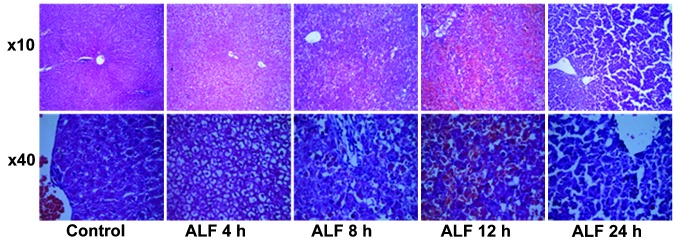
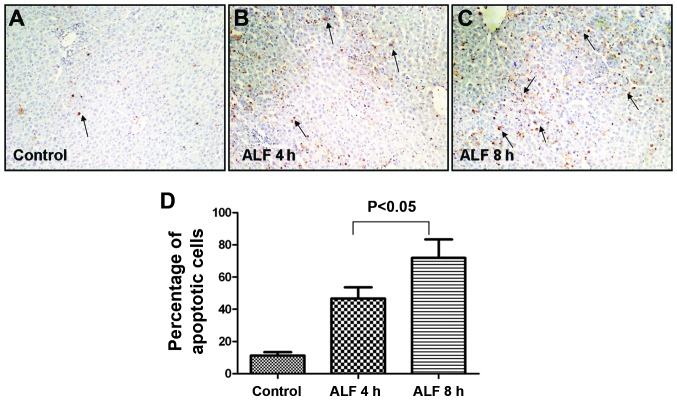
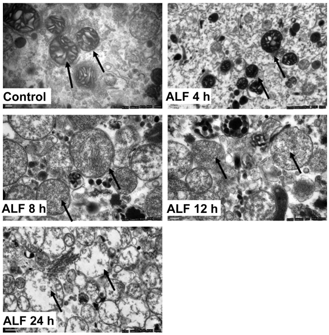
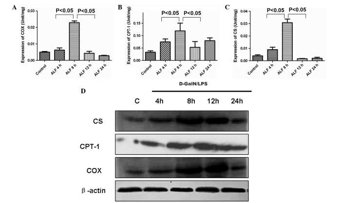
Hepatocyte apoptosis and energy metabolism in mitochondria have an important role in the mechanism of acute liver failure (ALF). However, data on the association between apoptosis and the energy metabolism of hepatocytes are lacking. The current study assessed the activity of several key enzymes in mitochondria during ALF, including citrate synthase (CS), carnitine palmitoyltransferase‑1 (CPT‑1) and cytochrome c oxidase (COX), which are involved in hepatocyte energy metabolism. A total of 40 male Sprague‑Dawley rats were divided into five groups and administered D‑galactosamine and lipopolysaccharide to induce ALF. Hepatic pathology and terminal deoxynucleotidyl transferase‑mediated dUTP nick end labeling examinations indicated that hepatocyte apoptosis was observed at 4 h and increased 8 h after ALF. Hepatocyte necrosis appeared at 12 h and was significantly higher at 24 h with inflammatory cell invasion. The results measured by electron microscopy indicated that ultrastructural changes in mitochondria began at 4 h and the mitochondrial outer membrane was completely disrupted at 24 h resulting in mitochondrial collapse. The expression of CS, CPT‑1 and COX was measured and analyzed using assay kits. The activity and protein expression of CS, CPT‑1 and COX began to increase at 4 h, reached a peak at 8 h and decreased at 12 h during ALF. The activities of CS, CPT‑1 and COX were enhanced during hepatocyte apoptosis suggesting that these enzymes are involved in the initiation and development of ALF. Therefore, these results demonstrated that energy metabolism is important in hepatocyte apoptosis during ALF and hepatocyte apoptosis is an active and energy‑consuming procedure. The current study on how hepatocyte energy metabolism affects the transmission of death signals may provide a basis for the early diagnosis and development of an improved therapeutic strategy for ALF.
Figures
Similar articles
-
[Mitochondrial activities of citrate synthase, carnitine palmitoyltransferase-1 and cytochrome C oxidase are increased during the apoptotic process in hepatocytes of a rat model of acute liver failure].Zhonghua Gan Zang Bing Za Zhi. 2014 Jun;22(6):456-61. doi: 10.3760/cma.j.issn.1007-3418.2014.06.012. Zhonghua Gan Zang Bing Za Zhi. 2014. PMID: 25203711 Chinese.
-
Inhibitory effect of oxymatrine on hepatocyte apoptosis via TLR4/PI3K/Akt/GSK-3β signaling pathway.World J Gastroenterol. 2017 Jun 7;23(21):3839-3849. doi: 10.3748/wjg.v23.i21.3839. World J Gastroenterol. 2017. PMID: 28638224 Free PMC article.
-
Endoplasmic reticulum stress-activated glycogen synthase kinase 3β aggravates liver inflammation and hepatotoxicity in mice with acute liver failure.Inflammation. 2015;38(3):1151-65. doi: 10.1007/s10753-014-0080-2. Inflammation. 2015. PMID: 25630719
-
Acute liver failure: mechanisms of hepatocyte injury and regeneration.Semin Liver Dis. 2008 May;28(2):167-74. doi: 10.1055/s-2008-1073116. Semin Liver Dis. 2008. PMID: 18452116 Review.
-
AMPK-associated signaling to bridge the gap between fuel metabolism and hepatocyte viability.World J Gastroenterol. 2010 Aug 14;16(30):3731-42. doi: 10.3748/wjg.v16.i30.3731. World J Gastroenterol. 2010. PMID: 20698033 Free PMC article. Review.
Cited by
-
Comparative analysis of gene expression profiles of OPN signaling pathway in four kinds of liver diseases.J Genet. 2016 Sep;95(3):741-50. doi: 10.1007/s12041-016-0673-7. J Genet. 2016. PMID: 27659347 Review.
-
BCR, not TCR, repertoire diversity is associated with favorable COVID-19 prognosis.Front Immunol. 2024 Oct 28;15:1405013. doi: 10.3389/fimmu.2024.1405013. eCollection 2024. Front Immunol. 2024. PMID: 39530088 Free PMC article.
-
Modulations of Histone Deacetylase 2 Offer a Protective Effect through the Mitochondrial Apoptosis Pathway in Acute Liver Failure.Oxid Med Cell Longev. 2019 Apr 28;2019:8173016. doi: 10.1155/2019/8173016. eCollection 2019. Oxid Med Cell Longev. 2019. PMID: 31183000 Free PMC article.
-
CDK9 inhibition blocks the initiation of PINK1-PRKN-mediated mitophagy by regulating the SIRT1-FOXO3-BNIP3 axis and enhances the therapeutic effects involving mitochondrial dysfunction in hepatocellular carcinoma.Autophagy. 2022 Aug;18(8):1879-1897. doi: 10.1080/15548627.2021.2007027. Epub 2021 Dec 10. Autophagy. 2022. PMID: 34890308 Free PMC article.
-
Antioxidant and Anti-Inflammatory Activity of Cynanchum acutum L. Isolated Flavonoids Using Experimentally Induced Type 2 Diabetes Mellitus: Biological and In Silico Investigation for NF-κB Pathway/miR-146a Expression Modulation.Antioxidants (Basel). 2021 Oct 27;10(11):1713. doi: 10.3390/antiox10111713. Antioxidants (Basel). 2021. PMID: 34829584 Free PMC article.
References
-
- Liver Failure and Artificial Liver Group. Chinese Society of Infectious Diseases. Chinese Medical Association. Severe Liver Diseases and Artificial Liver Group Chinese Society of Hepatology, Chinese Medical Association: Diagnostic and treatment guidelines for liver failure. Zhonghua Gan Zang Bing Za Zhi. 2006;14:643–646. In Chinese. - PubMed
Publication types
MeSH terms
Substances
LinkOut - more resources
Full Text Sources
Other Literature Sources
