

Frequent FOS Gene Rearrangements in Epithelioid Hemangioma: A Molecular Study of 58 Cases With Morphologic Reappraisal
- PMID: 26135557
- PMCID: PMC4567921
- DOI: 10.1097/PAS.0000000000000469
Frequent FOS Gene Rearrangements in Epithelioid Hemangioma: A Molecular Study of 58 Cases With Morphologic Reappraisal
Abstract
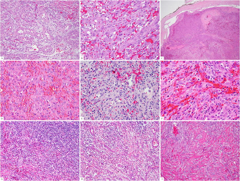
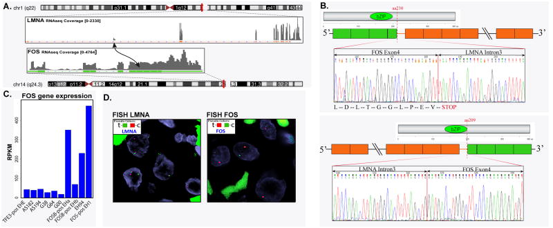
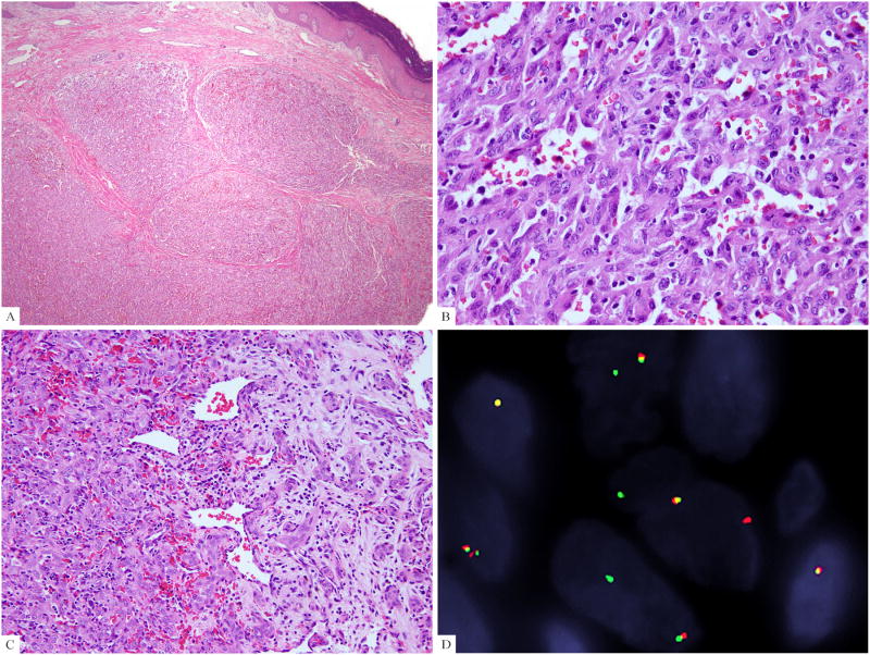
Epithelioid hemangioma (EH) is a unique benign vasoformative tumor composed of epithelioid endothelial cells. Although a small subset of EHs with atypical features harbor ZFP36-FOSB fusions, no additional genetic abnormalities have been found to date in the remaining cases. On the basis of a novel FOS-LMNA gene fusion identified by RNA sequencing in an index case of a skeletal EH with typical morphology, we sought to investigate the prevalence of FOS rearrangement in a large cohort of EHs. Thus 57 additional EH cases lacking FOSB rearrangements were studied for FOS gene abnormalities by fluorescence in situ hybridization, and results were correlated with morphologic appearance and clinical presentation. The EHs were subclassified as typical (n=25), cellular (n=21), and angiolymphoid hyperplasia with eosinophilia (ALHE) (n=12) variants. The ALHE was defined as an EH with a vascular "blow-out" pattern associated with a variable degree of inflammation. There were 17 (29%) cases bearing FOS gene rearrangements among 58 cases tested, including 12 male and 5 female patients, with a mean age of 42 years. Most FOS-rearranged EHs occurred in the bone (10) and soft tissue (6), whereas only 1 case was cutaneous. The predominant anatomic site was the extremity (12), followed by trunk (3), head and neck (1), and penis (1). The incidence of FOS rearrangement was significantly higher in bone (59%, P=0.006) and lower in head and neck (5%, P=0.009). Twelve of the FOS-rearranged cases were cellular EH (P=0.001) associated with moderate mitotic activity (2 to 5/10 HPF) and milder inflammatory background. All 12 ALHE cases lacked FOS gene abnormalities, suggesting different pathogenesis. In conclusion, FOS rearrangement was present in a third of EHs across different locations and histologic variants; however, it was more prevalent in cellular EH and intraosseous lesions, compared with those in skin, soft tissue, and head and neck. This genetic abnormality can be useful in challenging cases, to distinguish cellular EHs from malignant epithelioid vascular tumors. These results also suggest that dysregulation of the FOS family of transcription factors through chromosomal translocation is as a key event in the tumorigenesis of EH except for the ALHE variant.
Conflict of interest statement
Conflict of interest: none
Figures
References
-
- Fetsch JF, Sesterhenn IA, Miettinen M, et al. Epithelioid hemangioma of the penis: a clinicopathologic and immunohistochemical analysis of 19 cases, with special reference to exuberant examples often confused with epithelioid hemangioendothelioma and epithelioid angiosarcoma. Am J Surg Pathol. 2004;28:523–533. - PubMed
-
- Nielsen GP, Srivastava A, Kattapuram S, et al. Epithelioid hemangioma of bone revisited: a study of 50 cases. Am J Surg Pathol. 2009;33:270–277. - PubMed
-
- Rosai J, Akerman LR. Intravenous atypical vascular proliferation. A cutaneous lesion simulating a malignant blood vessel tumor. Arch Dermatol. 1974;109:714–717. - PubMed
-
- Castro C, Winkelmann RK. Angiolymphoid hyperplasia with eosinophilia in the skin. Cancer. 1974;34:1696–1705. - PubMed
Publication types
MeSH terms
Substances
Grants and funding
LinkOut - more resources
Full Text Sources
Medical
Miscellaneous
