. 2015 Jun;6(2):181-91.
doi: 10.1002/jcsm.12019. Epub 2015 Apr 30.

Gender-specific differences in the development of sarcopenia in the rodent model of the ageing high-fat rat

Affiliations

Gender-specific differences in the development of sarcopenia in the rodent model of the ageing high-fat rat

Robert Kob et al. J Cachexia Sarcopenia Muscle. 2015 Jun.

Abstract

Background: Sarcopenia is linked to functional impairments, loss of independence, and mortality. In the past few years, obesity has been established as being a risk factor for a decline in muscle mass and function. There are several molecular pathological mechanisms, which have been under discussion that might explain this relationship. However, most studies were conducted using male animals and for a short period of time.

Methods: In this study, the gender-specific effect of long-term, high-fat content feeding in Sprague-Dawley rats was examined. Development of the quadriceps muscle of the animals was monitored in vivo using magnetic resonance. The results of these measurements and of the biochemical analysis of the aged muscle were compared.

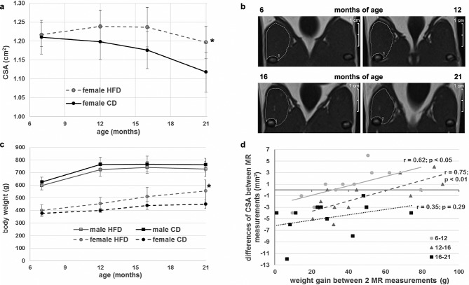
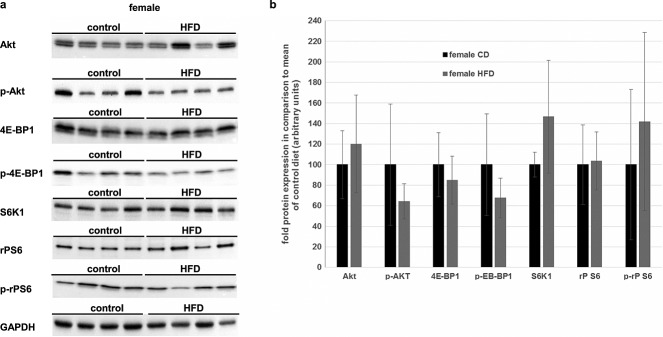
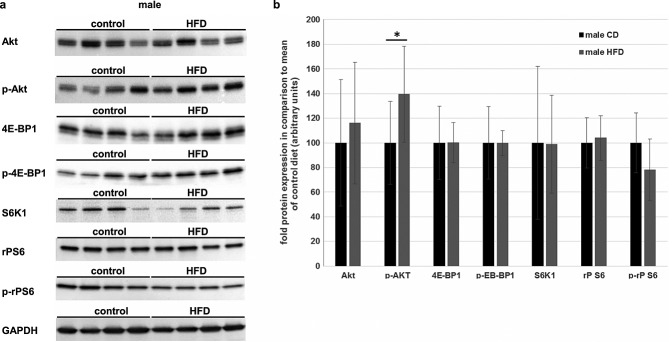
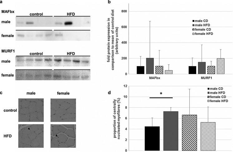
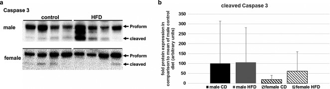
Results: Surprisingly, only male but not female rats showed a decline in muscle cross-sectional area at 16 months of age as a result of a chronic oversupply of dietary fats. This loss of muscle mass could not be explained by either de-regulation of the anabolic Akt pathway or by up-regulation of the main ubiquitin ligases of muscle, MAFbx and MuRF-1. However, fusion of satellite cells to myotubes was induced by the high-fat diet in male rats, possibly as a result of an increased need for compensatory regeneration processes. Caspase-3-dependent apoptosis induction, irrespective of diet, seems to be the major determinant of muscle decline during ageing in male but not female rats.

Conclusion: Taken together, activation of the apoptosis-inducing Caspase-3 seems to be the most important trigger for the age-related muscle loss. Male rats were more prone to the decline of muscle during ageing than female animals, which was further enforced by a long-term, high fat diet.

Keywords: Ageing skeletal muscle; Gender‐specificity; Lipotoxicity; Metabolism; Sarcopenic obesity.


Figures

Figure 1
Figure 1
Weight and cross-sectional area (CSA) development of the rats. (A) Mean CSA of the female rats during the study. The first measurement at 7 months of age was performed before the rats were divided into the control (black circles) and high-fat diet (HFD) (grey open circles) groups. At 21 months of age, the female HFD (fHFD) group showed a significantly higher CSA than the female control diet (fCD) group. (B) Representative images of the quadriceps muscle of one fHFD rat at 7, 12, 16, and 21 months of age. (C) Development of body weight of male CD (black squares and solid line), male HFD (grey, open squares, and solid lines), fCD (black circles and dashed line) and fHFD (grey open circles and dashed line). While the body mass of the male animals reached a plateau at 12 months, the weight of the female rats increased throughout the whole lifetime. Only the fHFD group at 21 months of age was significantly heavier than the age-matched fCD group. (D) Correlation between total body weight gain and CSA changes between two magnetic resonance (MR) measurements of all female animals. Between 7–12 and 12–16 months of age, CSA was significantly correlated with increased body mass. This relationship was abolished between 16 and 21 months of age. Only animals that survived the whole study period were analysed. All data are presented as mean ± SD. * Significant difference between dietary groups within gender.
Figure 2
Figure 2
Effects of a long-term high-fat diet (HFD) on regulators of muscular protein biosynthesis in female rats. (A) Protein-lysates of the M. vastus lateralis dexter of female rats were used for immunoblot analysis of total cellular protein level of Akt, 4E-BP1, S6K1, rP S6, and GAPDH (glyceraldehyde 3-phosphate dehydrogenase). Additionally, the phosphorylated forms (Ser473P-Akt, Ser65P-4E-BP1, and Ser240/244P-rpS6) were measured. For comparison, GAPDH, as a house-keeper, is added. Representative blots are shown. (B) Densitometric analysis of the immunoblots of all female animals that survived the whole study period (female CD: n = 5, black bars; female HFD:  = 6, grey bars). There were no significant changes detectable between the two dietary groups. Intensity of the bands was normalized to total protein content on the western blot membrane using the Stain-Free™ gel system (BioRad). All data are presented as mean ± SD.
Figure 3
Figure 3
Effects of a long-term high-fat diet (HFD) on regulators of muscular protein biosynthesis in male rats. (A) M. vastus lateralis dexter of male rats was lysed and analysed by immunoblotting. Representative blots of total cellular protein level of Akt, 4E-BP1, S6K1, rP S6, and GAPDH and additionally of the phosphorylated forms (Ser473P-Akt, Ser65P-4E-BP1, and Ser240/244P-rpS6) are shown. (B) Densitometric analysis of the immunoblots of all male animals that survived the whole study period (male control diet (CD): n = 12, black bars; male HFD: n = 7, grey bars). The total protein contents of all analysed regulators did not differ between the CD and the HFD group. Ser473P-Akt was significantly elevated in the HFD group. However, Ser65P-4E-BP1 and Ser240/244P-rpS6, which are the downstream effectors of P-Akt, revealed no difference between the dietary groups Total protein content on the western blot membrane, measured by the Stain-Free™ gel system (BioRad), was used for normalization. All data are presented as mean ± SD. * Significant difference between dietary groups.
Figure 4
Figure 4
Influence of chronically high-fat diet (HFD) on proteasomal protein decay and muscular regeneration. (A) Expression of the two ubiquitin ligases MAFbx and MuRF-1 in the M. vastus lateralis was visualized with immunoblotting. Most of the animals showed no or only little expression of MAFbx, albeit some rats showed a strong induction of this ligase. MuRF-1 protein levels differed only slightly in females, while male rats showed greater inter-individual variance. (B) Mean expressions of MAFbx and MURF-1 were determined by densitometric analysis. The control group of each gender was arbitrary set as 100. There were no significant differences detectable between the high-fat and the control diet group. Mean normalized protein level + SD is shown in the diagram. (C) Paraffin-embedded sections of the quadriceps muscle were haematoxylin-eosin stained. About 400 fibres of each animal were evaluated and those with at least one nucleus, which was not located at the sarcolemma, were counted as centrally nucleated. Exemplary pictures of each gender and dietary group are shown, and small arrows mark centrally located nuclei. (D) Statistical analysis of histology of all animals (male CD: n = 4; male HFD: n = 8; female CD: n = 4; female HFD: n = 5). Mean percent of centrally nucleated fibres per total number of myofibres + SD is depicted. While male rats receiving an HFD had a significantly greater number of regenerated fibres, there were no differences between the dietary groups amongst the female rats. * Significant difference between dietary groups.
Figure 5
Figure 5
Apoptose induction in skeletal muscle of aged rats. (A) Total protein level of Pro-Caspase 3 and cleaved Caspase 3 was measured using immunoblotting. Female rats had no or very low amounts of activated Caspase 3. In contrast, nearly all male rats exhibited a certain level of cleaved Caspase 3, and some animals showed strong apoptosis induction. (B) Summary of the densitometric analysis of the cleaved Caspase 3. Male rats showed considerable apoptose induction, whereas females had low levels of cleaved Caspase 3. Differences between dietary groups were not significant for both genders. Data are presented as mean + SD.

References

    1. Cruz-Jentoft AJ, Baeyens JP, Bauer JM, Boirie Y, Cederholm T, Landi F, et al. Sarcopenia: European consensus on definition and diagnosis. Age Ageing. 2010;39:412–23. - PMC - PubMed
    1. Biolo G, Cederholm T, Muscaritoli M. Muscle contractile and metabolic dysfunction is a common feature of sarcopenia of aging and chronic diseases: from sarcopenic obesity to cachexia. Clin Nutr. 2014;33:737–48. - PubMed
    1. Baumgartner RN. Body composition in healthy aging. Ann N Y Acad Sci. 2000;904:437–48. - PubMed
    1. Bouchard DR, Dionne IJ, Brochu M. Sarcopenic/obesity and physical capacity in older men and women: data from the Nutrition as a Determinant of Successful Aging (NuAge)—the Quebec longitudinal study. Obesity. 2009;17:2082–8. - PubMed
    1. Volkert D. The role of nutrition in the prevention of sarcopenia. Wien Med Wochenschr. 2011;161:409–15. - PubMed