. 2015 Jul 2:13:209.
doi: 10.1186/s12967-015-0578-x.

Febuxostat pretreatment attenuates myocardial ischemia/reperfusion injury via mitochondrial apoptosis

Affiliations

Febuxostat pretreatment attenuates myocardial ischemia/reperfusion injury via mitochondrial apoptosis

Shulin Wang et al. J Transl Med. .

Abstract

Background: Febuxostat is a selective inhibitor of xanthine oxidase (XO). XO is a critical source of reactive oxygen species (ROS) during myocardial ischemia/reperfusion (I/R) injury. Inhibition of XO is therapeutically effective in I/R injury. Evidence suggests that febuxostat exerts antioxidant effects by directly scavenging ROS. The present study was performed to investigate the effects of febuxostat on myocardial I/R injury and its underlying mechanisms.

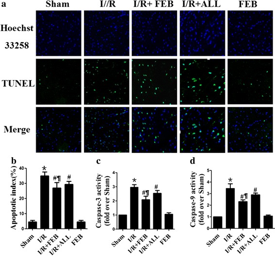
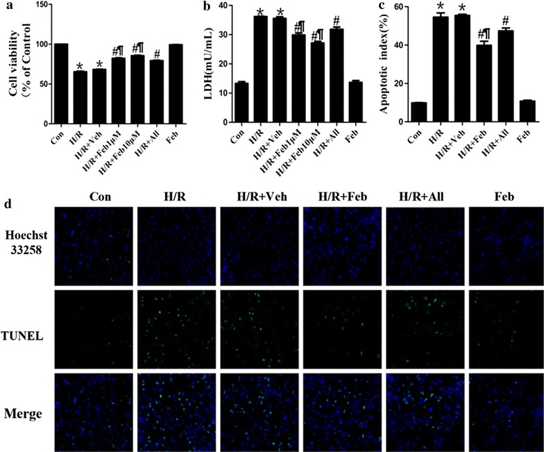
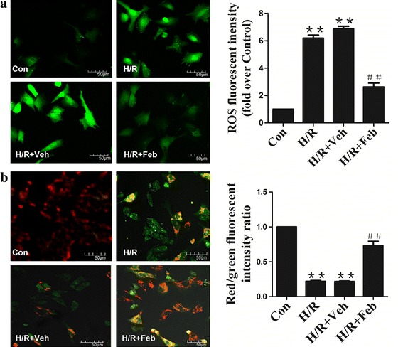
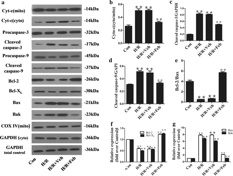
Methods: We utilized an in vivo mouse model of myocardial I/R injury and an in vitro neonatal rat cardiomyocyte (NRC) model of hypoxia/reoxygenation (H/R) injury. Mice were randomized into five groups: Sham, I/R (I/R + Vehicle), I/R + FEB (I/R + febuxostat), AL + I/R (I/R + allopurinol) and FEB (febuxostat), respectively. The I/R + FEB mice were pretreated with febuxostat (5 mg/kg; i.p.) 24 and 1 h prior to I/R. NRCs received febuxostat (1 and 10 µM) at 24 and 1 h before exposure to hypoxia for 3 h followed by reoxygenation for 3 h. Cardiac function, myocardial infarct size, serum levels of creatine kinase (CK) and lactate dehydrogenase (LDH), and myocardial apoptotic index (AI) were measured in order to ascertain the effects of febuxostat on myocardial I/R injury. Hypoxia/reperfusion (H/R) injury in NRCs was examined using MTT, LDH leakage assay and terminal deoxynucleotidyl transferase dUTP nick end labeling (TUNEL) assay. The underlying mechanisms were determined by measuring ROS production, mitochondrial membrane potential (ΔΨm), and expression of cytochrome c, cleaved caspases as well as Bcl-2 protein levels.

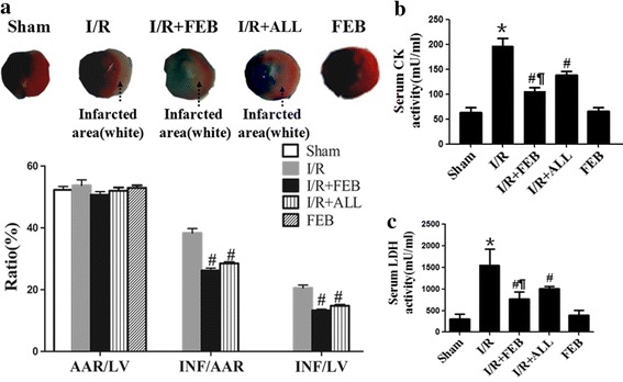
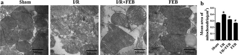
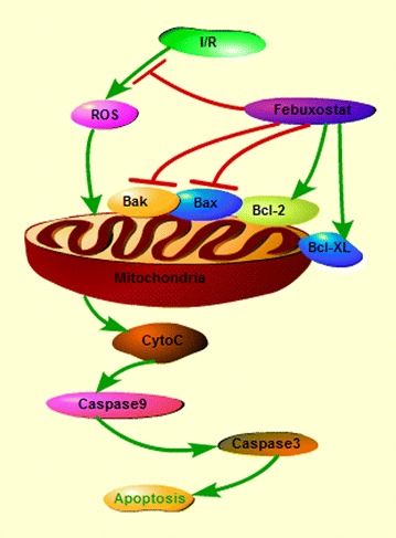
Results: Myocardial I/R led to an elevation in the myocardial infarct size, serum levels of CK and LDH, cell death and AI. Furthermore, I/R reduced cardiac function. These changes were significantly attenuated by pretreatment with febuxostat and allopurinol, especially by febuxostat. Febuxostat also protected the mitochondrial structure following myocardial I/R, inhibited H/R-induced ROS generation, stabilized the ΔΨm, alleviated cytosolic translocation of mitochondrial cytochrome C, inhibited activation of caspase-3 and -9, upregulated antiapoptotic proteins and downregulated proapoptotic proteins.

Conclusions: This study revealed that febuxostat pretreatment mediates the cardioprotective effects against I/R and H/R injury by inhibiting mitochondrial-dependent apoptosis.


Figures

Figure 1
Figure 1
Effects of febuxostat on infarct size and necrosis in mice. a Representative images of myocardial infarct size and the ratio of AAR/LV, INF/AAR, and INF/LV. b Serum CK levels. c Serum LDH levels. The results were expressed as mean ± SD. (n = 7). *P < 0.01 vs. Sham, # P < 0.01, vs. I/R, P < 0.01 vs. I/R + ALL.
Figure 2
Figure 2
Effects of febuxostat on apoptosis in mice. a Representative photomicrographs (×200) of TUNEL staining in myocardial I/R tissue. Green fluorescence indicates TUNEL-positive apoptotic nuclei; blue fluorescence indicates total cardiomyocyte nuclei. b Apoptotic index. c Myocardial caspase-3 activity. d Myocardial caspase-9 activity. The results were expressed as mean ± SD (n = 7). The results were expressed as mean ± SD (n = 7). *P < 0.01 vs. Sham, # P < 0.01, vs. I/R, P < 0.01 vs. I/R + ALL.
Figure 3
Figure 3
Effects of febuxostat on H/R injury in NRCs. a MTT assay following H/R. b Leakage of LDH in culture medium. c Apoptotic index in NRCs. d Typical morphological changes in apoptosis under fluorescence microscope (×200). All values indicate mean ± SD. *P < 0.01 vs. Sham, # P < 0.01, vs. I/R, P < 0.01 vs. H/R + All. These experiments were performed in quintuplicate with similar results.
Figure 4
Figure 4
Effect of febuxostat on mitochondrial structure in mice. a Representative electron micrographs (×50,000) of mitochondrial structures. Sham the structure of mitochondria was generally very good. I/R I/R-induced mitochondrial structural disruption (swelling, rupture and loss of cristae). I/R + FEB febuxostat treatment ameliorated I/R-induced mitochondrial impairment. FEB the structure of mitochondria was basically intact. b Mean area of mitochondria. All values represent mean ± SD. *P < 0.01 vs. Sham, # P < 0.01 vs. I/R, n = 4/group, these experiments were performed with similar results.
Figure 5
Figure 5
Effect of febuxostat treatment on ROS production and mitochondrial membrane potential (ΔΨm). a ROS staining was performed using DCFH-DA. Left: representative images of ROS staining; right: ROS fluorescence intensity. b Changes in mitochondrial membrane potential (ΔΨm) are indicated by JC-1 staining; left: Red fluorescence represents the mitochondrial aggregate form of JC-1, indicating intact mitochondrial membrane potential. Green fluorescence represents the monomeric form of JC-1, indicating dissipation of ΔΨm. Right: ratio of red/green fluorescence intensity. Both ROS and ΔΨm staining were observed with a laser confocal microscope (×400) (Bar 50 µm). All values indicate mean ± SD. **P < 0.01 vs. Con, ## P < 0.01 vs. H/R.
Figure 6
Figure 6
Effect of febuxostat treatment on the expression of proteins regulating mitochondria-mediated apoptosis. a Representative Western blots from each group. b The levels of Cyto-C (cyto/mito).c The levels of cleaved caspase-3. d The levels of cleaved caspase-9. e The ratio of Bcl-2/Bax. f The levels of Bcl-2 and Bcl-XL. g The levels of Bax and Bak. Levels of proteins were quantified using densitometry and normalized against COX IV or GADPH. Data are presented as mean ± SD. **P < 0.01 vs. Con, ## P < 0.01 vs. H/R. These experiments were performed in quintuplicate with similar results.
Figure 7
Figure 7
Schematic depicting protective signaling of febuxostat in I/R-induced apoptosis. Febuxostat inhibits the mitochondrial apoptotic pathway by reducing ROS production and modulating Bcl-2 family proteins.

Similar articles

Cited by

References

    1. Kalogeris T, Baines CP, Krenz M, Korthuis RJ. Cell biology of ischemia/reperfusion injury. Int Rev Cell Mol Biol. 2012;298:229–317. doi: 10.1016/B978-0-12-394309-5.00006-7. - DOI - PMC - PubMed
    1. Gottlieb RA. Cell death pathways in acute ischemia/reperfusion injury. J Cardiovasc Pharmacol Ther. 2011;16:233–238. doi: 10.1177/1074248411409581. - DOI - PMC - PubMed
    1. Windecker S, Bax JJ, Myat A, Stone GW, Marber MS. Future treatment strategies in ST-segment elevation myocardial infarction. Lancet. 2013;382:644–657. doi: 10.1016/S0140-6736(13)61452-X. - DOI - PubMed
    1. Sinha K, Das J, Pal PB, Sil PC. Oxidative stress: the mitochondria-dependent and mitochondria-independent pathways of apoptosis. Arch Toxicol. 2013;87:1157–1180. doi: 10.1007/s00204-013-1034-4. - DOI - PubMed
    1. Li C, Jackson RM. Reactive species mechanisms of cellular hypoxia-reoxygenation injury. AJP Cell Physiol. 2002;282:C227–C241. doi: 10.1152/ajpcell.00112.2001. - DOI - PubMed

Publication types

MeSH terms