. 2015 Jul 10;6(19):17178-91.
doi: 10.18632/oncotarget.3935.

Radiosensitization and downregulation of heterogeneous nuclear ribonucleoprotein K (hnRNP K) upon inhibition of mitogen/extracellular signal-regulated kinase (MEK) in malignant melanoma cells

Affiliations

Radiosensitization and downregulation of heterogeneous nuclear ribonucleoprotein K (hnRNP K) upon inhibition of mitogen/extracellular signal-regulated kinase (MEK) in malignant melanoma cells

Stefan Eder et al. Oncotarget. .

Abstract

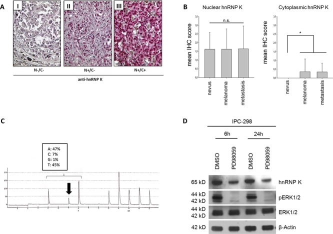
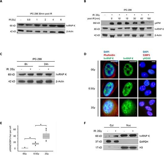
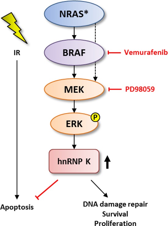
Background: Heterogeneous nuclear ribonucleoprotein K (hnRNP K) is an important cofactor in the p53-mediated DNA damage response pathway upon ionizing radiation (IR) and exerts anti-apoptotic effects also independent of p53 pathway activation. Furthermore, hnRNP K is overexpressed in various neoplasms including malignant melanoma (MM). Here, we investigate the role of hnRNP K in the radioresistance of MM cells.

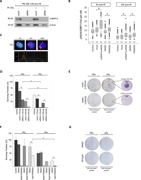
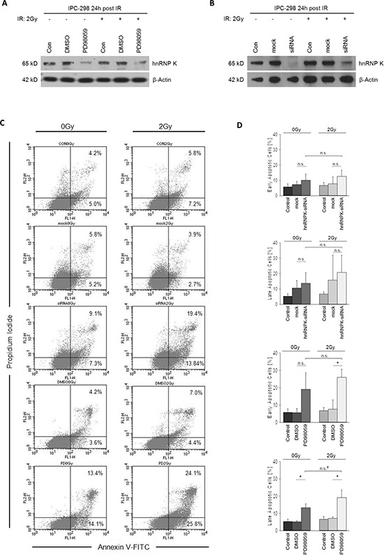
Methods and results: Our results show cytoplasmic expression of hnRNP K in human MM surgical specimens, but not in benign nevi, and a quick dose- and time-dependent upregulation in response to IR accompanied by cytoplasmic redistribution of the protein in the IPC-298 cellular tumor model carrying an activating NRAS mutation (p.Q61L). SiRNA-based knockdown of hnRNP K induced a delayed decline in γH2AX/53BP1-positive DNA repair foci upon IR. Pharmacological interference with MAPK signaling abrogated ERK phosphorylation, diminished cellular hnRNP K levels, impaired γH2AX/53BP1-foci repair and proliferative capability and increased apoptosis comparable to the observed hnRNP K knockdown phenotype in IPC-298 cells.

Conclusions: Our results indicate that pharmacological interference with MAPK signaling increases vulnerability of NRAS-mutant malignant melanoma cells to ionizing radiation along with downregulation of endogenous hnRNP K and point towards a possible use for combined MEK inhibition and localized radiation therapy of MM in the NRAS-mutant setting where BRAF inhibitors offer no clinical benefit.

Keywords: MEK inhibition; NRAS; melanoma; nRNP K; radiotherapy.


Conflict of interest statement

CONFLICTS OF INTEREST

The authors declare that no competing interests exist.

Figures

Figure 1
Figure 1
A. Expression of hnRNP K in malignant melanoma tissue specimens. Criteria for classifying nuclei (N) and cytoplasm (C) as positive or negative for hnRNP K are illustrated by representative immunohistochemistry for hnRNP K. B. IHC scoring was done by separately analyzing hnRNP K staining intensity for nuclear and cytoplasmic hnRNP K expression. Mean nuclear IHC scores of benign nevi, malignant melanoma (MM) or MM metastasis showed no significant difference. 25% of MM and MM metastases stained positively for cytoplasmic hnRNP K whereas nevi were completely devoid of cytoplasmic hnRNP K expression (n: nevi = 18, melanoma = 62, metastasis = 20; *p < 0.05 versus nevi). C. Verification of NRAS mutation in IPC-298 cells by DNA pyrosequencing. The CAA to CTA transition leads to a p.Q61L amino acid substitution, comprising constitutive signaling activity along the MAPK pathway. D. Inhibition of MEK signaling by 50 μM PD98059 abrogates phosphorylation of ERK1/2 and reduces endogenous hnRNP K levels.
Figure 2
Figure 2
A. Immunoblotting of hnRNP K shows dose-dependent upregulation within 30 minutes after IR. B. Immunoblotting of IPC-298 cell lysate showing time-dependent increase in intensity of the hnRNP K-double band with a maximum at 30 – 60 minutes and a subsequent normalization after 180 minutes, parallel to the detection of phospho-ATM. C. 6 hours and 24 hours after applying IR (2 Gy), there is no difference in hnRNP K expression between irradiated and non-irradiated samples. D. Immunofluorescence microscopy of IPC-298 cells shows strict nuclear localization of hnRNP K under control conditions. Within 30 minutes, exposure to IR causes perinuclear accumulation of hnRNP K in a dose-dependent manner. Scale bars represent 30μm. E. DNA damage repair as illustrated by dose-dependent increase in γH2AX/53BP1-Foci (n = 100; p < 0.05). F. Subcellular fractionation and immunoblotting reveals cytoplasmic (Cyt) accumulation of hnRNP K 30 min post-IR (2 Gy). Nuclear (Nuc) hnRNP K levels remain unchanged.
Figure 3
Figure 3
A. Immunoblotting shows effective knockdown of hnRNP K. B. Knockdown of hnRNP K and MEK inhibition result in a significantly reduced decline in γH2AX/53BP1 foci (*p < 0.05). C. Time-dependent DNA-damage response (DDR) shown by γH2AX/53BP1 foci upon 2 Gy IR. Co-localization of γH2AX/53BP1 was verified by staining intensity profile. D. Knockdown of hnRNP K evokes radiosensitization of IPC-298 cells (n = 4). F. MEK inhibition with PD98059 impairs clonogenic viability of IPC-298 melanoma cells in a dose-dependent manner (n = 4). G. Representative colony formation assays show partial depletion of clonogenic cell survival by treatment with 5 μM PD98059 and complete abrogation by additional irradiation with 2 Gy (n = 4).
Figure 4
Figure 4
A and B. MEK inhibition with PD98059 for 24 hours post-IR impairs normalization of cellular hnRNP K levels in irradiated and non-irradiated IPC-298 cells comparable to the hnRNP K knockdown phenotype. C. Effects of MEK inhibition and hnRNP K knockdown on apoptosis were analysed by flow cytometry using Annexin V-FITC/PI staining. Presented data show the results of a contiguous test series. Early apoptosis is represented by Annexin V+/PI events, whereas Annexin V+/PI+ counts demonstrate late apoptosis/necrosis. D. Statistical analyses of flow cytometry results. Experiments were performed in triplicate. Untreated cells served as control. The presented columns are given as the means ± S.D. (*p < 0.05, n.s. = not significant, *p = 0.05).
Figure 5
Figure 5
Schematic model for the role of MAPK-mediated upregulation of hnRNP K in the DDR pathway and possible targets for pharmacological interference.

References

    1. Rao NG, Yu HH, Trotti A, 3rd, Sondak VK. The role of radiation therapy in the management of cutaneous melanoma. Surg Oncol Clin N Am. 2011;20:115–31. - PubMed
    1. Pflugfelder A, Kochs C, Blum AM, Capellaro M, Czeschik C, Dettenborn T, Dill D, Dippel E, Eigentler T, Feyer P, Follmann M, Frerich B, Ganten MK, Gartner J, Gutzmer R, et al. Malignant melanoma S3-guideline “diagnosis, therapy and follow-up of melanoma”. J Dtsch Dermatol Ges. 2013;11:1–116. 1–126. - PubMed
    1. Burmeister BH, Henderson MA, Ainslie J, Fisher R, Di Iulio J, Smithers BM, Hong A, Shannon K, Scolyer RA, Carruthers S, Coventry BJ, Babington S, Duprat J, Hoekstra HJ, Thompson JF. Adjuvant radiotherapy versus observation alone for patients at risk of lymph-node field relapse after therapeutic lymphadenectomy for melanoma: a randomised trial. Lancet Oncol. 2012;13:589–97. - PubMed
    1. Bibault JE, Dewas S, Mirabel X, Mortier L, Penel N, Vanseymortier L, Lartigau E. Adjuvant radiation therapy in metastatic lymph nodes from melanoma. Radiat Oncol. 2011;6:12. - PMC - PubMed
    1. Moumen A, Masterson P, O'Connor MJ, Jackson SP, hnRNP K. an HDM2 target and transcriptional coactivator of p53 in response to DNA damage. Cell. 2005;123:1065–78. - PubMed

Substances